. 2017 Mar 7:7:43954.
doi: 10.1038/srep43954.

Single-molecule studies reveal reciprocating of WRN helicase core along ssDNA during DNA unwinding

Affiliations

Single-molecule studies reveal reciprocating of WRN helicase core along ssDNA during DNA unwinding

Wen-Qiang Wu et al. Sci Rep. .

Abstract

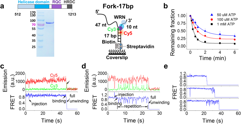
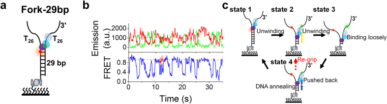
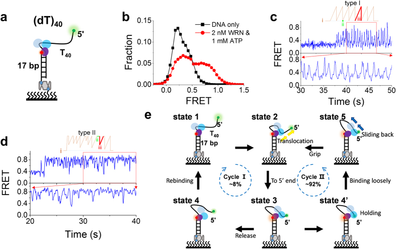
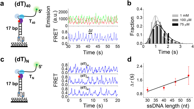
Werner syndrome is caused by mutations in the WRN gene encoding WRN helicase. A knowledge of WRN helicase's DNA unwinding mechanism in vitro is helpful for predicting its behaviors in vivo, and then understanding their biological functions. In the present study, for deeply understanding the DNA unwinding mechanism of WRN, we comprehensively characterized the DNA unwinding properties of chicken WRN helicase core in details, by taking advantages of single-molecule fluorescence resonance energy transfer (smFRET) method. We showed that WRN exhibits repetitive DNA unwinding and translocation behaviors on different DNA structures, including forked, overhanging and G-quadruplex-containing DNAs with an apparently limited unwinding processivity. It was further revealed that the repetitive behaviors were caused by reciprocating of WRN along the same ssDNA, rather than by complete dissociation from and rebinding to substrates or by strand switching. The present study sheds new light on the mechanism for WRN functioning.


Conflict of interest statement

The authors declare no competing financial interests.

Figures

Figure 1
Figure 1. WRN-catalyzed unwinding of Fork-17bp in smFRET assay.
(a) Analysis of the purified WRN helicase by SDS PAGE (left panel) and structure of the Cy3- and Cy5-labelled substrate Fork-17bp (right panel). DNA is immobilized on the streptavidin-coated coverslip surface through biotin. (b) Fractions of DNA molecules remaining on the coverslip surface versus time after addition of 1 nM WRN and different concentrations of ATP (Error bar = s.d.; n = 3). Fittings of the data to a single-exponential decay yielded the corresponding unwinding rates at different ATP concentrations (Supplementary Table S2). (c,d) Two typical time traces of fluorescence intensities of Cy3 and Cy5 (upper panel) and the corresponding FRET traces (lower panel) at 1 nM WRN and 1 mM ATP. (c) One-step full unwinding; (d) Repetitive oscillation before full unwinding. Full unwinding is indicated by an arrow. (e) Time traces showing the time at which full unwinding occurs lies in a wide range.
Figure 2
Figure 2. WRN-catalyzed unwinding of Fork-29bp and the proposed model.
(a) Schematic diagram of substrate Fork-29bp. (b) Typical time traces of fluorescence intensities of Cy3 and Cy5 (upper panel) and the corresponding FRET trace (lower panel). The new interaction phases are indicated by red arrows, corresponding to the ‘re-grip’ process in (c) (i.e., state 4 to 2). (c) Proposed model for WRN-catalyzed unwinding of Fork-29bp. State 1, WRN binding at the fork and starting to unwind the duplex. The two RecA-like domains bind and translocate along the tracking ssDNA while RQC interacts with and melts the DNA duplex at the ss/dsDNA junction. State 2, WRN has unwound some base pairs. State 3, in a certain nucleotide state during DNA unwinding, the two RecA-like domains loosen their strong binding to the tracking ssDNA. State 4, WRN is pushed backwards along the tracking strand by DNA annealing, while RQC remains contacting with the retreating ss/dsDNA junction. WRN can be pushed back until DNA annealing is completed (state 1), or, alternatively, WRN may re-grip the tracking strand and resume unwinding of the partially annealed duplex (state 2).
Figure 3
Figure 3. WRN induces looping of the 5′-ssDNA overhang.
(a) Schematic diagram of substrate (dT)40, which contains a duplex DNA with a 40-nt poly(dT) 5′-overhang, labelled with Cy3 at the 5′-tail end and Cy5 at the ss/dsDNA junction. (b) FRET histograms obtained before and 20 s after adding 2 nM WRN and 1 mM ATP. (c,d) Two typical patterns of FRET changes: (c) periodic sawtooth-shaped bursting, (d) irregular bursting. (e) Our proposed model to explain how WRN induces looping of the 5′-ssDNA overhang (see more details in the text).
Figure 4
Figure 4. WRN loops 5′ ssDNA periodically.
(a) Typical time traces of fluorescence intensities of Cy3 and Cy5 and FRET obtained with (dT)40. Δt is defined as the time between two successive FRET peaks. (b) Histograms of Δt at different ATP concentrations were best fitted by a γ-distribution with time constant Δτ = 1.12, 1.31 and 1.42 s for 1 mM (27 traces), 100 μM (30 traces) and 75 μM ATP (20 traces), respectively. (c) Representative time traces of FRET showing repetitive looping of 5′-ssDNA tails with different lengths. (d) Time constant Δτ versus 5′-tail length at 1 mM ATP. A WRN translocation rate of 30 ± 6 nt/s was obtained by a linear fit of the data (Error bar = s.e.m.).
Figure 5
Figure 5. Type-II FRET bursting for DNA substrates with different 5′-tail lengths.
(a) Representative FRET trace showing type-II bursting. (b) Histograms of the bursting FRET signal for different 5′-tail lengths. Solid lines are Gaussian fittings with the same high-FRET peak positions at FRET = 0.87. (c) The high-FRET population, obtained from the peak area in (b), versus ssDNA length (Error bar = s.e.m.).
Figure 6
Figure 6. G4 hinders WRN translocation.
(a) Schematic diagram of the G4-containing structure DG4S. (b) Representative FRET trace for WRN-catalyzed unwinding of DG4S in the presence of 2 nM WRN and 1 mM ATP. (c) Remaining fractions of DNA molecules on the coverslip surface at different times after addition of 10 nM WRN and 1 mM ATP under various buffer conditions. Light gray, gray and dark gray indicate, respectively, imaging buffer, imaging buffer containing 20 nM CS, and imaging buffer with KCl being replaced by LiCl (Error bar = s.d.; n = 3).
Figure 7
Figure 7. G4 hinders WRN translocation and an additional anchoring site (ss/dsDNA junction) accelerates duplex unwinding.
(a) Schematic diagrams of substrates DS, DSD, DG4SD, DSH and DG4SH. (b,c) Fractions of DNA molecules remaining on the coverslip surface versus time after 10 nM WRN and 1 mM ATP were added (Error bar = s.d.; n = 3).

References

    1. Singleton M. R., Dillingham M. S. & Wigley D. B. Structure and mechanism of helicases and nucleic acid translocases. Annu. Rev. Biochem. 76, 23–50 (2007). - PubMed
    1. van Brabant A. J., Stan R. & Ellis N. A. DNA helicases, genomic instability, and human genetic disease. Annu. Rev. Genomics Hum. Genet. 1, 409–459 (2000). - PubMed
    1. Bernstein K. A., Gangloff S. & Rothstein R. The RecQ DNA helicases in DNA repair. Annu. Rev. Genet. 44, 393–417 (2010). - PMC - PubMed
    1. Croteau D. L., Popuri V., Opresko P. L. & Bohr V. A. Human RecQ helicases in DNA repair, recombination, and replication. Annu. Rev. Biochem. 83, 519–552 (2014). - PMC - PubMed
    1. Brosh R. M. Jr. & Bohr V. A. Human premature aging, DNA repair and RecQ helicases. Nucleic Acids Res. 35, 7527–7544 (2007). - PMC - PubMed

Publication types

LinkOut - more resources