. 2017 Apr 12:646:62-67.
doi: 10.1016/j.neulet.2017.03.002. Epub 2017 Mar 4.

Spectral properties of the zebrafish visual motor response

Affiliations

Spectral properties of the zebrafish visual motor response

Charles E Burton et al. Neurosci Lett. .

Abstract

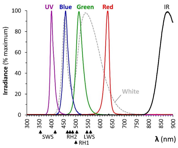
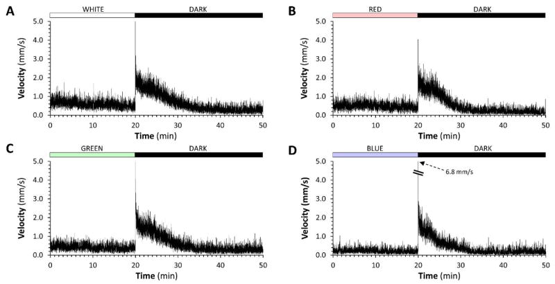
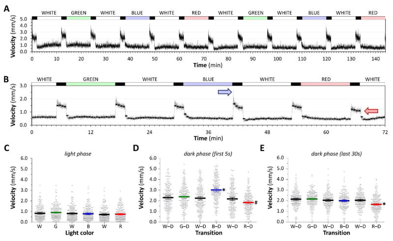
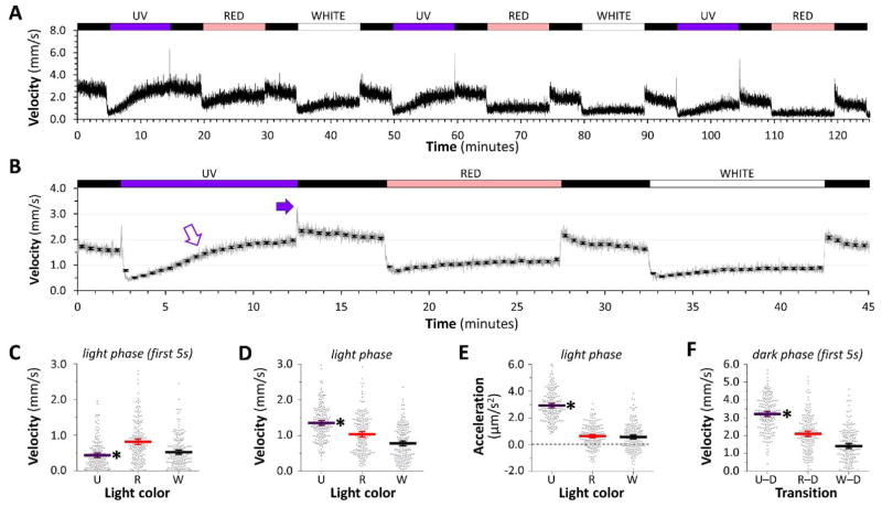
Larval zebrafish react to changes in ambient illumination with a series of stereotyped motor responses, called the visual motor response (VMR). The VMR has been used widely in zebrafish models to analyze how genetic or environmental manipulations alter neurological function. Prior studies elicited the VMR using white light. In order to elucidate the underlying afferent pathways and to identify light wavelengths that elicit the VMR without also activating optogenetic reagents, we employed calibrated narrow-waveband light sources to analyze the spectral properties of the response. Narrow light wavebands with peaks between 399nm and 632nm triggered the characteristic phases of the VMR, but there were quantitative differences between responses to different light wavelengths at the same irradiant flux density. The O-bend component of the VMR was elicited readily at dark onset following illumination in 399nm or 458nm light, but was less prominent at the transition from 632nm light to dark. Conversely, stable motor activity in light was observed at 458nm, 514nm, and 632nm, but not at 399nm. The differential effect of discrete light wavebands on components of the VMR suggests they are driven by distinct photoreceptor populations. Furthermore, these data enable the selection of light wavebands to drive the VMR in a separate channel to the activation of optogenetic reagents and photosensitizers.

Keywords: Non-visual opsins; Optogenetics; Reflex; Retina; Visual motor response; Zebrafish.


Conflict of interest statement

Contributions:

CEB conceived the idea for the study, built equipment, designed and carried out experiments and contributed to writing the paper; YZ designed and developed analysis methods; QB designed and carried out experiments; EAB built equipment, designed experiments, analyzed data and wrote the paper.

Figures

Figure 1
Figure 1. Spectral properties of lights used in the study
Irradiance spectra are shown for the LED light sources used in this study, measured at the position occupied by the zebrafish within the behavioral apparatus. Spectra from the four single-color LED sources are shown as solid colored lines; the white LED source is shown by a dotted line; the infrared light source used for video recording is labeled ‘IR’. For comparison, absorption maxima are shown for opsins expressed in short (SWS1) and long (SWS2) single cones, principal (LWS1 and 2) and accessory (RH2 1 – 4) double cones and rods (RH1).
Figure 2
Figure 2. The zebrafish visual motor response elicited with red, green or blue light is similar to the white light response
In each panel, 96 zebrafish larvae at 5dpf were recorded swimming in the wells of a 96-well plate at 4 frames/sec using infrared macrovideography. The y-axis shows mean displacement of the 96 larvae at each frame-to-frame transition, scaled to show instantaneous mean velocity in mm/s as a measure of group motor activity. The traces show the final 20 minutes of a 40 minute acclimatization period to light (A: white; B: red; C: green; D: blue; all lights were calibrated to 450mW/cm2), followed by the first 30 minutes of the response to an abrupt light-dark transition.
Figure 3
Figure 3. Quantitative differences in the visual motor response elicited with light of different wavelengths
A: Group motor activity of 96 zebrafish in response to changes in illumination indicated above the trace. All light channels were calibrated to 450mW/m2. B: Responses were averaged over two experiments of three complete stimulus cycles (white – green – white – blue – white – red). The gray line shows mean frame-by-frame activity; the black markers show mean ± 2SE values for 30-second time bins. Colored arrows indicate the prominent O-bend response after blue light and the attenuated dark response following red light. C, D, E: Scatter plots showing 192 individual zebrafish responses, each averaged over three stimulus cycles. Large colored bars show population mean ± 2SE. C: motor activity during minutes 2 – 8 of illumination in each light segment of the cycle; D: motor activity in the first 5 seconds after each light-dark transition of the cycle; E: motor activity in the final 30 seconds of each dark segment of the cycle. *p<0.0001, #p<0.01 compared with all other groups, 1-way ANOVA with Tukey post hoc test.
Figure 4
Figure 4. Visual motor response elicited by UV light
A: Group motor activity of 96 zebrafish in response to changes in illumination indicated above the trace. UV and red channels were calibrated to 300mW/m2; for comparison the white channel was calibrated to 150mW/m2. B: Responses were averaged as shown in figure 3B. Closed arrow indicates the prominent O-bend response after UV light; open arrow shows increasing motor activity during UV illumination. C, D, E, F: Scatter plots showing 192 individual zebrafish responses, each averaged over three cycles of light stimuli. The large bars show the population mean ± 2SE. C: motor activity in the first 5 seconds after each dark-light transition; D: motor activity during minutes 2 – 8 of illumination in each light segment of the cycle; E: mean acceleration during each light segment of the cycle; F: motor activity in the first 5 seconds after each light-dark transition. *p<0.0001 compared with both other groups, 1-way ANOVA with Tukey post hoc test.

References

    1. Arrenberg AB, Del Bene F, Baier H. Optical control of zebrafish behavior with halorhodopsin. Proc Natl Acad Sci U S A. 2009;106:17968–17973. - PMC - PubMed
    1. Bulina ME, Chudakov DM, Britanova OV, Yanushevich YG, Staroverov DB, Chepurnykh TV, Merzlyak EM, Shkrob MA, Lukyanov S, Lukyanov KA. A genetically encoded photosensitizer. Nat Biotechnol. 2006;24:95–99. - PubMed
    1. Burgess HA, Granato M. Modulation of locomotor activity in larval zebrafish during light adaptation. J Exp Biol. 2007;210:2526–2539. - PubMed
    1. Cario CL, Farrell TC, Milanese C, Burton EA. Automated measurement of zebrafish larval movement. J Physiol. 2011;589:3703–3708. - PMC - PubMed
    1. Chinen A, Hamaoka T, Yamada Y, Kawamura S. Gene duplication and spectral diversification of cone visual pigments of zebrafish. Genetics. 2003;163:663–675. - PMC - PubMed

Publication types

LinkOut - more resources