

Maternal exposure to low levels of corticosterone during lactation protects adult rat progeny against TNBS-induced colitis: A study on GR-mediated anti-inflammatory effect and prokineticin system
- PMID: 28267767
- PMCID: PMC5340375
- DOI: 10.1371/journal.pone.0173484
Maternal exposure to low levels of corticosterone during lactation protects adult rat progeny against TNBS-induced colitis: A study on GR-mediated anti-inflammatory effect and prokineticin system
Abstract
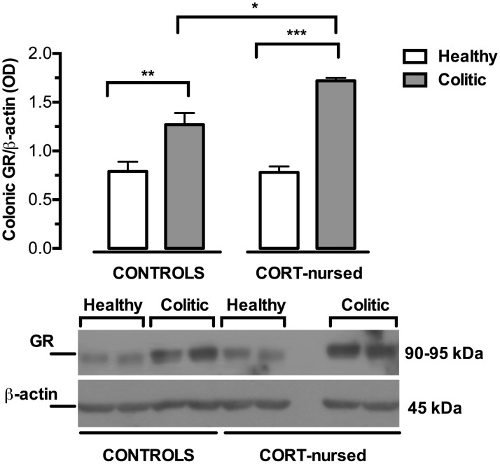
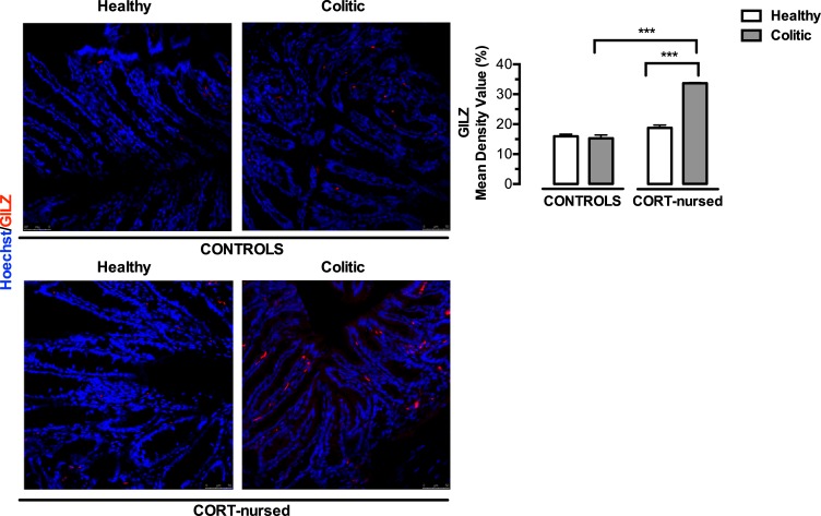
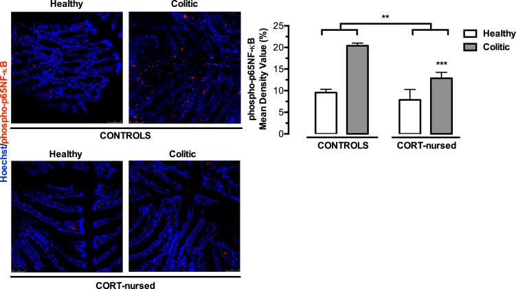
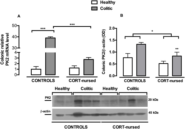
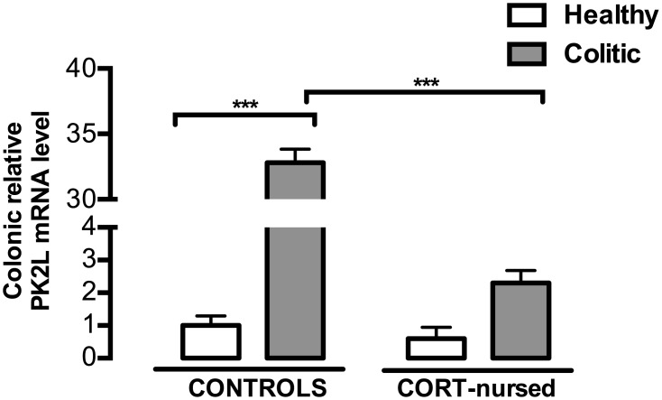
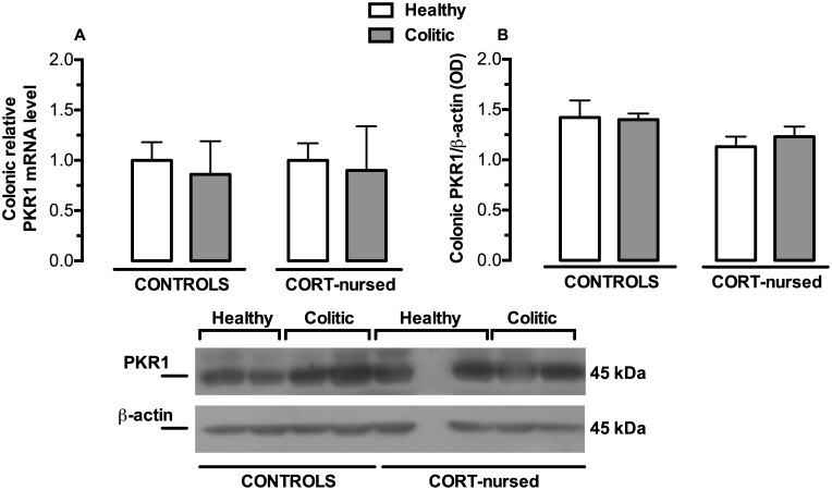
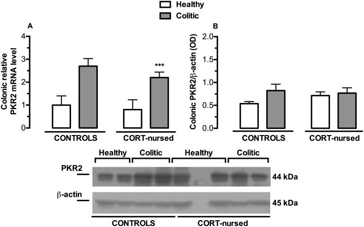
The early phase of life represents a critical period for the development of an organism. Interestingly, early life experiences are able to influence the development of the gastrointestinal tract and the reactivity to colonic inflammatory stress. We recently demonstrated that adult male rats exposed to low doses of corticosterone during lactation (CORT-nursed rats) are protected against experimental colitis induced by the intracolonic infusion of 2,4,6-trinitrobenzenesulfonic acid (TNBS). Based on these interesting results, we wanted to better investigate which cellular actors could be involved in the protection of CORT-nursed rats from TNBS-induced experimental colitis. Therefore, in the present work, we focused our attention on different factors implicated in GR-mediated anti-inflammatory effect. To address this issue, colonic tissues, collected from control and CORT-nursed healthy animals and from control and CORT-nursed colitic rats, were processed and the following inflammatory factors were evaluated: the expression of (i) glucocorticoid receptors (GR), (ii) glucocorticoid-induced leucine zipper (GILZ), (iii) phospho-p65NF-κB, (iv) the pro-inflammatory cytokines IL-1β and TNF-α, (v) the prokineticins PK2 and PK2L and (vi) their receptors PKR1 and PKR2. We found that adult CORT-nursed rats, in comparison to controls, showed increased expression of colonic GR and reduced expression of pro-inflammatory molecules (IL-1β, TNF-α, PK2 and PK2L) in response to inflammatory colitis. The observed changes were associated with an increase in GILZ colonic expression and with a reduction in phospo-p65NF-κB colonic expression.
Conflict of interest statement
Figures

Similar articles
-
Maternal exposure to low levels of corticosterone during lactation protects against experimental inflammatory colitis-induced damage in adult rat offspring.PLoS One. 2014 Nov 18;9(11):e113389. doi: 10.1371/journal.pone.0113389. eCollection 2014. PLoS One. 2014. PMID: 25405993 Free PMC article.
-
Efficacy of thalidomide on trinitrobenzene sulfonate-induced colitis in young rats and its mechanism.Chin Med J (Engl). 2014;127(12):2368-75. Chin Med J (Engl). 2014. PMID: 24931258
-
Lessons on the Sigma-1 Receptor in TNBS-Induced Rat Colitis: Modulation of the UCHL-1, IL-6 Pathway.Int J Mol Sci. 2020 Jun 5;21(11):4046. doi: 10.3390/ijms21114046. Int J Mol Sci. 2020. PMID: 32516975 Free PMC article.
-
Versatile Role of Prokineticins and Prokineticin Receptors in Neuroinflammation.Biomedicines. 2021 Nov 9;9(11):1648. doi: 10.3390/biomedicines9111648. Biomedicines. 2021. PMID: 34829877 Free PMC article. Review.
-
Prokineticin Is a New Linker between Obesity and Cardiovascular Diseases.Front Cardiovasc Med. 2017 Apr 12;4:20. doi: 10.3389/fcvm.2017.00020. eCollection 2017. Front Cardiovasc Med. 2017. PMID: 28447033 Free PMC article. Review.
Cited by
-
Chemokines in Alzheimer's Disease: New Insights Into Prokineticins, Chemokine-Like Proteins.Front Pharmacol. 2019 May 29;10:622. doi: 10.3389/fphar.2019.00622. eCollection 2019. Front Pharmacol. 2019. PMID: 31231219 Free PMC article. Review.
-
Alzheimer's disease-associated inflammatory pathways might contribute to osteoporosis through the interaction between PROK2 and CSF3.Front Neurol. 2022 Sep 20;13:990779. doi: 10.3389/fneur.2022.990779. eCollection 2022. Front Neurol. 2022. PMID: 36203970 Free PMC article.
-
A Healthy Gut for a Healthy Brain: Preclinical, Clinical and Regulatory Aspects.Curr Neuropharmacol. 2021;19(5):610-628. doi: 10.2174/1570159X18666200730111528. Curr Neuropharmacol. 2021. PMID: 32744976 Free PMC article. Review.
-
A link between gastrointestinal disorders and migraine: Insights into the gut-brain connection.Headache. 2021 Apr;61(4):576-589. doi: 10.1111/head.14099. Epub 2021 Apr 1. Headache. 2021. PMID: 33793965 Free PMC article. Review.
-
Corticosterone effects induced by stress and immunity and inflammation: mechanisms of communication.Front Endocrinol (Lausanne). 2025 Mar 20;16:1448750. doi: 10.3389/fendo.2025.1448750. eCollection 2025. Front Endocrinol (Lausanne). 2025. PMID: 40182637 Free PMC article. Review.
References
-
- Ahmad T, Tamboli CP, Jewell D, Colombel J-F. Clinical relevance of advances in genetics and pharmacogenetics of IBD. Gastroenterology. 2004. May;126(6):1533–49. - PubMed
-
- Shanahan F. Inflammatory bowel disease: Immunodiagnostics, immunotherapeutics, and ecotherapeutics. Gastroenterology. 2001. February;120(3):622–35. - PubMed
-
- Collins SM. Stress and the Gastrointestinal Tract IV. Modulation of intestinal inflammation by stress: basic mechanisms and clinical relevance. Am J Physiol Gastrointest Liver Physiol. 2001. March;280(3):G315–318. - PubMed
-
- Fava GA, Pavan L. Large bowel disorders. I. Illness configuration and life events. Psychother Psychosom. 1976 1977;27(2):93–9. - PubMed
MeSH terms
Substances
LinkOut - more resources
Full Text Sources
Other Literature Sources
Medical
