. 2017 Mar 7;13(3):e1006253.
doi: 10.1371/journal.ppat.1006253. eCollection 2017 Mar.

A self-perpetuating repressive state of a viral replication protein blocks superinfection by the same virus

Affiliations

A self-perpetuating repressive state of a viral replication protein blocks superinfection by the same virus

Xiao-Feng Zhang et al. PLoS Pathog. .

Abstract

Diverse animal and plant viruses block the re-infection of host cells by the same or highly similar viruses through superinfection exclusion (SIE), a widely observed, yet poorly understood phenomenon. Here we demonstrate that SIE of turnip crinkle virus (TCV) is exclusively determined by p28, one of the two replication proteins encoded by this virus. p28 expressed from a TCV replicon exerts strong SIE to a different TCV replicon. Transiently expressed p28, delivered simultaneously with, or ahead of, a TCV replicon, largely recapitulates this repressive activity. Interestingly, p28-mediated SIE is dramatically enhanced by C-terminally fused epitope tags or fluorescent proteins, but weakened by N-terminal modifications, and it inversely correlates with the ability of p28 to complement the replication of a p28-defective TCV replicon. Strikingly, p28 in SIE-positive cells forms large, mobile punctate inclusions that trans-aggregate a non-coalescing, SIE-defective, yet replication-competent p28 mutant. These results support a model postulating that TCV SIE is caused by the formation of multimeric p28 complexes capable of intercepting fresh p28 monomers translated from superinfector genomes, thereby abolishing superinfector replication. This model could prove to be applicable to other RNA viruses, and offer novel targets for antiviral therapy.


Conflict of interest statement

The authors have declared that no competing interests exist.

Figures

Fig 1
Fig 1. Two TCV-derived replicons exclude each other at the cellular level.
See S1 Fig for additional information. (A) Schematic depiction of TCV genome structure. Note that the three 3’ proximal open reading frames (ORFs) encoding p8, p9, and p38 are translated from two subgenomic (sg) RNAs (sgRNA1 and 2). (B) Diagrams of TCV and CarMV replicon constructs expressing GFP and mCherry, used experiments depicted in (C) and (D). (C) Mutual exclusion occurs between TCV_sg2G and TCV_sg2R but not between TCV_sg2G and CarMV_sg2R. Note that a p19-expressing construct was included in this and subsequent experiments to protect the transcribed RNAs from RNA silencing-mediated degradation. (D) Sequential delivery (16-hour interval) allows primary replicons to exclude the superinfecting ones in most cells. See S1D Fig for additional controls. Numbers under the panels represent the percentages of cells fluorescing GFP (G), mCherry (R), or both (G+R) averaged from estimates of at least four different viewing fields. The ranges are standard deviations (SD).
Fig 2
Fig 2. TCV-encoded p28 alone is sufficient to cause the repression of co-introduced TCV replicons.
See S2 Fig for additional information. (A) Transiently expressed p28 protein exerts a potent repression on co-introduced TCV_sg2G replicon. Agro-infiltrated leaves were photographed under UV illumination to visualize GFP fluorescence. Names of various constructs tested are shown on respectively panels. Note that all transiently expressed proteins except p19 contain a C-terminal double HA tag. (B) Verification of results in (A) with Northern and Western blottings (NB and WB). EB: ethidium bromide staining of the RNA gel; Coomassie: Coomassie blue staining of the protein gel. (C) Δp88ΔMP_sg2R, a TCV replicon encoding p28 as the sole viral protein, once replicationally rescued by another replicon (ΔMP_sg2G), in turn represses the replication of the latter. Cells were stained with DAPI to show nuclei as well as cell boundaries. Only merged images were shown. See S2B Fig for single channel images of the bottom right panel.
Fig 3
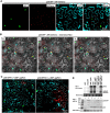
Fig 3. p28-GFP fusion protein form large, mobile intracellular inclusions that are not associated with cell nuclei or mitochondria, and repress the replication of a co-introduced TCV replicon.
Also see S3 Fig for the spatial separation of p28-GFP foci relative from ER. A p19-expressing construct was included in all infiltrations. (A) p28-GFP forms dense foci that rival nuclei (DAPI-stained) in size, and spatially separate from mitochondria and cell nuclei. MT-mCherry: a construct expressing mCherry tagged with an N-terminal mitochondrion-localizing signal. Note that DAPI stains cell nuclei as well as cell wall. (B) Mobility of p28-GFP foci. ER-mCherry: a construct expressing mCherry tagged with an N-terminal endoplasmic reticulum-localizing signal. mCerulean-NLS: a construct expressing mCerulean tagged with a C-terminal nuclear localization signal (NLS). The images additionally contained a bright field layer to help define cell boundaries. The three panels denote the same leaf area photographed at three time points: 0, 1, and 2 minutes. Three lower case letters, a, b, and c, denote three mobile p28-GFP foci, with orange and white arrowheads marking the starting and ending points, respectively. Also see a time lapse movie (S1 Video) in Supporting Information for more details. (C) p28-GFP foci do not occur in the same cells with mCherry expressed from a co-introduced replicon (ΔMP_sg2R). The two constructs were mixed at either 1:1 (left) or 0.1:1 ratios. Only merged images were presented. (D) NB and WB verifications of results in (C). Note in lane 5 that the wild-type p28 translated from the ΔMP_sg2R replicon was readily detectable despite of its inability to replicate.
Fig 4
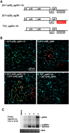
Fig 4. Compared to the N-terminally tagged G11-p28, untagged p28 is less competent at complementing the replication of a p28-defective replicon, but more potent at repressing the replication of another replicon encoding wt p28.
(A) Diagrams of the N-terminally tagged G11-p28 construct, and the p28-defective [p28fs]_sg2R replicon. (B) Complementation of [p28fs]_sg2R (top row), and repression of TCV_sg2R, by various p28 derivatives (p28-GFP, untagged p28, and G11-p28). As in earlier experiments, all treatments included a p19-expressing construct. Within each row, the left four panels represent leaf patches infiltrated with transiently expressed p28 derivatives and replicon constructs simultaneously. By contrast, the right two panels represent leaf patches that were first infiltrated with p28 derivatives, and then with replicons, with a 16 hour delay. The numbers on the panels represent the averaged percentages of cells with mCherry fluorescence indicating active replication, plus SDs.(C) NB confirmation of results in (B). The relative accumulation levels of replicon genomic RNA were estimated with ImageJ. Note that the readings of lane 8 and 10 were set at 1 for the left and right blots, respectively.
Fig 5
Fig 5. The G11-p28 derivative of p28 does not form intracellular inclusions, but is trans-coalesced by untagged p28 and p28-mCherry.
As in previous experiments, a p19-expressing construct was present in all infiltrations. (A) Diagram of the constructs used. Note that the N-terminal G11 tag of G11-p28 does not fluoresce by itself, but becomes fluorescent in the presence of G1-10 expressed from a separate construct. (B) Confocal microscopy of various constructs expressed in N. benthamiana cells. Only merged images were presented. Bar = 50 μm. See time lapse movies in S2 and S3 Videos for the intracellular migration of the p28 inclusions induced by replicon-borne p28.
Fig 6
Fig 6. TCV replicons encoding G11-p28 abolish SIE among themselves, but are dominantly repressed by those expressing wt p28.
(A) Diagrams of three replicon constructs used in the current set of experiments. Note that GFP fluorescence reconstitution by the top construct is dependent on its successful replication, and fluorescence signals co-localize with G11-p28. By contrast, mCherry expressed from the middle construct, while dependent on replication as well, should not associate with G11-p28. (B) Confocal microscopy of the replicon constructs expressed either separately or in combination in N. benthamiana cells. Only merged images were presented. (C) Verification of replication of various TCV replicons with NB.
Fig 7
Fig 7. A model of TCV p28-mediated SIE.
Step 1: the concentration of p28 translated from the genome of primary invader (top right, black dots) is relatively low. They interact with each other relatively slowly, allowing the incorporation of p88, and enclosure of the complex by mitochondrion outer membrane to form VRCs. Step 2: genomic RNA of the primary invader enters VRCs to initiate replication. We hypothesize that some VRCs stay vacant and await the entry of newly synthesized viral RNA. However, the majority of progeny RNA will probably not have the chance to repeat the replication cycle in the same cell. Also beginning with this step, large amounts of progeny RNA become template for p28 translation, drastically increasing the intracellular concentration of p28. Step 3: higher p28 concentration propels faster p28 polymerization, leading to quick formation of oversized p28 polymers that escape the membrane enclosure. Note that the forms of polymeric p28 complexes are hypothetical as we do not yet know the specific mode of p28-p28 interactions. Step 4: since the p28 polymers are by definition capable of nucleating homologous monomers, they are expected to capture the p28 monomers (blue dots) translated from superinfector genomes, preventing them from forming new VRCs.

Similar articles

Cited by

References

    1. Nethe M, Berkhout B, van der Kuyl AC (2005) Retroviral superinfection resistance. Retrovirology 2:52 10.1186/1742-4690-2-52 - DOI - PMC - PubMed
    1. Schaller T, et al. (2007) Analysis of hepatitis C virus superinfection exclusion by using novel fluorochrome gene-tagged viral genomes. J Virol 81:4591–4603. 10.1128/JVI.02144-06 - DOI - PMC - PubMed
    1. Tscherne DM, et al. (2007) Superinfection exclusion in cells infected with hepatitis C virus. J Virol 81:3693–3703. 10.1128/JVI.01748-06 - DOI - PMC - PubMed
    1. Zou G, et al. (2009) Exclusion of West Nile virus superinfection through RNA replication. J Virol 83:11765–11776. 10.1128/JVI.01205-09 - DOI - PMC - PubMed
    1. Simon KO, Cardamone JJ Jr, Whitaker-Dowling A, Youngner JS, Widnell CC (1990) Cellular mechanisms in the superinfection exclusion of vesicular stomatitis virus. Virology 177:375–379. - PubMed

Publication types

LinkOut - more resources