. 2017 Mar 7;12(3):e0172825.
doi: 10.1371/journal.pone.0172825. eCollection 2017.

Wfs1 is expressed in dopaminoceptive regions of the amniote brain and modulates levels of D1-like receptors

Affiliations

Wfs1 is expressed in dopaminoceptive regions of the amniote brain and modulates levels of D1-like receptors

Triin Tekko et al. PLoS One. .

Abstract

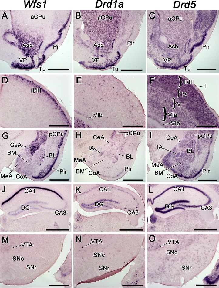
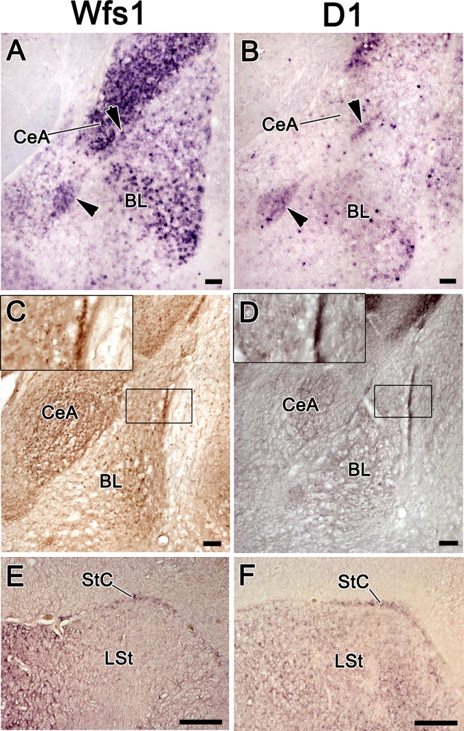
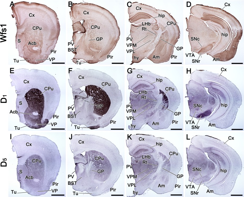
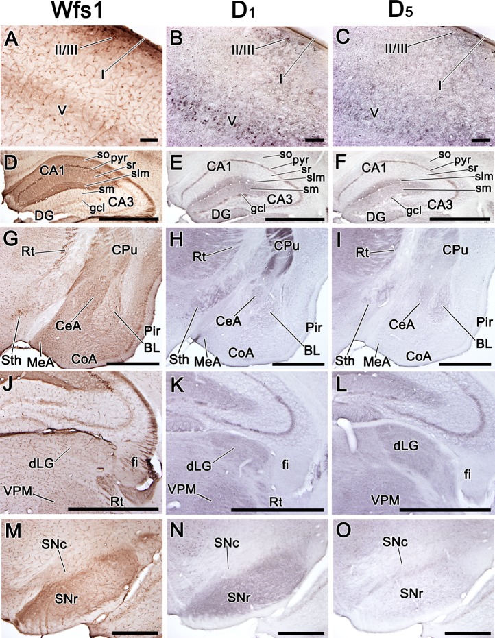
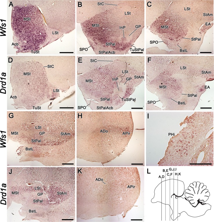
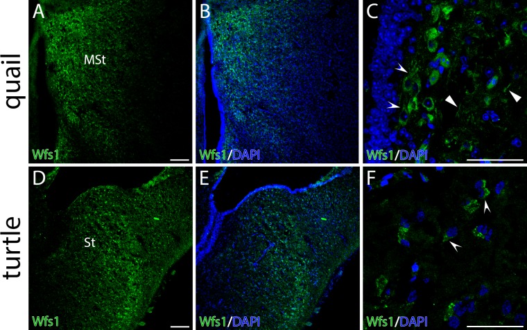
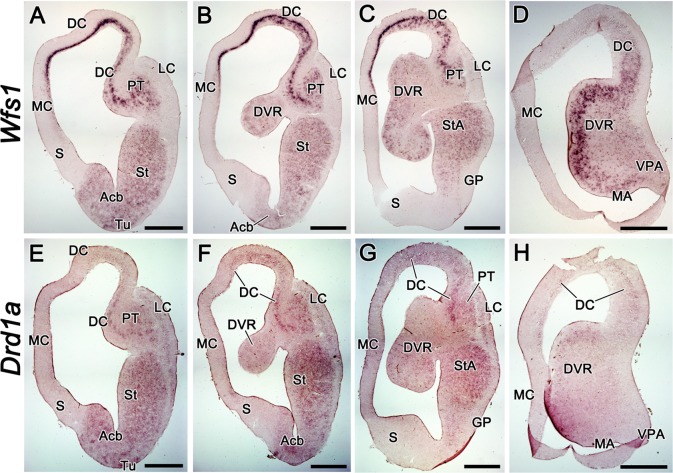
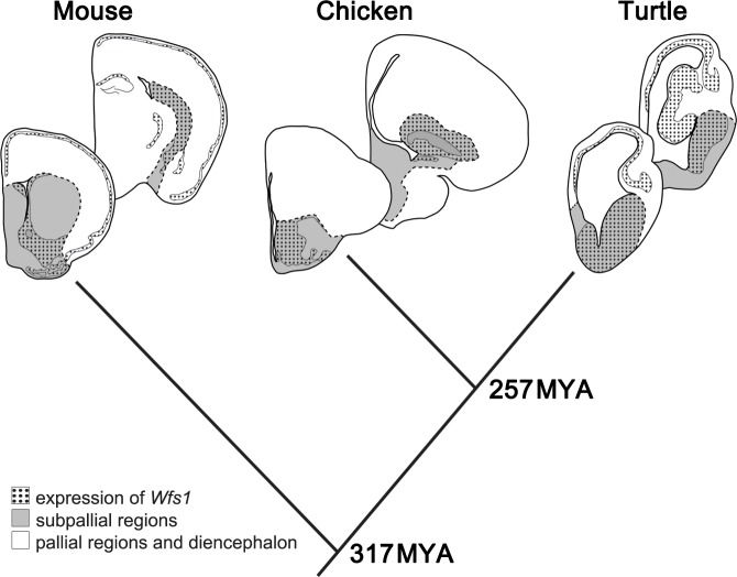
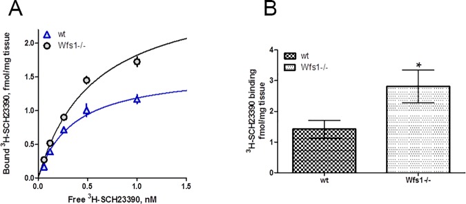
During amniote evolution, the construction of the forebrain has diverged across different lineages, and accompanying the structural changes, functional diversification of the homologous brain regions has occurred. This can be assessed by studying the expression patterns of marker genes that are relevant in particular functional circuits. In all vertebrates, the dopaminergic system is responsible for the behavioral responses to environmental stimuli. Here we show that the brain regions that receive dopaminergic input through dopamine receptor D1 are relatively conserved, but with some important variations between three evolutionarily distant vertebrate lines-house mouse (Mus musculus), domestic chick (Gallus gallus domesticus) / common quail (Coturnix coturnix) and red-eared slider turtle (Trachemys scripta). Moreover, we find that in almost all instances, those brain regions expressing D1-like dopamine receptor genes also express Wfs1. Wfs1 has been studied primarily in the pancreas, where it regulates the endoplasmic reticulum (ER) stress response, cellular Ca2+ homeostasis, and insulin production and secretion. Using radioligand binding assays in wild type and Wfs1-/- mouse brains, we show that the number of binding sites of D1-like dopamine receptors is increased in the hippocampus of the mutant mice. We propose that the functional link between Wfs1 and D1-like dopamine receptors is evolutionarily conserved and plays an important role in adjusting behavioral reactions to environmental stimuli.


Conflict of interest statement

Competing Interests: The authors have declared that no competing interests exist.

Figures

Fig 1
Fig 1. The mRNA expression pattern of Wfs1, Drd1a, and Drd5 in the adult mouse brain.
The mRNA expression pattern of Wfs1, Drd1a, and Drd5 in the adult mouse brain, shown by in situ hybridization. In this and all subsequent figures, the medial side of the coronal sections is on the left and the lateral side on the right. The probes are indicated above. The expression in CPu and ventral striatum (A-C), in somatosensory cortex at the level of bregma 0.74 (D-F), in the amygdala at the level of central and basolateral nuclei (G-I), in the hippocampus (J-L), in the substantia nigra and VTA (M-O). For abbreviations, see list. Scale bar is 1 mm.
Fig 2
Fig 2. The expression of Wfs1 and Drd1a in the mouse intercalated amygdala and in its putative avian homologue, StC, in chick.
The expression of Wfs1 and Drd1a in the mouse intercalated amygdala and in its putative avian homologue, StC, in chick. A, B–in situ hybridization on coronal sections of the mouse brain. C, D–immunohistochemistry on coronal sections of the mouse brain. E, F–in situ hybridization on the coronal sections of the chick brain. The intercalated nuclei of the amygdala (arrowheads) are expressing both Wfs1 and Drd1a in mouse brain (A, B). Wfs1 and D1 proteins are both strongly expressed in the intercalated nuclei (C, D). The insets in C and D show closer view on the intercalated nucleus between the BL and claustrum-endopiriform formation. In chick brain, the StC is expressing both Wfs1 and Drd1a. For abbreviations, see list. Scale bar is 100 μm in A-D and 1 mm in E-F.
Fig 3
Fig 3. Distribution of Wfs1, D1, and D5 proteins in the adult mouse brain.
Distribution of Wfs1, D1, and D5 proteins in the adult mouse brain, shown by immunohistochemistry on coronal sections. The sections are in anterio-posterior order from left to right. The detected proteins are indicated on the left side of the figure. Wfs1 is present in the cerebral cortex, in CA1 of hippocampus, CPu, Acb, Tu, amygdala, Rt, PV, VPM, hypothalamus and SNr (A-D). D1 is strongly present in CPu, Acb, Tu, hip, thalamus and SNr (E-H). D5 is present in CPu, Acb, S, hip, thalamus and SNc (I-L). Note that in Tu and SNr the distribution of D1, but not D5, is similar to Wfs1. For abbreviations, see list. Scale bar is 1 mm.
Fig 4
Fig 4. Distribution of Wfs1, D1, and D5 proteins in selected regions of the adult mouse brain.
Distribution of Wfs1, D1, and D5 proteins in selected regions of the adult mouse brain. Immunohistochemistry on coronal sections. The detected proteins are indicated above. In the cortex Wfs1, D1, and D5 are all present in layer I, upper part of layer II/III, and in layer V (images show somatosensory cortex; A-C); in the hippocampus Wfs1, D1, and D5 are simultaneously present in pyr and slm of CA1 (D-F); Wfs1, D1 and D5 are all present in Rt and Sth; in amygdala Wfs1 is strongly present in CeA, the lateral edge of BL, and medial and cortical nuclei (G-I), whereas D1 and D5 show only weak signal in CeA and BL (G-I); in the dorsolateral thalamus Wfs1, D1, and D5 are delineating dLG and VPM, note that strongly Wfs1-positive fibers are present in fi, but no D1 or D5 is seen there (J-L), in the substantia nigra Wfs1 has similar distribution with D1, but not with D5 (M-O). For abbreviations, see list. Scale bar is 100 μm in A-C, 1 mm in D-L, 500 μm in M-O.
Fig 5
Fig 5. The expression of Wfs1 and Drd1a in the adult chick brain.
The expression of Wfs1 and Drd1a in the adult chick brain, shown by mRNA in situ hybridization on coronal brain sections. The section plane is shown on image L. The probes are indicated on the left side of the figure. Both, Wfs1 and Drd1a show strong expression in rostral to medial MSt (A-F). In Acb and StPalAcb, the expression of Wfs1 is substantially weaker than in the surrounding striatal structures (A-B). The expression of Drd1a is weak in Acb and StPalAcb, and is missing in SPO (D-F). In LSt, both Wfs1 and Drd1a expression show strengthening gradient in rostrocaudal direction (A-G,J). Wfs1-expressing cells in PHi are shown in higher magnification (I). In the adult brain, ADo and APir are delineated with Wfs1 expression, but remain hardly distinguishable by Drd1a expression (H,K). Note that GP is devoid of both Drd1a and Wfs1 (B,E,G,J). For abbreviations, see list. Scale bar is 1 mm in A-H and J-K and 500 μm in I.
Fig 6
Fig 6. Distribution of Wfs1 protein in the striata of common quail (Coturnix coturnix) and red-eared slider turtle (Trachemys scripta) brains.
Distribution of Wfs1 protein in the striata of quail (Coturnix coturnix) and red-eared slider turtle (Trachemys scripta) brains. The panel shows fluorescent immunohistochemistry on coronal brain sections. Wfs1 expression (green) is seen in medial striatum of quail (A, MSt) and turtle (D, St). Concave arrowheads point the expression in soma in both species (C, F). Wfs1 is detectable in neuronal processes of quail (C, concave arrowheads). Nuclei are counterstained with DAPI (blue). Scale bar is 100 μm.
Fig 7
Fig 7. The expression of Wfs1 and Drd1a in the adult red-eared slider turtle (Trachemys scripta) brain.
The expression of Wfs1 and Drd1a in the adult red-eared slider turtle (Trachemys scripta) brain, shown by mRNA in situ hybridization on coronal brain sections. The sections are in anterio-posterior order from left to right. The probes are indicated on the left side of the figure. Wfs1 expression is widespread in the brain of T.scripta, being distinguishedly strong in MC, DC, PT and near the ventricular surface in the caudal DVR (A-D). Drd1a expression occupies the same regions as that of Wfs1, but is missing in MC and very weak in DC and caudal DVR (E-H). Unlike Wfs1, Drd1a is present in LC (E-G). For abbreviations, see list. Scale bar is 1 mm.
Fig 8
Fig 8. The expression of Wfs1 in pallial and subpallial regions of mouse, chick and red-eared slider turtle brain.
Schematic depiction of the expression of Wfs1 (dotted area) in pallial (white) and subpallial (grey) regions of mouse, chick and red-eared slider turtle brain. Coronal sections. Times of evolutionary divergence are based on [37]. Dashed line—border delineating subpallial regions.
Fig 9
Fig 9. Binding of D1/D5 specific ligand [3H]SCH23390 to hippocampal membranes of wt and Wfs1 knockout mice.
Comparison of specific binding of radioligand [3H]SCH23390 to hippocampal membranes of wt and Wfs1 knockout mice. (A) Binding curve of [3H]SCH23390 binding to pooled samples of wt (triangle) and Wfs1 knockout (circle) mice. The membrane suspensions (3 mg/well) were incubated with different concentrations of [3H]SCH23390 for 60 min and bound radioactivity was measured. Data are presented as mean ± SEM from experiments (n = 3) performed in duplicates. (B) The level of [3H]SCH23390 binding sites of individual wt and Wfs1 knockout mice determined in hippocampal membrane suspensions (6.7 mg/ml.) incubated with 4 nM radioligand. Data presented as mean ± SEM of all the mice tested. *P < 0.05. Data of individual mice are presented in S1 Table.

References

    1. Gabreëls BA, Swaab DF, de Kleijn DP. The vasopressin precursor is not processed in the hypothalamus of Wolfram syndrome patients with diabetes insipidus: evidence for the involvement of PC2 and 7B2. J Clin Endocrinol Metab. 1998;83(11): 4026–4033. 10.1210/jcem.83.11.5158 - DOI - PubMed
    1. Osman AA, Saito M, Makepeace C, Permutt MA, Schlesinger P, Mueckler M. Wolframin expression induces novel ion channel activity in endoplasmic reticulum membranes and increases intracellular calcium. J Biol Chem. 2003;278(52): 52755–52762. 10.1074/jbc.M310331200 - DOI - PubMed
    1. Ishihara H, Takeda S, Tamura A, Takahashi R, Yamaguchi S, Takei D, et al. Disruption of the WFS1 gene in mice causes progressive beta-cell loss and impaired stimulus-secretion coupling in insulin secretion. Hum Mol Genet. 2004; 13: 1159–1170. 10.1093/hmg/ddh125 - DOI - PubMed
    1. Ueda K, Kawano J, Takeda K, Yujiri T, Tanabe K, Anno T, et al. Endoplasmic reticulum stress induces Wfs1 gene expression in pancreatic beta-cells via transcriptional activation. Eur J Endocrinol. 2005;153(1): 167–176. 10.1530/eje.1.01945 - DOI - PubMed
    1. Fonseca SG, Fukuma M, Lipson KL, Nguyen LX, Allen JR, Oka Y, et al. WFS1 is a novel component of the unfolded protein response and maintains homeostasis of the endoplasmic reticulum in pancreatic beta-cells. J Biol Chem. 2005;280(47): 39609–39615. 10.1074/jbc.M507426200 - DOI - PubMed