. 2017 Mar 7;12(3):e0173040.
doi: 10.1371/journal.pone.0173040. eCollection 2017.

Decellularized scaffold of cryopreserved rat kidney retains its recellularization potential

Affiliations

Decellularized scaffold of cryopreserved rat kidney retains its recellularization potential

Baldeep Chani et al. PLoS One. .

Abstract

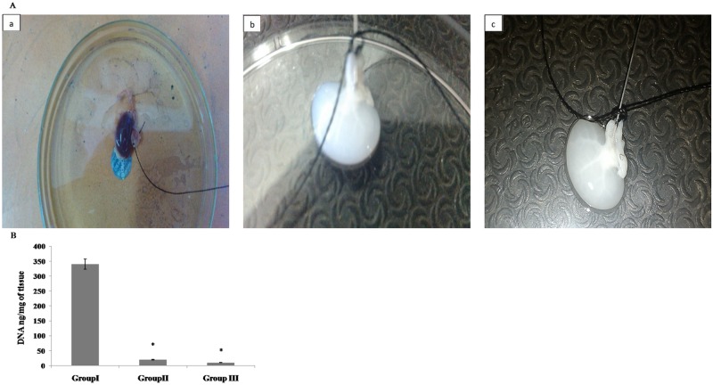
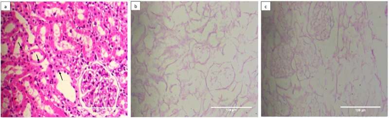
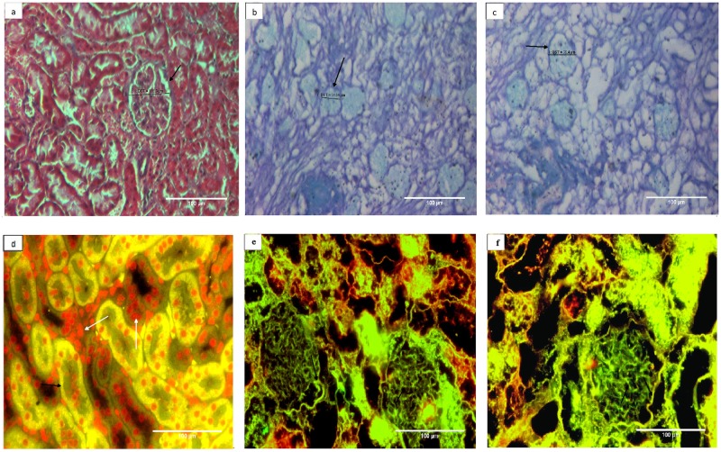
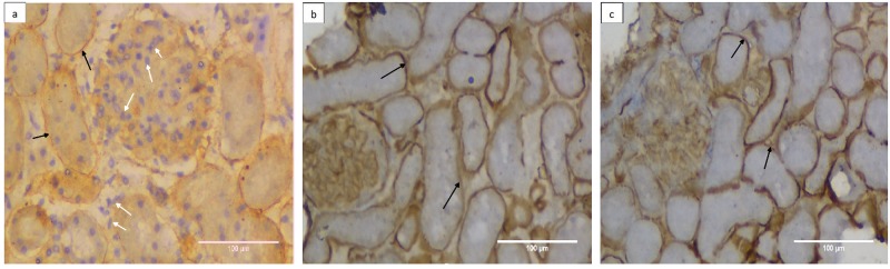
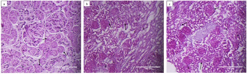
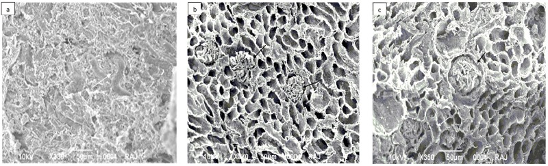
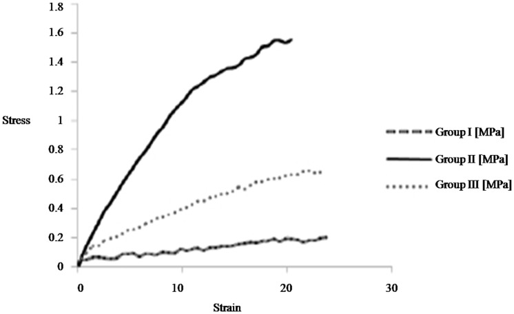
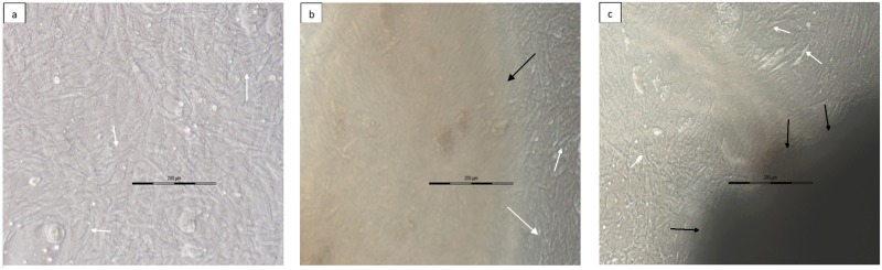
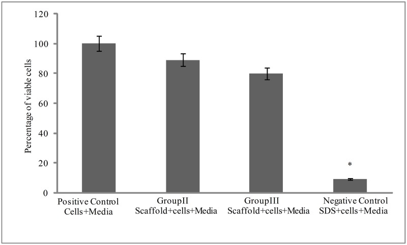
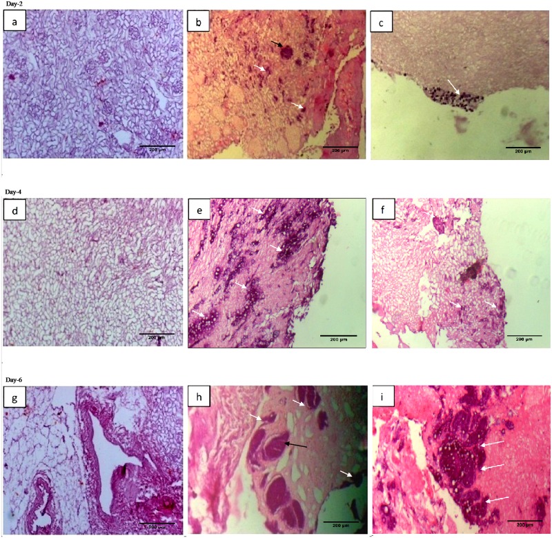
The multi-cellular nature of renal tissue makes it the most challenging organ for regeneration. Therefore, till date whole organ transplantations remain the definitive treatment for the end stage renal disease (ESRD). The shortage of available organs for the transplantation has, thus, remained a major concern as well as an unsolved problem. In this regard generation of whole organ scaffold through decellularization followed by regeneration of the whole organ by recellularization is being viewed as a potential alternative for generating functional tissues. Despite its growing interest, the optimal processing to achieve functional organ still remains unsolved. The biggest challenge remains is the time line for obtaining kidney. Keeping these facts in mind, we have assessed the effects of cryostorage (3 months) on renal tissue architecture and its potential for decellularization and recellularization in comparison to the freshly isolated kidneys. The light microscopy exploiting different microscopic stains as well as immuno-histochemistry and Scanning electron microscopy (SEM) demonstrated that ECM framework is well retained following kidney cryopreservation. The strength of these structures was reinforced by calculating mechanical stress which confirmed the similarity between the freshly isolated and cryopreserved tissue. The recellularization of these bio-scaffolds, with mesenchymal stem cells quickly repopulated the decellularized structures irrespective of the kidneys status, i.e. freshly isolated or the cryopreserved. The growth pattern employing mesenchymal stem cells demonstrated their equivalent recellularization potential. Based on these observations, it may be concluded that cryopreserved kidneys can be exploited as scaffolds for future development of functional organ.


Conflict of interest statement

Competing Interests: The authors have declared that no competing interests exist.

Figures

Fig 1
Fig 1. A. Perfusion with 1% SDS completely decellularized the kidney structures.
The collateral kidneys from rats were isolated and perfused with normal saline prior to perfusion with 1% SDS diluted in distilled water at room temperature for a period of 48 hours. The kidneys were then washed with a solution of deoxyribonuclease I (0.2 mg/ml) and 10 mM MgCl2 in PBS at room temperature for a period of 16 h to ensure complete removal of detergents and nuclear material. Finally the structures were rinsed with PBS to remove deoxyribonuclease 1 and MgCl2 The photomicrographs in Panel-A represents a) control freshly isolated kidney representing group I, b) decellularized freshly isolated kidney representing group II, and c) decellularized kidney from 3 months cryopreserved kidneys representing group III. White transparent appearance of both kidneys from groups I & II shows that kidneys have undergone complete loss of parenchyma in comparison to freshly isolated kidneys group I. Panel B represents a graph of total DNA content analyzed in control kidneys, group I, following decellularization of freshly isolated kidneys, group II and cryostroed kidneys, group III. Based on the DNA quantification it was observed that 1% SDS was sufficient to decellularize the kidneys to similar extent whether the kidneys were freshly isolated or cryostored. Data are expressed as mean ±SEM. Based on ANOVA, significant differences among groups II, III w.r.t. control group I are indicated at *p<0.05.
Fig 2
Fig 2. Cryostorage retains the basic kidney micro-architecture and undergoes decellularization to a similar level as in freshly isolated kidneys.
Photomicrographs represented H&E staining of a) control kidneys from group, I b) decellularized structure from freshly isolated kidney, group II, c) decellularized structure from cryostored kidney, group III at 200X. Well preserved ECM architecture with no evidence of residual nuclei or intact cells as resolved by Hematoxylin (blue stained nuclei) and Eosin (pink stained intracellular proteins), could be seen in both the cryostored kidneys (group III and freshly isolated kidneys group II). Black arrow represents renal cells.
Fig 3
Fig 3. Collagen content of basement membrane is well retained whether or not the kidneys were cryostored prior to decellularization.
Both Masson’s trichrome staining (photomicrogrpahs a-c) and immunohistochemistry using anti collagen IV antisera (photomicrograph d-f) were used to analyze the effect of cryostorage on collagen status of the decellularized structures. Masson’s trichrome stains collagen blue, cellular component red and nuclei black. The blue stained structures (black arrows) showed well retained collagen component of the ECM from a) represented control kidney from group I, b) decellularized kidney from group II, c) decellularized kidney from group III. Almost similar extent of staining patterns among group II and III demonstrated that decellularization process of kidneys did not affect the collagen content whether the tissues were cryopreserved or not. Absence of red and black stain re-confirms the absence of any cellular material and nucleus in these decellularized kidneys. With respect to immunohistochemistry (fig d-f) green fluorescence represented collagen IV and the red fluorescence indicated cell nucleus (Propidium iodide, PI stain). Photomicrographs represented d) control kidney from group I, e) decellularized structures from freshly isolated kidney, group II and f) decellularized structure from cryostored kidney, group III at 200X. Where-ever shown, the white arrows represent renal cells (red fluorescence) and black arrows represent collagen 1V (green fluorescence). It is clear from the Figs that after decellularization overall presence of ECM component collagen IV in glomerulus and kidney structure (black arrow) is preserved and cells are completely removed as no red fluorescence observed when compared with control.
Fig 4
Fig 4. Cryopreserevd kidneys retain their laminin content even after decellularization.
Laminin immunohistostaining was carried out using anti laminin sera followed by FITC conjugated secondary IgG representing a) control kidney from group I, b) decellularized structures from freshly isolated kidney, group II and c) decellularized structure from cryostored kidney, group III at 200X. Where ever shown, the white arrows represented renal cells (blue stain) with black arrows representing laminin (brown stain). The cryostorage for 3 month minimally affected the histological appearance and overall presence of ECM component in the decellularized structure, group III in comparison to freshly de-celularized structure, group II.
Fig 5
Fig 5. Cellular level of Glycosaminoglaycans (GAGs) underwent a loss following decellularization and not the GAGs present in Extracellular matrix.
Alcian blue staining was used to localize the GAGs as shown in photomicrographs a) control kidney from group I, b) decellularized structures from freshly isolated kidney, group II and c) decellularized structure from cryostored kidney, group III at 200X. Where ever shown, the white arrows represented renal cells (black stain) and black arrows represented GAG staining (blue stain). A low content of staining was observed in the decellularized structures from group II and III in comparison to wild type kidneys, group I.
Fig 6
Fig 6. Glycoproteins of tubular basement membrane remained well preserved in decellularized structures from cryostored or freshly isolated kidneys.
The glycoproteins were analyzed using PAS staining from a) control kidney from group I, b) decellularized kidney from fresh kidneys, group II and c) decellularized kidney from cryostored kidneys, group III at 200X. Black arrows represent glycoproteins, which remained uniformly stained whether the decellularized structures were obtained from freshly isolated kidneys or cryostored kidneys.
Fig 7
Fig 7. Scanning electron micrograph confirmed complete removal of cells in both cases.
Photomicrographs represented a) control kidney from group I, b) decellularized kidney from freshly isolated kidneys, group II and c) decellularized kidney from cryostored kidneys, group III. The acellular glomerulus (black arrow) and adjacent tubules (white arrow) show continuous basement membrane architecture after SDS protocol of decellularization in both the groups i.e. group II & III as compared to control.
Fig 8
Fig 8. The well preserved ECM maintains the mechanical strength of the decellularized structure from cryostored kidneys.
Stress & Strain curve demonstrate progressive rise in tensile strength as Young’s modulus increase after decellularization in both group II, III kidneys when compared with control kidneys from group I. The tensile strength is function of the ECM components of the decellularized structures. Higher tensile strength of the decellularized structures from cryostored kidneys (group III) in comparison to freshly isolated decellularized kidneys (group) is indicative of well preserved ECM following cryopreservation.
Fig 9
Fig 9. Decellularized renal scaffold from cryostored kidneys retained the cells growth characteristics.
Photomicrographs following phase contrast microscopy represented a) control kidney from group I, b) decellularized kidney from freshly isolated kidneys, group II and c) decellularized kidney from cryostored kidneys, group III. As shown the acellular matrix appeared to be biocompatible when murine C3H10T½ cells were seeded in vitro. The cells showed potential for the attachment and underwent proliferation and remained viable when cultured with the decellularized structures whether kidneys were obtained following 3 months of cryostorage or freshly isolated kidneys. Black arrows represent kidney scaffold, white arrows represent attached cells.
Fig 10
Fig 10. Decellularized scaffold were found to be nontoxic to murine C3H10T½ cells.
The MTT assay was performed to check if the SDS based decellularization of the kidneys bioscaffolds remained nontoxic to mesenchymal stem cells. The assay was performed under different conditions i.e. using only cells and media (i.e. without scaffold) as a positive control, cells with SDS in media as negative control, cells incubated with freshly isolated decellularized kidnyes (group II), and decellularized structures from cryostored kidnyes (group III). As shown in the graph, the SDS inhibited the growth of cells and for negative control the cell viability was only 9.29% while its was found to be 89.2% for the group II) and approximately 80% for the group III). Absorbance was measured in replicates of six and the calculated standard error of the mean (SEM) plotted error bars. Data are expressed as mean ±SEM. Based on ANOVA, significant differences among groups II, III w.r.t. control group I are indicated at *p<0.05.
Fig 11
Fig 11. The decellularized structures from cryostored kidneys retained the recellularization potential similar to that of freshly decellularized kidneys.
Approximately 2 million C3H10T½ cells were used for the recellularization. The recellularization of the scaffolds was analyzed at different time intervals Photomicrographs a), d), g) represented decellularized kidney scaffolds without recellularization as control, photomicrographs b), e), h) represented recellularized kidney scaffolds from group II and photomicrographs c), f), i) represented recellularized kidney scaffolds from group III for day 2, 4 and 6, respectively. The cells movement was found to be increased to more deeper areas at different time points following recellularization. Black arrows represented cells repopulating in glomerulus and white arrows represented cells repopulating in different renal tissue areas.

References

    1. Shin'oka T, Imai Y, Ikada Y. Transplantation of a tissue-engineered pulmonary artery. N Engl J Med. 2001; 344: 532–533. 10.1056/NEJM200102153440717 - DOI - PubMed
    1. Atala A, Bauer SB, Soker S, Yoo JJ, Retik AB. Tissue-engineered autologous bladders for patients needing cystoplasty. Lancet. 2006; 367: 1241–1246. 10.1016/S0140-6736(06)68438-9 - DOI - PubMed
    1. Macchiarini P, Jungebluth P, Go T, Asnaghi MA, Rees LE, Cogan TA, et al.Clinical transplantation of a tissue-engineered airway. Lancet. 2008; 372: 2023–2030. 10.1016/S0140-6736(08)61598-6 - DOI - PubMed
    1. Ott HC, Matthiesen TS, Goh SK, Black LD, Kren SM, Netoff TI, et al. Perfusion-Decellularized matrix: using nature's platform to engineer a bioartificial heart. Nat Med 2008;14: 213–221. 10.1038/nm1684 - DOI - PubMed
    1. Freytes DO, Stoner RM, Badylak SF. Uniaxial and biaxial properties of terminally sterilized porcine urinary bladder matrix scaffolds. J Biomed Mater Res B Appl Biomater. 2008; 84: 408–414. 10.1002/jbm.b.30885 - DOI - PubMed

LinkOut - more resources