. 2016 Aug:2016:1955-1958.
doi: 10.1109/EMBC.2016.7591106.

Nanoporous membrane robustness / stability in small form factor microfluidic filtration system

Nanoporous membrane robustness / stability in small form factor microfluidic filtration system

Dean G Johnson et al. Annu Int Conf IEEE Eng Med Biol Soc. 2016 Aug.

Abstract

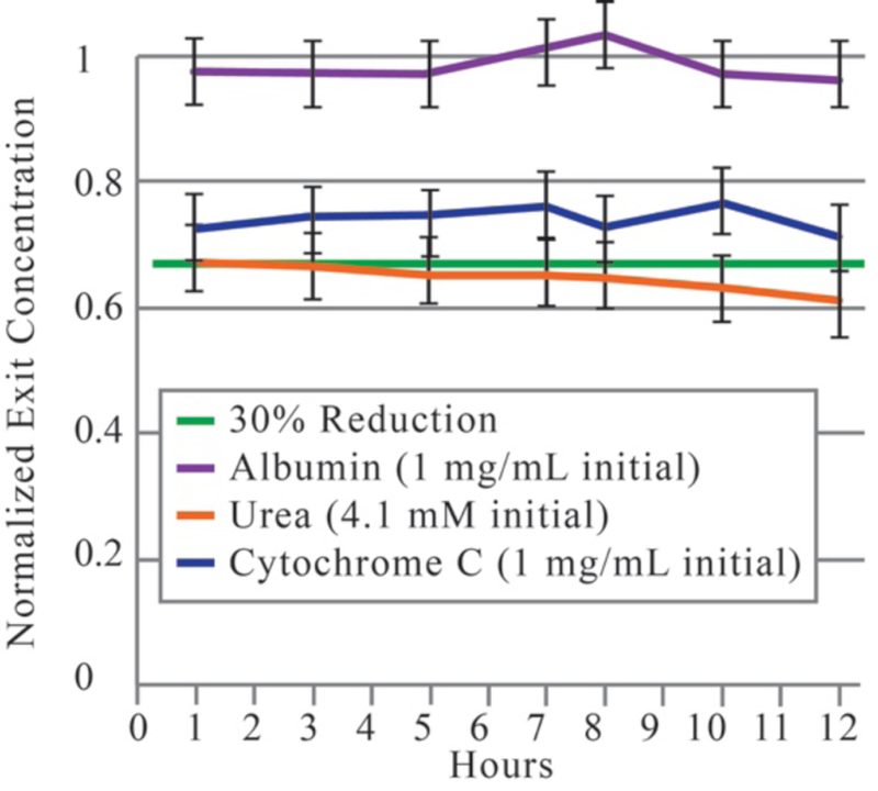
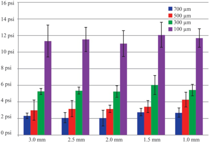
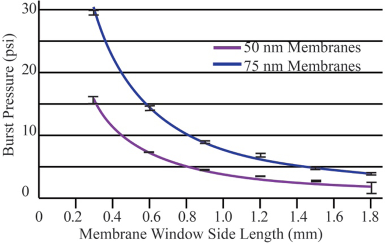
The development of wearable hemodialysis (HD) devices that replace center-based HD holds the promise to improve both outcomes and quality-of-life for patients with end-stage-renal disease (ERD). A prerequisite for these devices is the development of highly efficient membranes that can achieve high toxin clearance in small footprints. The ultrathin nanoporous membrane material developed by our group is orders of magnitude more permeable than conventional HD membranes. We report on our progress making a prototype wearable dialysis unit. First, we present data from benchtop studies confirming that clinical levels of urea clearance can be obtained in a small animal model with low blood flow rates. Second, we report on efforts to improve the mechanical robustness of high membrane area dialysis devices.


Figures

Figure 1:
Figure 1:
Long-term dialysis clearance set up. Syringe pump: 5 mL of 4.1 mM urea (or 1mg/mL Cytochrome-C and 1mg/mL Albumin), µL/min. Single channel dialyzer, 10 mm long, 0.5 mm wide. Fraction Collector programed to rotate every hour. Setup in refrigerator at 40 °C.
Figure 2:
Figure 2:
Two-chip dialyzer. ‘Blood-side’ shown, inlet from left divides fluid to both chips then evenly into the channels in the surface of the chips. Fluid was then collected from the channels and exits the single outlet on the right. Similar fluids route the dialysate across the smooth underside of the chip.
Figure 3:
Figure 3:
Flow pressure burst test setup. Peristaltic pump: 0.5 mL/min pumps fluid to and from beaker (at atmosphere) through ‘blood-side’ of the single chip dialyzer. ‘Dialysate-side’ is supplied via gravity feed.
Figure 4:
Figure 4:
Static burst pressure setup. 5.4 mm chips were placed on the rubber o-ring over the compressed gas inlet port. The chip is held in place with an aluminum plate with an access hole that allows visual inspection of the membrane and acts as an outlet for the compressed gas after the membrane has burst. An electronic manometer recorded the burst pressure.
Figure 5:
Figure 5:
12-hour clearance study. Exit concentrations normalized to inlet concentrations. Urea (4.1 mM), Cytochrome-C (1 mg/mL), and Albumin (1 mg/mL) in 100% serum were pumped through a single pass counter flow device. While the filter retained albumin, urea met its expected 30% reduction. Cytochrome-C (13 kDa) was also reduced.
Figure 6:
Figure 6:
Burst pressures of rectangular membranes, lengths of 1.0 mm to 3 mm, and widths of 0.1 mm to 0.7 mm. Clearly, membrane width plays a more important role in determining the burst pressure than does lengths, for these dimension ranges.
Figure 7:
Figure 7:
Static burst pressure results of 75 nm membranes vs. 50 nm membranes (square membranes). Burst pressures are doubled for a 1.5 fold increase in membrane thickness.

Similar articles

Cited by

References

    1. Blagg CR, “The 50th anniversary of long-term hemodialysis: University of Washington Hospital, March 9th, 1960,” in J Nephrol vol. 24 Suppl 17, ed Italy, 2011, pp. S84–8. - PubMed
    1. Striemer CC, Gaborski TR, McGrath JL, and Fauchet PM, “Charge- and size-based separation of macromolecules using ultrathin silicon membranes,” Nature, vol. 445, pp. 749–53, February 2007. - PubMed
    1. Snyder JL, Clark A, Fang DZ, Gaborski TR, Striemer CC, Fauchet PM, et al., “An experimental and theoretical analysis of molecular separations by diffusion through ultrathin nanoporous membranes,” Journal of Membrane Science, vol. 369, pp. 119–129, 2011. - PMC - PubMed
    1. Agrawal AA, Nehilla BJ, Reisig KV, Gaborski TR, Fang DZ, Striemer CC, et al., “Porous nanocrystalline silicon membranes as highly permeable and molecularly thin substrates for cell culture,” Biomaterials, vol. 31, pp. 5408–17, July 2010. - PubMed
    1. Getpreecharsawas J, McGrath JL, and Borkholder DA, “The electric field strength in orifice-like nanopores of ultrathin membranes,” Nanotechnology, vol. 26, p. 045704, January 2015. - PubMed

LinkOut - more resources