. 2017 Mar 7;14(1):16.
doi: 10.1186/s12977-017-0341-x.

Prototype foamy virus elicits complete autophagy involving the ER stress-related UPR pathway

Affiliations

Prototype foamy virus elicits complete autophagy involving the ER stress-related UPR pathway

Peipei Yuan et al. Retrovirology. .

Abstract

Background: Prototype foamy virus (PFV) is a member of the Spumaretrovirinae subfamily of retroviruses, which maintains lifelong latent infection while being nonpathogenic to their natural hosts. Autophagy is a cell-programmed mechanism that plays a pivotal role in controlling homeostasis and defense against exotic pathogens. However, whether autophagy is the mechanism for host defense in PFV infection has not been investigated.

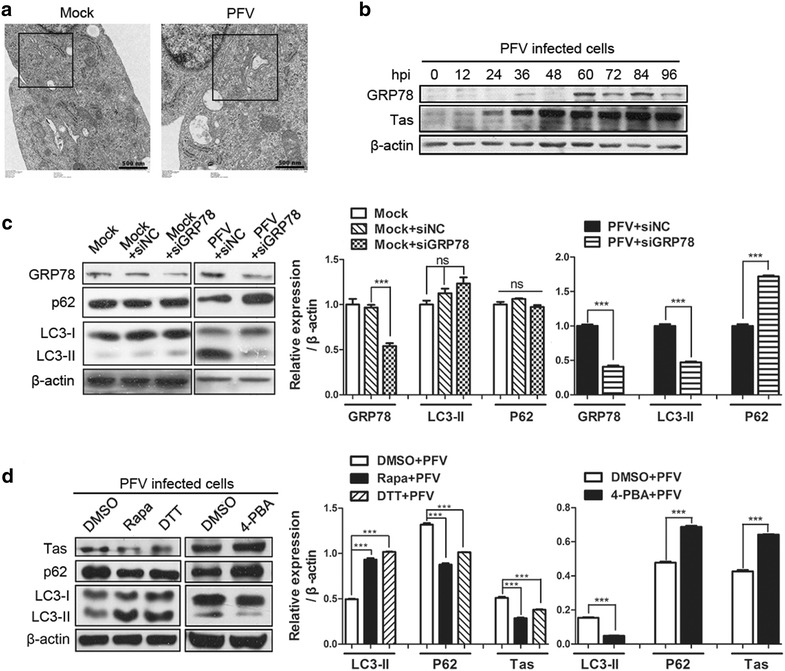
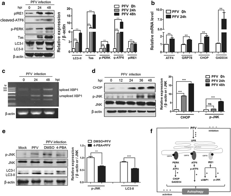
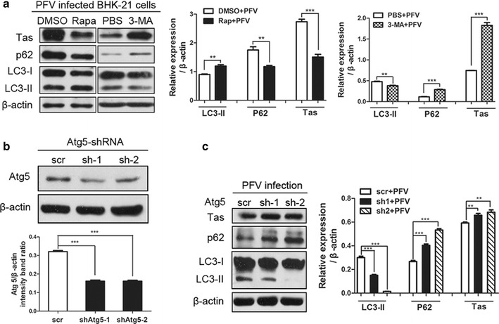
Findings: Our results revealed that PFV infection induced the accumulation of autophagosomes and triggered complete autophagic flux in BHK-21 cells. PFV infection also altered endoplasmic reticulum (ER) homeostasis. The PERK, IRE1 and ATF6 pathways, all of which are components of the ER stress-related unfolded protein response (UPR), were activated in PFV-infected cells. In addition, accelerating autophagy suppressed PFV replication, and inhibition of autophagy promoted viral replication.

Conclusions: Our data indicate that PFV infection can induce complete autophagy through activating the ER stress-related UPR pathway in BHK-21 cells. In turn, autophagy negatively regulates PFV replication.

Keywords: Autophagy; ER stress; PFV; Viral replication.


Figures

Fig. 1
Fig. 1
PFV infection promotes the accumulation of autophagosomes. a BHK-21 cells were infected with PFV to analyze LC3 protein expression by western blotting. After 1.5 h of virus absorption at 37 °C, the cells were further cultured in maintenance medium. The cells were infected with mock or PFV at an MOI of 0.5. After PFV infection, cell samples were harvested at 0, 12, 24, 48, 72 and 96 hpi, and cell extracts were blotted with anti-LC3 and anti-Tas antibodies. b GFP-LC3 dots were visualized via confocal microscopy. BHK-21 cells were transfected with GFP-LC3 plasmids for 24 h, followed by PFV infection (MOI = 0.5) or mock infection for 24 h, and the GFP-LC3 aggregates in the cells were assessed via confocal microscopy. Scale bars 10 μm. The graph shows the quantification of autophagosomes by calculating the average number of dots in 20 cells. c Autophagic vacuoles were detected via TEM. BHK-21 cells infected with PFV at an MOI of 0.5 were processed and analyzed at 24 hpi for the accumulation of autophagosomes via transmission electron microscopy. Black frame indicated representative autophagosomes and arrow indicated representative PFV capsid structures during PFV infection. Scale bars 500 and 100 nm. d BHK-21 cells were infected with UV-inactivated PFV, and the inactivated virus infectivity was confirmed by examining the viral structure protein Gag via western blot. Before infection, PFV were radiated by UV for 0, 0.5, 1.0, 1.5 and 2.0 h respectively. Mock-infected supernatant was also radiated with UV for 2.0 h before infecting cells. Then cells were infected with these UV-inactivated mock supernatant or PFV supernatant for 24 h. e BHK-21 cells were inoculated with normal PFV or UV-inactivated PFV (MOI = 0.5) for 24 h. Before infection, PFV and Mock supernatant were treated with UV for 1.5 h. Then, the cell samples were processed and blotted with anti-LC3 antibody. Quantitation of protein levels from the western blot by using Quantity one software (Bio-Rad); all data are representative of three independent experiments with triplicate samples. Significance was analyzed with a two-tailed Student’s t test. ns P > 0.05, ***P < 0.001
Fig. 2
Fig. 2
Measurement of autophagic flux in BHK-21 cells infected with PFV. a BHK-21 cells were infected with PFV to analyze p62 protein expression by western blotting. After 1.5 h of virus absorption at 37 °C, the cells were further cultured in maintenance medium. The cells were infected with PFV at an MOI of 0.5. After PFV infection, cell samples were harvested at 0, 12, 24, 48, 72 and 96 hpi, and cell extracts were blotted with anti-p62 antibody. b BHK-21 cells were pretreated with the autophagy inhibitor CQ (50 μM) for 4 h, followed by infection with mock or PFV at an MOI of 0.5. After 1.5 h of virus absorption at 37 °C, the cells were further cultured in maintenance medium in the absence or presence of CQ. At 24 h of infection with mock or PFV, the cells were subjected to western blotting using anti-LC3 and anti-p62 antibodies. c BHK-21 cells transfected with ptfLC3 were infected with mock or PFV (MOI = 0.5). The cells were collected, fixed, and visualized at 24 hpi using a confocal microscope. Scale bars 100 μm. d Co-localization of the GFP-LC3 and LAMP1 proteins in BHK-21 cells infected with PFV and treated with CQ or DMSO. The cells were cotransfected with GFP-LAMP1 and RFP-LC3 for 12 h and pretreated with CQ (50 μM) or DMSO control. Then cells were infected with mock or PFV at an MOI of 0.5 for 24 h. The protein localization was observed using a confocal microscope. Scale bars 10 μm. Significance was analyzed with a two-tailed Student’s t test. ns P > 0.05, *P < 0.05, **P < 0.01, ***P < 0.001
Fig. 3
Fig. 3
PFV triggers autophagy by activating ER stress. a Expansion of the ER was detected via transmission electron microscopy (TEM). BHK-21 cells infected with mock supernatant or PFV at an MOI of 0.5 were processed and analyzed at 24 hpi for the ER expansion via electron microscopy. Black frames indicated representative expanded ER lumen. Scale bars 500 nm. b BHK-21 cells were infected with PFV to analyze GRP78/Bip protein expression by western blotting. c BHK-21 cells were transfected with the siRNAs of GRP78 or siRNAs of Negative control for 36 h, followed by infection with mock supernatant or PFV at an MOI of 0.5 or mock infecting. After 1.5 h of virus absorption at 37 °C, the cells were further cultured in maintenance medium. At 24 h after infection with mock or PFV, the cells were subjected to western blotting using anti-GRP78, anti-P62 and anti-LC3 and antibodies. In lane 1, cells were only infected with mock supernatant and cultured in maintenance medium for 48 h. d Western blotting was used to analyze the expression of the viral protein Tas and LC3 in PFV-infected cells in the absence or presence of the ER stress inducer DTT (1 mM) or the ER stress inhibitor 4-PBA (1 mM). Rapa-treated (400 nM) cells were used as a positive control. Significance was analyzed with a two-tailed Student’s t test. ns P > 0.05, ***P < 0.001
Fig. 4
Fig. 4
ER stress-related UPR signaling was activated as PFV infection progressed. a BHK-21 cells were infected with PFV to analyze ATF6, JNK and PERK phosphorylation and IRE1 activation by western blotting. Cells were infected with PFV at an MOI of 0.5. After PFV infection, cell samples were harvested at 0, 24 and 48 h, and cell extracts were evaluated by western blotting. β-actin was used as the loading control. b BHK-21 cells were seeded in 6-well plates and infected with PFV for 24 h (MOI = 0.5). Then, the total RNA (2 μg) was reverse transcribed to cDNA. Q-PCR was used to examine the relative expression (normalized to β-actin) of ER stress sensors such as CHOP, GADD34, ATF4, and GRP78. c BHK-21 cells were seeded in 6-well plates and infected with PFV for 0, 24 and 48 h (MOI = 0.5). The mRNA levels of spliced XBP1 and unspliced XBP1 were measured by RT-PCR. DNA agarose gel electrophoresis revealed the mRNA levels of spliced XBP1 and unspliced XBP1 in the presence or absence of PFV infection. d Western blotting was used to analyze the expression of CHOP and JNK signaling in PFV-infected cells. At 0, 12, 24, 34, 48 h after infection with PFV (MOI = 0.5), the cells were subjected to western blotting with the indicated antibodies. e Western blotting was used to analyze the expression of LC3 and JNK signaling in PFV-infected cells in the absence or presence of the ER-stress inhibitor 4-PBA. BHK-21 cells were pretreated with 4-PBA for 4 h, followed by infection with PFV (MOI = 0.5). Then cells were further cultured in maintenance medium in the absence or presence of 4-PBA. At 48 h after infection with PFV, the cells were subjected to western blotting with the indicated antibodies. As the control groups, BHK-21 cells were infected with mock or PFV (MOI = 0.5). Then cells were further cultured in maintenance medium for 48 h. f The graphical illustrated principle signaling pathways involved in PFV-induced autophagy via the ER stress-related UPR and the summary of regulation between PFV replication and PFV-induced autophagy. Significance was analyzed with a two-tailed Student’s t test. ns P > 0.05, **P < 0.01, ***P < 0.001
Fig. 5
Fig. 5
Inhibition of autophagy promotes PFV replication, and activation of autophagy reduces its replication. a Western blotting was used to analyze the expression of the viral protein Tas in PFV-infected cells in the absence or presence of 3-MA or Rapa. BHK-21 cells were pre-treated with 3-MA (10 mM) or Rapa (400 nM) for 4 h, followed by infection with PFV at an MOI of 0.5. The intracellular virus yields were determined by measuring viral protein at 24 hpi. b Western blotting was used to analyze the expression of Atg5 in cells that down-regulated Atg5 in response to specific shRNAs. BHK-21 cells were transfected with Atg5-specific shRNA1 or shRNA2 for 48 h. The cell samples were harvested and lysed, and cell extracts were measured by immunoblotting with anti-Atg5 antibody. c BHK-21 cells were transfected with Atg5-specific shRNA1 or shRNA2 for 48 h and then infected with PFV (MOI = 0.5) for another 24 h to analyze the expression of the viral protein Tas in PFV-infected cells that down-regulated Atg5 in response to specific shRNAs. Significance was analyzed with a two-tailed Student’s t test. **P < 0.01, ***P < 0.001

References

    1. Rethwilm A. Molecular biology of foamy viruses. Med Microbiol Immunol. 2010;199:197–207. doi: 10.1007/s00430-010-0158-x. - DOI - PubMed
    1. Bodem J, Löchelt M, Yang P, Flügel RM. Regulation of gene expression by human foamy virus and potentials of foamy viral vectors. Stem Cells. 1997;15:141–147. doi: 10.1002/stem.5530150818. - DOI - PubMed
    1. Russell DW, Miller AD. Foamy virus vectors. J Virol. 1996;70:217–222. - PMC - PubMed
    1. Yap MW, Lindemann D, Stanke N, Reh J, Westphal D, Hanenberg H, et al. Restriction of foamy viruses by primate Trim5α. J Virol. 2008;82:5429–5439. doi: 10.1128/JVI.02462-07. - DOI - PMC - PubMed
    1. Delebecque F, Suspène R, Calattini S, Casartelli N, Saïb A, Froment A, et al. Restriction of foamy viruses by APOBEC cytidine deaminases. J Virol. 2006;80:605–614. doi: 10.1128/JVI.80.2.605-614.2006. - DOI - PMC - PubMed

Publication types

LinkOut - more resources