. 2017 Aug 1;23(15):4450-4461.
doi: 10.1158/1078-0432.CCR-16-2426. Epub 2017 Mar 7.

Jab1/Csn5-Thioredoxin Signaling in Relapsed Acute Monocytic Leukemia under Oxidative Stress

Affiliations

Jab1/Csn5-Thioredoxin Signaling in Relapsed Acute Monocytic Leukemia under Oxidative Stress

Fuling Zhou et al. Clin Cancer Res. .

Abstract

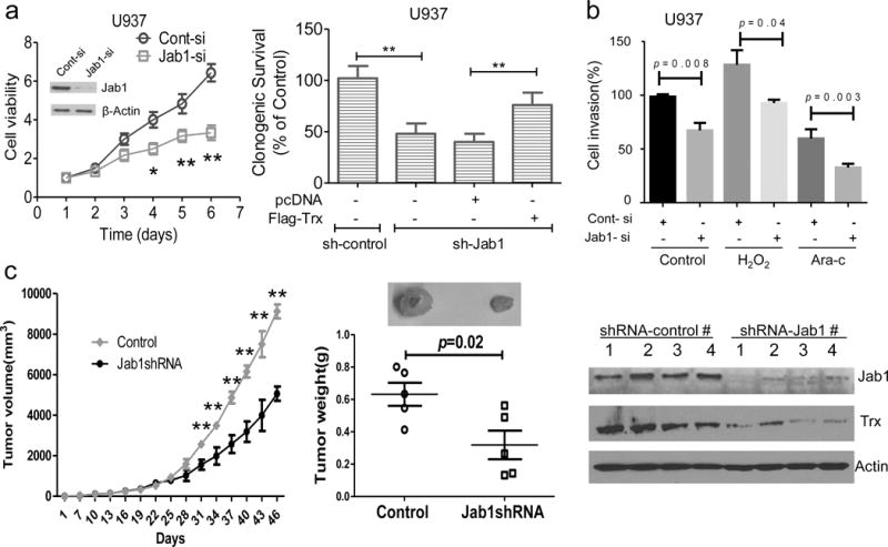
Purpose: High levels of ROS and ineffective antioxidant systems contribute to oxidative stress, which affects the function of hematopoietic cells in acute myeloid leukemia (AML); however, the mechanisms by which ROS lead to malignant transformation in relapsed AML-M5 are not completely understood. We hypothesized that alterations in intracellular ROS would trigger AML-M5 relapse by activating the intrinsic pathway.Experimental Design: We studied ROS levels and conducted c-Jun activation domain-binding protein-1 (JAB1/COPS5) and thioredoxin (TRX) gene expression analyses with blood samples obtained from 60 matched AML-M5 patients at diagnosis and relapse and conducted mechanism studies of Jab1's regulation of Trx in leukemia cell lines.Results: Our data showed that increased production of ROS and a low capacity of antioxidant enzymes were characteristics of AML-M5, both at diagnosis and at relapse. Consistently, increased gene expression levels of TRX and JAB1/COPS5 were associated with low overall survival rates in patients with AML-M5. In addition, stimulating AML-M5 cells with low concentrations of hydrogen peroxide led to increased Jab1 and Trx expression. Consistently, transfection of ectopic Jab1 into leukemia cells increased Trx expression, whereas silencing of Jab1 in leukemia cells reduced Trx expression. Mechanistically, Jab1 interacted with Trx and stabilized Trx protein. Moreover, Jab1 transcriptionally regulated Trx. Furthermore, depletion of Jab1 inhibited leukemia cell growth both in vitro and in vivoConclusions: We identified a novel Jab1-Trx axis that is a key cellular process in the pathobiologic characteristics of AML-M5. Targeting the ROS/Jab1/Trx pathway could be beneficial in the treatment of AML-M5. Clin Cancer Res; 23(15); 4450-61. ©2017 AACR.


Conflict of interest statement

Conflict of interest

The authors declare no competing interests.

Figures

Figure 1
Figure 1. Comparison of ROS levels, antioxidant capacity, LDH levels, and accumulated oxidative damage in AML-M5 patients
(a) Left, intracellular ROS levels were measured by flow cytometry after the cells had been incubated with the redox-sensitive fluorescent dye DCF-DA. The AML-M5 subtype is the most susceptible to oxidative stress, and these cells commonly express high intracellular ROS levels. Relative ROS levels in AML-M5 patients are indicated by the fluorescence intensities of DCF, expressed as a mean ± SD. The levels of oxidative stress were significantly higher at diagnosis and relapse (10.7- and 21.5-fold, respectively) than in healthy donors. ADA and XOD levels were measured using chemical colorimetric assays and used to measure ROS production. ADA and XOD activity in patients with newly diagnosed and relapsed AML-M5 was higher than that in healthy controls: 5.8-fold and 6.4-fold for ADA and 1.6-fold and 2.1-fold for XOD, respectively. Right, the antioxidant capacity levels of T-AOC, SOD, and GSH-Px were lower at diagnosis and relapse than in healthy donors. (b) Left, the antioxidant capacity levels of CAT were lower at diagnosis and relapse than in healthy donors. Disease progression led to decreased regulation of antioxidant enzyme capacity in response to changing levels of oxidation. Elevated LDH levels were found in AML-M5 patients. Patients had higher LDH levels at relapse than did healthy donors. However, there were no significant difference in Cyt-c levels between healthy individuals and AML-M5 patients. Right, the accumulation of ROS in cells damages the resident proteins, lipids, and DNA. It is evident that the accumulation of AOPP, MDA, and 8-OhdG levels was characteristic of AML and reflected the AML-M5 stage. Data are means ± s.d.,*P < 0.05, **P < 0.01, ***P < 0.001, ns = not significant.
Figure 2
Figure 2. High levels of Jab1 and Trx in AML-M5
(a) Left, three groups were compared: patients at diagnosis, patients at relapse, and healthy donors (controls). As determined by quantitative RT-PCR, Jab1 and Trx were overexpressed at diagnosis and relapse compared with their levels in CD34+ cells from healthy donors. GAPDH was used as a control. Values here are means ± the standard error (SE). (b) Left, the expression levels of Jab1 and Trx were correlated in AML-M5 patients. Right, the expression levels of ROS and Jab1 were correlated in AML-M5 patients. The R and P values based on the Pearson Correlation. (c) Left, Overview of the two genes across online data sets (http://r2.amc.nl). Right, Jab1 and Trx were also overexpressed in AML-M5 cell lines compared with their levels in CD34+ cells from healthy donors, as determined by quantitative RT-PCR. Data are means ± s.d. (d) Jab1 and Trx protein expression levels in AML-M5 patients, and healthy donors were analyzed by Western blot analysis. The protein levels were quantified by ImageJ software.
Figure 3
Figure 3. Relationship between ROS/Jab1/Trx levels and overall patient survival
AML-M5 patients with high ROS (above the median) (a), Jab1 (above the median) (b), Trx (above the median) (c), and XOD (above the median) (d) and low T-AOC (below the median) (e) at diagnosis had a poor prognosis. Abnormal cytogenetic (cyto) (f) findings were associated with a poor prognosis.
Figure 4
Figure 4. Jab1 regulates Trx expression in AML
(a) Regulation of Jab1 and Trx expression in AML cells treated with H2O2 (10, 25, and 50 μM) (left) and β-ME (right). (b) Ectopic Jab1 increased Trx protein expression (top), whereas Jab1 knockdown remarkably reduced Trx levels (bottom). (c) Ectopic Jab1 increased Trx RNA levels in U937 and THP-1 cells. (d) The levels of Trx and Jab1 in the cytoplasmic (Cyto) fraction were increased significantly in U937 cells and THP-1 cells after 24 h of H2O2 exposure compared with their levels in the control cells, as indicated. In contrast, only a slight increase in Trx levels was found in the nucleus, and the amount of nuclear Jab1 was unchanged. PARP and tubulin were used as nuclear and cytoplasmic protein markers, respectively. (e) H2O2 did not increase Trx expression in Jab1-deficient U937 cells. Cont-si, control siRNA; Jab1-si, Jab1 siRNA. Data are means ± s.d.,*P < 0.05, **P < 0.01. The protein levels were quantified by ImageJ software.
Figure 5
Figure 5. Jab1 specifically interacts with Trx and regulates Trx stability
(a) U937 and THP-1 cell lysates were immunoprecipitated (IP) with non-immune mouse serum immunoglobulin or Jab1 and immunoblotted with anti-Trx antibodies. Immunoglobulin G (IgG) pull-down assays and total (Tot.) cell lysates were analyzed by Western blotting. Cell lysates were immunoprecipitated with anti-Jab1 antibodies and immunoblotted with anti-Trx. Jab1 specifically interacted with Trx and vice versa. Immunoglobulin G heavy chains (HC) and light chains (LC) are indicated. (b) 293T cells were transfected with His-Jab1, HA/Flag-Trx, or both for 48 h. Cell lysates were immunoprecipitated with anti-His or anti-Flag antibodies and immunoblotted with anti-His, anti-HA and anti-Flag antibodies and other antibodies as indicated. (c) U937 cells were transfected with or without His-Jab1 and treated with 100 ng/ml cycloheximide (CHX) for the indicated times. Cell lysates were collected for Western blot analysis using antibodies targeting Trx, with β-actin used as a control. Bottom, quantification of Trx intensity, as measured by Image J software.
Figure 6
Figure 6. Jab1 positively regulated Trx promoter activity
(a) 293T cells were transfected with either Jab1 siRNA or Jab1 plasmid DNA, along with Trx-luciferase reporter constructs and pRL as an internal control. Trx promoter luciferase activity was measured and normalized to Renilla luciferase activity. Trx promoter activity decreased following Jab1 silencing and increased in a dose-dependent manner with the level of Jab1 plasmid DNA. Rel., relative. (b) Deletion analysis of the Trx gene AP-1. Trx-Luc reporter constructs showing the wild-type (WT) Trx (−975/+1) construct, a deletion (Del.) of AP-1 Trx-Luc, a shorter promoter (−527/+1) that contains only the AP-1 sites (Up AP-1) of Trx, and deletion of AP-1 (Down AP-1). Jab1-induced activation of the Trx gene requires AP-1 binding sites. 293T cells were co-transfected with the indicated Trx-Luc reporter constructs, with or without an increased concentration of Jab1 (μg). Luciferase assays revealed that Trx promoter transactivation by Jab1 occurred, in a dose-dependent manner, only when the AP-1 binding sites were present. Deletion or mutation of the AP-1 site in the Trx promoter abrogated Jab1 transactivation. The result is representative of three independent experiments performed in triplicate. (c) Nuclear extracts of U937 cells were immunoprecipitated by IgG or anti-Jab1. Co-precipitated DNAs were detected by quantitative reverse transcriptase PCR using primers for Trx (including the AP-1 site) or negative control primer (2311 bp downstream of the AP-1 site). The results are expressed relative to the corresponding values for input DNAs. Data are means ± s.d.,*P < 0.05, **P < 0.01.
Figure 7
Figure 7. Depletion of Jab1 inhibits AML-M5 cell viability both in vitro and in vivo
(a) Left, Jab1 knockdown inhibited cell proliferation. U937 cells were transfected with control or Jab1 siRNA, and cell viability was determined by an MTT assay. Right, loss of Jab1 inhibited U937 cell colony formation but transfection of ectopic Trx into Jab1-deficient cells rescued the colony formation. Each well was seeded with 5,000 live cells in medium containing soft agar, and colonies were allowed to form for 2 weeks. (b) The invasiveness of U937 cells was enhanced by H2O2 treatment, inhibited by Ara-c treatment, and further decreased by Jab1 knockdown. Equal numbers of cells were added to the upper wells of Matrigel-coated chambers in serum-free RPMI-1640 medium and treated, as indicated, with H2O2 (50 μM) or Ara-c (0.5 μM). After 24 h of culture, invasive cells were counted using the CyQUANT cell proliferation assay. (c) Left, the tumor-suppressive effects of Jab1 depletion in NOD-SCID mice, as evidenced by tumor growth curves; representative photographs of harvested tumors (inset in panel); and tumor weights measured at the indicated time points (middle). Right, the expression levels of Jab1 and Trx proteins in tissues resected from the same mice were examined via Western blot analysis. Cont-si, control siRNA; Jab1-si, Jab1 siRNA. Data are means ± s.d.,*P < 0.05, **P < 0.01.

References

    1. Zhou FL, Zhang WG, Wei YC, Meng S, Bai GG, Wang BY, et al. Involvement of oxidative stress in the relapse of acute myeloid leukemia. The Journal of biological chemistry. 2010;285(20):15010–5. doi: 10.1074/jbc.M110.103713. - DOI - PMC - PubMed
    1. Jayavelu AK, Muller JP, Bauer R, Bohmer SA, Lassig J, Cerny-Reiterer S, et al. NOX4-driven ROS formation mediates PTP inactivation and cell transformation in FLT3ITD-positive AML cells. Leukemia. 2016;30(2):473–83. doi: 10.1038/leu.2015.234. - DOI - PubMed
    1. Bossis G, Sarry JE, Kifagi C, Ristic M, Saland E, Vergez F, et al. The ROS/SUMO axis contributes to the response of acute myeloid leukemia cells to chemotherapeutic drugs. Cell reports. 2014;7(6):1815–23. doi: 10.1016/j.celrep.2014.05.016. - DOI - PubMed
    1. Sabharwal SS, Schumacker PT. Mitochondrial ROS in cancer: initiators, amplifiers or an Achilles’ heel? Nature reviews Cancer. 2014;14(11):709–21. doi: 10.1038/nrc3803. - DOI - PMC - PubMed
    1. Werlenius O, Aurelius J, Hallner A, Akhiani AA, Simpanen M, Martner A, et al. Reactive oxygen species induced by therapeutic CD20 antibodies inhibit natural killer cell-mediated antibody-dependent cellular cytotoxicity against primary CLL cells. Oncotarget. 2016;7(22):32046–53. doi: 10.18632/oncotarget.8769. - DOI - PMC - PubMed

MeSH terms