. 2017 Apr 21;292(16):6667-6679.
doi: 10.1074/jbc.M117.775858. Epub 2017 Mar 7.

Essential role of the flexible linker on the conformational equilibrium of bacterial peroxiredoxin reductase for effective regeneration of peroxiredoxin

Affiliations

Essential role of the flexible linker on the conformational equilibrium of bacterial peroxiredoxin reductase for effective regeneration of peroxiredoxin

Neelagandan Kamariah et al. J Biol Chem. .

Abstract

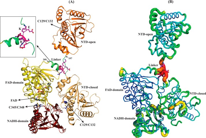
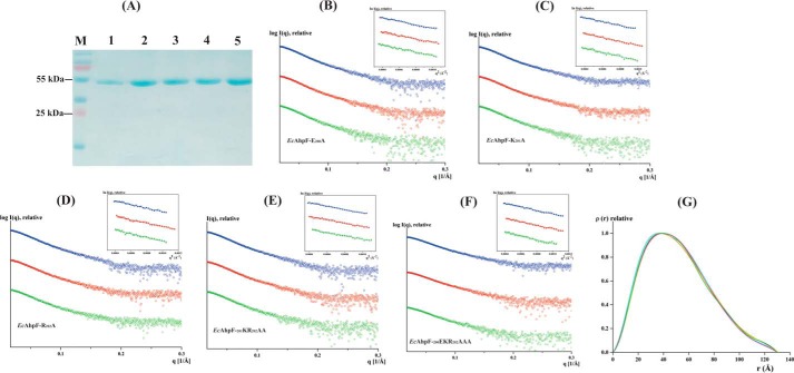
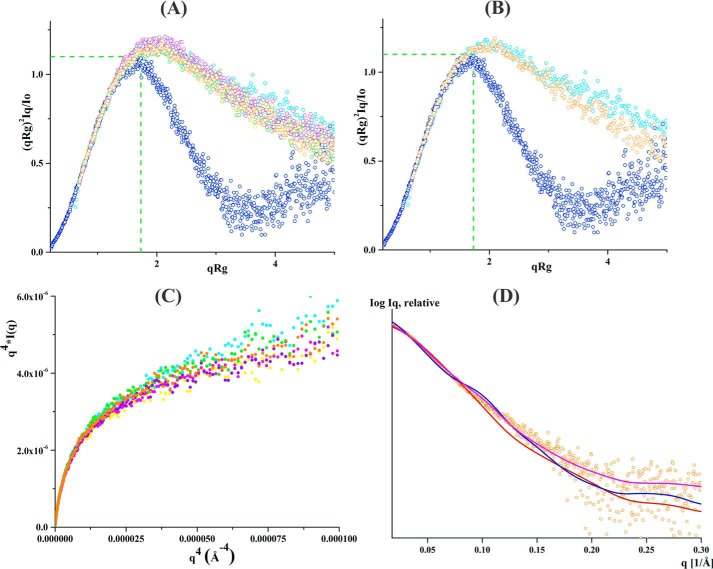
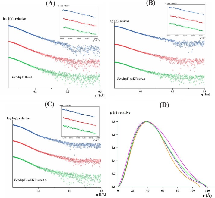
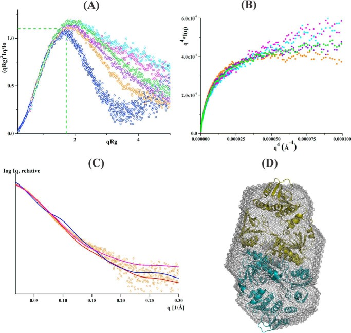
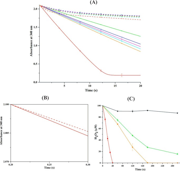
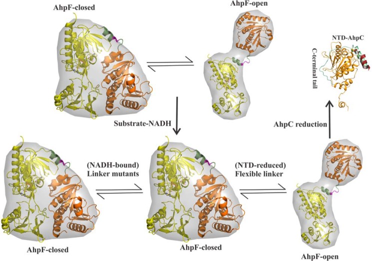
Reactive oxygen species (ROS) can damage DNA, proteins, and lipids, so cells have antioxidant systems that regulate ROS. In many bacteria, a dedicated peroxiredoxin reductase, alkyl hydroperoxide reductase subunit F (AhpF), catalyzes the rapid reduction of the redox-active disulfide center of the antioxidant protein peroxiredoxin (AhpC) to detoxify ROS such as hydrogen peroxide, organic hydroperoxide, and peroxynitrite. AhpF is a flexible multidomain protein that enables a series of electron transfers among the redox centers by accepting reducing equivalents from NADH. A flexible linker connecting the N-terminal domain (NTD) and C-terminal domain (CTD) of AhpF suggests that the enzyme adopts a large-scale domain motion that alternates between the closed and open states to shuttle electrons from the CTD via the NTD to AhpC. Here, we conducted comprehensive mutational, biochemical, and biophysical analyses to gain insights into the role of the flexible linker and the residues critical for the domain motions of Escherichia coli AhpF (EcAhpF) during electron transfer. Small-angle X-ray scattering studies of linker mutants revealed that a group of charged residues, 200EKR202, is crucial for the swiveling motion of the NTD. Moreover, NADH binding significantly affected EcAhpF flexibility and the movement of the NTD relative to the CTD. The mutants also exhibited a decrease in H2O2 reduction by the AhpF-AhpC ensemble. We propose that a concerted movement involving the NTD, C-terminal NADH, and FAD domains, and the flexible linker between them is essential for optimal intra-domain cross-talk and for efficient electron transfer to the redox partner AhpC required for peroxidation.

Keywords: antioxidant; bioenergetics; electron transfer; enzyme mechanism; flavoprotein; protein structure; small-angle X-ray scattering (SAXS).


Conflict of interest statement

The authors declare that they have no conflicts of interest with the contents of this article

Figures

Figure 1.
Figure 1.
Structural comparison of EcAhpF and StAhpF. A, superimposition of the extended and compact conformations of monomeric EcAhpF (14) and StAhpF (16), respectively, reveals the most significant structural difference in the position of NTD. The linker region connects the NTD with the CTD, which contains the FAD- and NADH-binding sites. The linker adopts a loop-helix structure (inset) and the group of charged residues, 200EKR202, in the interface between the loop and helix region, might be involved in the large-scale domain movements of the NTD in solution. B, temperature factor (B-factor) putty representation of the crystal structures of EcAhpF and StAhpF colored by B-factor from low (blue) to high (red), shows the high B-factor for the linker region in both structures. For clarity, AhpF is shown as a monomer.
Figure 2.
Figure 2.
Solution X-ray scattering studies of EcAhpF linker mutants without NADH. A, the 17% SDS gel shows the high purity of the recombinant: lane 1, EcAhpF(E200A/K201A/R202A); lane 2, EcAhpF(K201A/R202A); lane 3, EcAhpF(E200A); lane 4, EcAhpF(K201A); and lane 5, EcAhpF(R202A). Lane M contains a molecular mass protein marker. Small angle X-ray scattering profiles with their corresponding Guinier plots (insets) are shown for the mutants EcAhpF(E200A) (B), EcAhpF(K201A) (C), EcAhpF(R202A) (D), EcAhpF(K201A/R202A) (E), and EcAhpF(E200A/K201A/R202A) (F) measured at various concentrations ranging from 2 to 6 mg/ml in buffer containing 50 mm Tris-HCl, pH 7.5, 200 mm NaCl. G, pair-distance distribution function P(r) shown for EcAhpF(E200A) (yellow), EcAhpF(K201A) (magenta), EcAhpF(R202A) (green), EcAhpF(K201A/R202A) (violet), and EcAhpF(E200A/K201A/R202A) (orange) along with the WT EcAhpF (cyan).
Figure 3.
Figure 3.
SAXS data analysis of linker mutants without NADH. A, normalized Kratky plot of lysozyme (blue) shows the bell-shaped profile. The bell-shapes profile vanishes for the EcAhpF(E200A) (yellow), EcAhpF(K201A) (magenta), EcAhpF(R202A) (green), EcAhpF(K201A/R202A) (violet), EcAhpF(E200A/K201A/R202A) (orange), and WT EcAhpF (cyan). B, normalized Kratky plots of EcAhpF(E200A/K201A/R202A) (orange) show a slightly altered profile compared with WT EcAhpF (cyan). C, the absence of plateau at low q angles of the Porod-Debye plots for EcAhpF(E200A) (yellow), EcAhpF(K201A) (magenta), EcAhpF(R202A) (green), EcAhpF(K201A/R202A) (violet), EcAhpF(E200A/K201A/R202A) (orange), and WT EcAhpF (cyan) supports the presence of flexibility. D, the fits between the experimental scattering data of EcAhpF(E200A/K201A/R202A) (○) and the theoretical scattering pattern (—) calculated using the dimers of compact (red) and extended (blue) crystal conformations. The mixture of both conformations (magenta) fits better to the experimental data.
Figure 4.
Figure 4.
SAXS studies of linker mutants in the presence of NADH. Experimental scattering profile of linker mutants in the presence of 1 mm NADH for: A, EcAhpF(R202A); B, EcAhpF(K201A/R202A); and C, EcAhpF(E200A/K201A/R202A) and their corresponding Guinier plots (insets) demonstrates linearity, indicating no aggregation. D, pair-distance distribution function P(r) for EcAhpF(R202A) (green), EcAhpF(K201A/R202A) (violet), and EcAhpF(E200A/K201A/R202A) (orange) is shown along with WT EcAhpF (magenta).
Figure 5.
Figure 5.
SAXS flexibility analysis of linker mutants in the presence of NADH. A, normalized Kratky plots of EcAhpF(R202A) (green), EcAhpF(K201A/R202A) (violet), EcAhpF(E200A/K201A/R202A) (orange), and WT EcAhpF (magenta) measured in the presence of NADH along with the substrate-free WT EcAhpF (cyan) and lysozyme (blue). B, Porod-Debye plots of EcAhpF(R202A) (green), EcAhpF(K201A/R202A) (violet), and EcAhpF(E200A/K201A/R202A) (orange) and WT EcAhpF (magenta) measured in the presence of NADH along with the substrate-free WT EcAhpF (cyan). C, the fits of the experimental scattering data of the NADH-bound form of EcAhpF(E200A/K201A/R202A) (○) with the theoretical scattering pattern (—) calculated using the compact (red), extended (blue) dimers and the mixture of both conformations. D, the averaged ab initio model (mesh) is overlaid with the compact dimer structure of StAhpF (16).
Figure 6.
Figure 6.
Peroxidase activity of the AhpF-AhpC complex. A, NADH-dependent peroxidase activity was measured for 0.4 μm WT EcAhpF (red) or the different linker mutants, EcAhpF(E200A) (cyan), EcAhpF(K201A) (magenta), EcAhpF(R202A) (orange), EcAhpF(K201A/R202A) (blue), and EcAhpF(E200A/K201A/R202A) (green) in the presence of 30 μm EcAhpC, 300 μm NADH with (straight lines) or without (dashed lines) 1 mm H2O2 in a reaction mixture containing 50 mm phosphate buffer, pH 7.0, 100 mm ammonium sulfate, and 0.5 mm EDTA. B, the initial rate of NADH oxidation for WT EcAhpF and EcAhpC with (straight lines) or without (dashed lines) 1 mm H2O2. C, to determine the H2O2 decomposition, 0.4 μm (■) WT EcAhpF, (●) EcAhpF(R202A), and (▴) EcAhpF(E200A/K201A/R202A) were added to the reaction mixture containing 30 μm EcAhpC, 750 μm NADH, and 100 μm H2O2. The residual H2O2 in the reaction mixtures was measured at the indicated times using ferrous oxidation xylenol reagent. The control was measured in the absence of EcAhpF (▾). The linker mutants significantly affect the EcAhpC-dependent peroxidase activity. The data presented were averaged of three independent measurements, and error bars represent the standard deviation.
Figure 7.
Figure 7.
EcAhpC reduction assay. Non-reducing SDS-PAGE analysis for the reduction of 20 μm oxidized EcAhpC by 50 nm WT EcAhpF (A) and EcAhpF(E200A/K201A/R202A) (B) in the presence of 100 μm NADH for different time periods (0.5 to 10 min) in 50 mm HEPES buffer, pH 7.0. The reaction without NADH was measured as control (denoted as C). Reduced AhpC runs as a monomer (M) and oxidized AhpC forms a dimer (D).
Figure 8.
Figure 8.
NTD conformational selection. The NTD (orange) of AhpF is proposed to be present in a rapid conformational equilibrium between the closed and open states. The flexible linker (green) enables the rapid equilibria of NTD motion, wherein the closed state enables the NTD to interact with the CTD (yellow) and the open conformation allows the NTD to bind the oxidized AhpC for regeneration. The 200EKR202 mutants (magenta) affect the conformational equilibria of NTD movement in solution. For clarity, AhpF is shown as a monomer.

References

    1. Imlay J. A. (2013) The molecular mechanisms and physiological consequences of oxidative stress: lessons from a model bacterium. Nat. Rev. Microbiol. 11, 443–454 - PMC - PubMed
    1. D'Autréaux B., and Toledano M. B. (2007) ROS as signalling molecules: mechanisms that generate specificity in ROS homeostasis. Nat. Rev. Mol. Cell Biol. 8, 813–824 - PubMed
    1. Winterbourn C. C. (2008) Reconciling the chemistry and biology of reactive oxygen species. Nat. Chem. Biol. 4, 278–286 - PubMed
    1. Rhee S. G., Woo H. A., Kil I. S., and Bae S. H. (2012) Peroxiredoxin functions as a peroxidase and a regulator and sensor of local peroxides. J. Biol. Chem. 287, 4403–4410 - PMC - PubMed
    1. Jacobson F. S., Morgan R. W., Christman M. F., and Ames B. N. (1989) An alkyl hydroperoxide reductase from Salmonella typhimurium involved in the defense of DNA against oxidative damage: purification and properties. J. Biol. Chem. 264, 1488–1496 - PubMed

LinkOut - more resources