. 2017 Apr 28;292(17):7244-7257.
doi: 10.1074/jbc.M117.782037. Epub 2017 Mar 7.

Structure and specificity of a new class of Ca2+-independent housekeeping sortase from Streptomyces avermitilis provide insights into its non-canonical substrate preference

Affiliations

Structure and specificity of a new class of Ca2+-independent housekeeping sortase from Streptomyces avermitilis provide insights into its non-canonical substrate preference

Sreetama Das et al. J Biol Chem. .

Abstract

Surface proteins in Gram-positive bacteria are incorporated into the cell wall through a peptide ligation reaction catalyzed by transpeptidase sortase. Six main classes (A-F) of sortase have been identified of which class A sortase is meant for housekeeping functions. The prototypic housekeeping sortase A (SaSrtA) from Staphylococcus aureus cleaves LPXTG-containing proteins at the scissile T-G peptide bond and ligates protein-LPXT to the terminal Gly residue of the nascent cross-bridge of peptidoglycan lipid II precursor. Sortase-mediated ligation ("sortagging") of LPXTG-containing substrates and Gly-terminated nucleophiles occurs in vitro as well as in cellulo in the presence of Ca2+ and has been applied extensively for protein conjugations. Although the majority of applications emanate from SaSrtA, low catalytic efficiency, LPXTG specificity restriction, and Ca2+ requirement (particularly for in cellulo applications) remain a drawback. Given that Gram-positive bacteria genomes encode a variety of sortases, natural sortase mining can be a viable complementary approach akin to engineering of wild-type SaSrtA. Here, we describe the structure and specificity of a new class E sortase (SavSrtE) annotated to perform housekeeping roles in Streptomyces avermitilis Biochemical experiments define the attributes of an optimum peptide substrate, demonstrate Ca2+-independent activity, and provide insights about contrasting functional characteristics of SavSrtE and SaSrtA. Crystal structure, substrate docking, and mutagenesis experiments have identified a critical residue that dictates the preference for a non-canonical LAXTG recognition motif over LPXTG. These results have implications for rational tailoring of substrate tolerance in sortases. Besides, Ca2+-independent orthogonal specificity of SavSrtE is likely to expand the sortagging toolkit.

Keywords: Streptomyces; crystal structure; enzyme catalysis; peptide; peptides; sortase A; substrate specificity; transpeptidase.


Conflict of interest statement

The authors declare that they have no conflicts of interest with the contents of this article

Figures

Figure 1.
Figure 1.
Characterization of recombinant SavSrtE. A, SDS-PAGE of purified recombinant SavSrtE comprising residues 51–230 expressed with an N-terminal hexa-His tag: lane 1, molecular weight marker; lane 2, SavSrtE. B, ES-MS of the purified protein. The deconvoluted spectrum produced two peaks (22,157 and 22,335 Da, respectively). A mass of 22,157 Da corresponds to the calculated mass of expressed sequence (22,289 Da) without a Met residue. The second peak, which is higher by 178 Da, presumably represents a gluconylated protein.
Figure 2.
Figure 2.
Residue specificity at the second position of the pentapeptide recognition motif. Transpeptidation assays were carried out using YALXNTGK (0.5 mm) as a model donor and GGGKY (1 mm) as an acceptor peptide in the presence of 50 μm SavSrtE at 20 °C for 6 h. A, chemical structure of residue integrated at X position in YALXNTGK. B, time course of the transpeptidation reaction of YALANTGK (X = 6) with GGGKY. Inset shows a representative RP-HPLC profile of transpeptidation reaction carried out for 6 h. C, transpeptidation yield obtained with various substrates after 6 h of reaction. Data represents mean ± S.D. of three independent experiments.
Figure 3.
Figure 3.
Influence of residues adjacent to the pentapeptide recognition motif. The SavSrtE-catalyzed transpeptidation reactions were carried with respective LAXTG or LPXTG motif peptide (0.5 mm) and GGGKY (1 mm) with 50 μm enzyme at 20 °C for the indicated time. Each reaction mixture was subjected to RP-HPLC analysis as described under “Experimental procedures.” A, time course of transpeptidation reaction of LANTG motif: YALANTGA (filled circle); YALANTGK (filled square); and YALANTGE (filled triangles). B, comparative reaction time course of LAETG and LPETG peptide substrates; YNLAETGA (filled circles) and YNLPETGA (filled squares).
Figure 4.
Figure 4.
Comparison of SavSrtE to sortases of classes A–D. A, structure-based sequence alignment of SavSrtE (SavE in the figure) to other sortases of known structure, namely S. aureus SrtA (SauA), S. pyogenes SrtA (SpyA), S. agalactiae SrtA (SagA), B. anthracis SrtA (BanA), S. pyogenes SrtB (SpyB), B. anthracis SrtB (BanB), S. aureus SrtB (SauB), C. difficile SrtB (CdfB), S. pneumoniae SrtC1 (SpnC1), S. pneumoniae SrtC2 (SpnC2), S. pneumoniae SrtC3 (SpnC3), C. perfringens SrtD (CprD), and B. anthracis SrtD (BanD). The corresponding PDB codes are mentioned. The numerals 1–5 refer to the five different classes. Conserved residues are marked in white on a red background, and similar residues are in red. The secondary structure of S. aureus SrtA is shown. The catalytic residues are marked by red triangles; Ca2+-binding residues in S. aureus SrtA are marked by blue triangles; and a Tyr residue in SavSrtE (Tyr-112), found to be conserved in all sortase E enzymes listed in the Sortase database, has been marked by an inverted brown triangle. B, structure of sortases (class A–E) depicting the eight-stranded β-barrel fold (bottom) and the corresponding topological diagram (top). Strands are in yellow; helices are in red, and loops are in green or black. The catalytic residues are shown as blue sticks. The structures depicted are S. aureus SrtA (PDB code 2kid), S. aureus SrtB (PDB code 1ng5), S. pneumoniae SrtC1 (PDB: 2w1j) with the Lid region that closes the active site in the absence of substrate colored in blue, B. anthracis SrtD (PDB code 2ln7), and S. avermitilis SrtE (PDB code 5GO5).
Figure 5.
Figure 5.
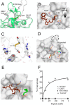
Analysis of SavSrtE structure. A, crystal structure of SavSrtE, with the catalytic residues shown as blue sticks (the dots represent part of the β7/β8 loop whose electron density is not observed). The insertion in the β6/β7 loop, containing a 310 helix, is highlighted by a dotted box. B, surface representation of SavSrtE, colored by B-factors from low (blue) to high (red). Active-site loops and catalytic residues are labeled. The first 310 helix in β6/β7 loop, which is expected to interact with the bound peptide, has low B-factors, whereas the portion of the β7/β8 loop with observed electron density has high B-factors (the dots represent part of the β7/β8 loop for which interpretable electron density is not observed). C, β6/β7 loop in the apo-structure of S. aureus SrtA (SaSrtA, PDB code 1ija, pink) is in open conformation, whereas a 310 helix is induced upon Ca2+ binding (green sphere), which closes over the bound substrate (not shown) in the holo-structure (PDB code 2kid, cyan). Comparison of apo-SavSrtE (blue) with holo-SaSrtA and apo-structure from B. anthracis SrtA (BaSrtA, PDB code 2kw8, orange) shows the 310 helix in β6/β7 loop and its closed conformation, resulting in a preformed binding pocket. D, SaSrtA contains a cluster of negatively charged residues which are stabilized by Ca2+ binding. The equivalent residues in SavSrtE do not form a charged cluster that requires neutralization by Ca2+ ion. Interactions of equivalent residues in BaSrtA involve a Lys in charge neutralization. E, electrostatic surface potential in SaSrtA showing the negatively charged residues, whereas SavSrtE has no such charged cluster. Surface electrostatics was calculated using Accelrys Discovery studio software. The color scheme ranges from blue (for electropositive regions) to red (for electronegative regions). F, effect of Ca2+ on transpeptidation reaction. The reactions were carried out at 20 °C using 50 μm SavSrtE with YNLAETGA (0.5 mm) and GGGKY (1 mm) in the absence and presence of Ca2+ (5 mm). The reaction mixture was processed by RP-HPLC, and the product was quantified as described under “Experimental procedures.”
Figure 6.
Figure 6.
Binding site of the second substrate. A, binding of the first substrate leads to the displacement of the β7/β8 loop (gray) from the position in the apo-protein (green) and the formation of a binding site for the second substrate in SaSrtA (PDB codes 1ija and 2kid) and BaSrtA (not shown). B, surface representation shows the active-site residues, bound-peptide (brown stick representation), and the second groove in SaSrtA. C, electron density (2FoFc omit map at 1σ level) for a Gly residue in the putative second pocket close to the active site in SavSrtE (catalytic residues are shown for reference; Cys-198 is modified to Came by β-mercaptoethanol); and D, surface representation of SavSrtE showing the second pocket (the dots represent part of the β7/β8 loop whose electron density is not observed) and the bound Gly. E, model of the second substrate GGG peptide (green sticks) docked into SavSrtE with bound ALANT peptide (shown as brown sticks; missing residues in the β7/β8 loop have been modeled). F, SavSrtE-mediated transpeptidation reaction of YNLAETGA (0.5 mm) was carried out with varying concentrations of each nucleophile at 20 °C for 6 h using 50 μm enzyme.
Figure 7.
Figure 7.
Modeling protein-substrate interactions shows the importance of a Tyr residue at the active site. The molecular surface of the protein has been rendered in gray, and the peptides are shown as sticks. Interactions of the peptides to the active site Arg are shown by black dots. Important active site residues have been labeled. A, ALPNT (cyan) peptide in the active site pocket of SaSrtA, modeled using the LPAT*-bound SaSrtA structure (PDB code 2kid). B, ALPNT (cyan), and C, ALANT (green) peptides docked into the active-site pocket of SavSrtE. The presence of Tyr-112 hinders better binding of the Pro-containing peptide and favors the Ala-containing peptide. D, percent identity matrix showing the variation in sequence identity (in shades of gray; black indicates identity of 100%, and white indicates low identity) among SrtE sequences in the Sortase database. Sequences sharing higher identity have been grouped together. The sequences are as follows: 1) NP_789137 and 2) NP_787692 (Tropheryma whipplei); 3) cory_diphtheriae.6302c (Corynebacterium diphtheriae); 4) NP_739396 (Corynebacterium efficiens); 5) NP_602126 (Corynebacterium glutamicum); 6) thermo_fusca.3646 (Thermobifida fusca); 7) bifido_longum_DJO10A.7548; 8) NP_695779 (Bifidobacterium longum); 9) NP_825510 (SAV4333); 10) NP_826383 (SAV5206); 11) NP_825514 (SAV4337); 13) NP_825513 (SAV4336) (Streptomyces avermitilis); 12) NP_628037; and 14) NP_628038 (Streptomyces coelicolor). E, multiple sequence alignment of class E sortases depicting the presence of a Tyr (inverted brown triangle) equivalent to Tyr-112 at the active site of SavSrtE. The catalytic His residue is marked by a red triangle.
Figure 8.
Figure 8.
Transpeptidation activity of Y112F mutant. Transpeptidation assay was carried out with 0.5 mm Ala substrate (YNLAETGA) or Pro substrate (YNLPETGA) and 1 mm GGGKY in the presence of 50 μm wild-type SavSrtE or Y112F mutant. A, representative RP-HPLC traces of reaction carried out for 6 h at 20 °C. The chromatographic profile of the wild-type SavSrtE and Y112F mutant are shown in top and bottom panel, respectively. B, comparative time course of reactions catalyzed by wild type and Y112F for Ala and Pro substrates. The time course profile of the wild type is shown by continuous curves, and curves drawn with broken lines represent data of the Y112F mutant. The Ala substrate and Pro substrate are indicated by circle and rectangle, respectively.

References

    1. Marraffini L. A., Dedent A. C., and Schneewind O. (2006) Sortases and the art of anchoring proteins to the envelopes of Gram-positive bacteria. Microbiol. Mol. Biol. Rev. 70, 192–221 - PMC - PubMed
    1. Mazmanian S. K., Ton-That H., and Schneewind O. (2001) Sortase-catalysed anchoring of surface proteins to the cell wall of Staphylococcus aureus. Mol. Microbiol. 40, 1049–1057 - PubMed
    1. Mazmanian S. K., Liu G., Ton-That H., and Schneewind O. (1999) Staphylococcus aureus sortase, an enzyme that anchors surface proteins to the cell wall. Science 285, 760–763 - PubMed
    1. Novick R. P. (2000) Sortase: the surface protein anchoring transpeptidase and the LPXTG motif. Trends Microbiol. 8, 148–151 - PubMed
    1. Lévesque C. M., Voronejskaia E., Huang Y. C., Mair R. W., Ellen R. P., and Cvitkovitch D. G. (2005) Involvement of sortase anchoring of cell wall proteins in biofilm formation by Streptococcus mutans. Infect. Immun. 73, 3773–3777 - PMC - PubMed

Publication types