. 2017 Jun;66(6):1470-1478.
doi: 10.2337/db16-1107. Epub 2017 Mar 7.

Functional Human Beige Adipocytes From Induced Pluripotent Stem Cells

Affiliations

Functional Human Beige Adipocytes From Induced Pluripotent Stem Cells

Anne-Claire Guénantin et al. Diabetes. 2017 Jun.

Abstract

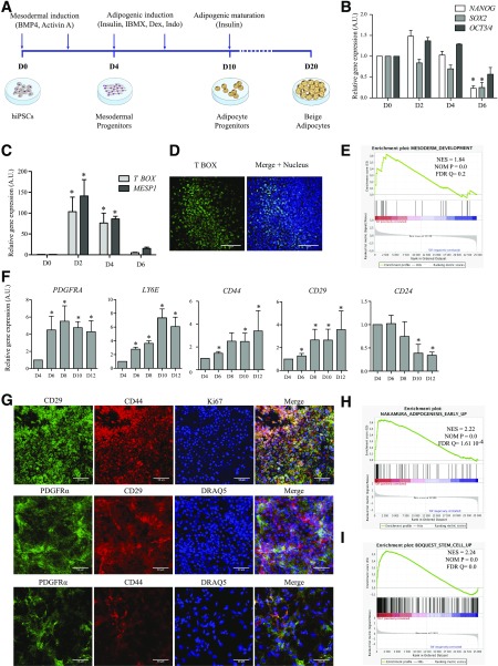
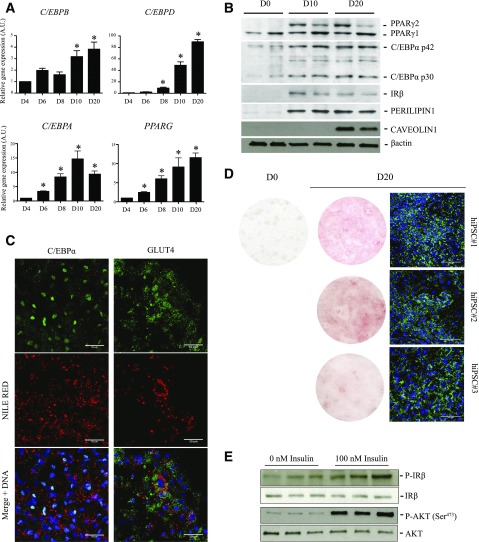
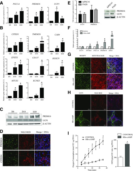
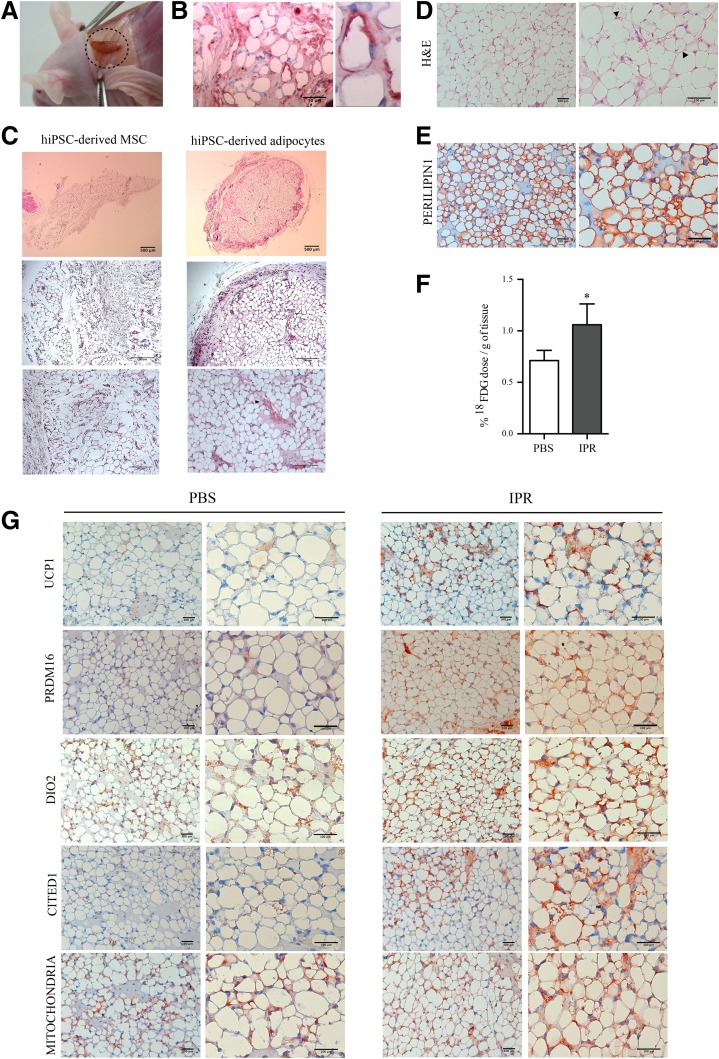
Activation of thermogenic beige adipocytes has recently emerged as a promising therapeutic target in obesity and diabetes. Relevant human models for beige adipocyte differentiation are essential to implement such therapeutic strategies. We report a straightforward and efficient protocol to generate functional human beige adipocytes from human induced pluripotent stem cells (hiPSCs). Without overexpression of exogenous adipogenic genes, our method recapitulates an adipogenic developmental pathway through successive mesodermal and adipogenic progenitor stages. hiPSC-derived adipocytes are insulin sensitive and display beige-specific markers and functional properties, including upregulation of thermogenic genes, increased mitochondrial content, and increased oxygen consumption upon activation with cAMP analogs. Engraftment of hiPSC-derived adipocytes in mice produces well-organized and vascularized adipose tissue, capable of β-adrenergic-responsive glucose uptake. Our model of human beige adipocyte development provides a new and scalable tool for disease modeling and therapeutic screening.


Figures

Figure 1
Figure 1
hiPSC-derived mesodermal progenitors efficiently differentiate into adipose progenitors. A: Experimental scheme for the differentiation of hiPSCs into beige adipocytes. RT-qPCR analysis of expression of indicated pluripotency markers (B) and mesodermal transcription factors (C) in hiPSC#1 clone from D0 to D6 (mean ± SEM arbitrary units [A.U.] relative to D0; n ≥ 3 independent experiments; *P < 0.05 relative to D0, Mann-Whitney U test). D: T BOX immunostaining of mesodermal progenitors issued from hiPSC#1 (D4). Nuclei were stained with DRAQ5 (blue). Scale bars = 100 µm. E: GSEA of hiPSC-derived cells on D4 vs. D0 using the mesoderm development Gene Ontology gene set (GO:0007498); n ≥ 3 experiments. F: Time-course RT-qPCR analysis of expression of indicated genes after adipogenic induction of hiPSC#1 (D4 to D12; mean ± SEM A.U. relative to D4; n ≥ 3 experiments; *P < 0.05 relative to D4; Mann-Whitney U test). G: Immunodetection of markers of adipose progenitors and of cell proliferation (Ki67) on D10. Nuclei were stained with DRAQ5. Scale bars = 50 μm. H and I: GSEA of hiPSC-derived cells using published data sets, with early adipogenesis transcriptomic signatures on D8 vs. D4 (H) and human adult adipose stromal cell signature on D12 vs. D4 (I) (n = 3 independent experiments).
Figure 2
Figure 2
hiPSCs differentiate into adipocytes. A: Time-course mRNA expression of indicated adipocyte transcription factors during differentiation of hiPSC#1 (mean ± SEM arbitrary units [A.U.] relative to D4; n ≥ 3 experiments; *P < 0.05 relative to D4, Mann-Whitney U test). B: Detection of PPARγ1, PPARγ2, C/EBPα p30/p42, IR-β, perilipin 1, and caveolin 1 in hiPSC#1 on D0, D10, and D20. β-Actin was used as loading control. Duplicate lanes shown are from duplicate differentiation wells. C: Immunodetection of C/EBPα and GLUT4 in lipid-containing (Nile Red) adipocytes (hiPSC#1) on D20. Scale bars = 50 µm. D: Oil Red O (left panels) and BODIPY (right panel) staining of adipocyte-differentiated hiPSC#1, hiPSC#2, and hiPSC#3 on D0 and/or D20. Cells were fixed in 3.2% paraformaldehyde for 20 min and incubated for 1 h in Oil Red O (O0625; Sigma-Aldrich) diluted in isopropanol. Scale bars = 100 μm. E: Short-term insulin-mediated phosphorylation of IR-β and AKT/PKB evaluated using phosphospecific and pan antibodies in hiPSC#1 on D20. Differentiated adipocytes were deprived in insulin and serum for 12 h before 10 min incubation in 100 nmol/L insulin (Sigma-Aldrich), and then immediately lysed and processed for Western blotting. Triplicate lanes are from three independent stimulation experiments.
Figure 3
Figure 3
hiPSC-derived adipocytes display beige properties. Time-course mRNA expression of beige/brown adipocyte markers (A) and beige adipocyte-specific genes (B) (mean ± SEM arbitrary units [A.U.] relative to D4; n ≥ 3 experiments; *P < 0.05 relative to D4, Mann-Whitney U test). C: PRDM16 and UCP1 protein expression on D0, D10, and D20. D: PRDM16 and CITED1 immunostaining on D20 in lipid-containing (Nile Red) adipocytes. Scale bars = 50 µm. E: Comparison of PRDM16 and UCP1 mRNA and protein expression in hiPSC-derived adipocytes and in PAZ6 cells, at baseline and/or after 20 days of differentiation. F: Relative levels of expression of indicated genes in hiPSC#1-derived adipocytes at D20 treated or not with 1 mmol/L 8Br-cAMP (Sigma-Aldrich) for 48 h (mean ± SEM A.U. relative to D4; n ≥ 3 experiments; *P < 0.05 relative to D4; Mann-Whitney U test). Mitochondrial content (MitoTracker) (G) and UCP1 immunostaining (H) in lipid-containing cells (BODIPY or Nile Red, as indicated) analyzed as in F. Cells were incubated with 1 μmol/L MitoTracker Red CMXRos (Life Technologies) for 45 min and fixed with 3.2% paraformaldehyde for 15 min. Scale bars = 50 µm. I: Time-course analysis of oxygen consumption (left) and oxygen consumption rates (OCR; right) in hiPSC#1-derived adipocytes at D20 stimulated or not with 8Br-cAMP (mean ± SEM; n ≥ 3; *P < 0.05 relative to control, Mann-Whitney U test). PO2 values were plotted over time to calculate OCR from the slope.
Figure 4
Figure 4
hiPSC-derived adipocytes generate adipose tissue in vivo. A: Macroscopic view of a neoformed fat pad developed from hiPSC-derived adipocytes. B: Human cytoplasmic staining of neoformed adipose tissue (n ≥ 3). C: Hematoxylin-eosin (H&E) staining of fat pads developed from hiPSC-derived MSCs or hiPSC-derived adipocytes. Arrowheads indicate blood vessels. Scale bars = 500 μm (top), 200 μm (middle), 100 μm (bottom) (n ≥ 3). Staining of a neoformed fat pad developed from hiPSC-derived adipocytes with H&E (D) or antibodies against perilipin 1 (E). Arrowheads indicate multilocular adipocytes. Scale bars = 100 μm (n ≥ 3 independent experiments). F: 18FDG uptake in neoformed fat pads after hiPSC-derived adipocyte engraftment in immunodeficient mice treated with vehicle (PBS) or isoproterenol (IPR; mean ± SEM; n ≥ 3 independent experiments; *P < 0.05 relative to PBS control, Wilcoxon signed rank test). G: Immunostaining of a neoformed fat pad developed from hiPSC-derived adipocytes for UCP1, PRDM16, DIO2, CITED1, and a human-specific mitochondrial marker (targeted by the MAB1273 antibody) in basal (PBS) and stimulated (IPR) conditions. Scale bars = 100 μm.

Comment in

References

    1. Cypess AM, Lehman S, Williams G, et al. . Identification and importance of brown adipose tissue in adult humans. N Engl J Med 2009;360:1509–1517 - PMC - PubMed
    1. Lidell ME, Betz MJ, Dahlqvist Leinhard O, et al. . Evidence for two types of brown adipose tissue in humans. Nat Med 2013;19:631–634 - PubMed
    1. Cereijo R, Giralt M, Villarroya F. Thermogenic brown and beige/brite adipogenesis in humans. Ann Med 2015;47:169–177 - PubMed
    1. Chondronikola M, Volpi E, Børsheim E, et al. . Brown adipose tissue improves whole-body glucose homeostasis and insulin sensitivity in humans. Diabetes 2014;63:4089–4099 - PMC - PubMed
    1. Kajimura S, Spiegelman BM, Seale P. Brown and beige fat: physiological roles beyond heat generation. Cell Metab 2015;22:546–559 - PMC - PubMed

Publication types

MeSH terms