Randomized Controlled Trial
. 2017 Mar 29;37(13):3610-3620.
doi: 10.1523/JNEUROSCI.3700-16.2017. Epub 2017 Mar 7.

Mild Cognitive Impairment Is Characterized by Deficient Brainstem and Cortical Representations of Speech

Affiliations
Randomized Controlled Trial

Mild Cognitive Impairment Is Characterized by Deficient Brainstem and Cortical Representations of Speech

Gavin M Bidelman et al. J Neurosci. .

Abstract

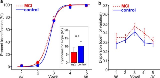
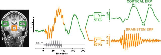
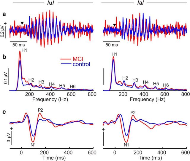
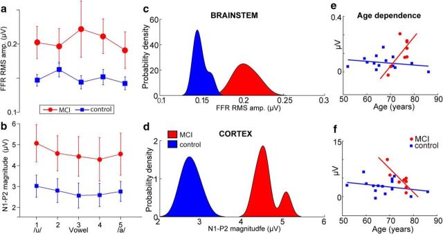
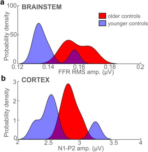
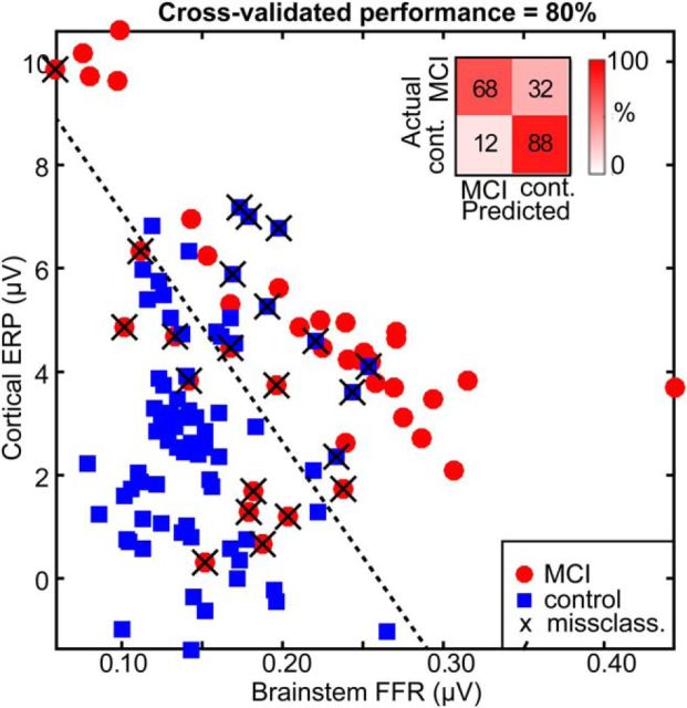
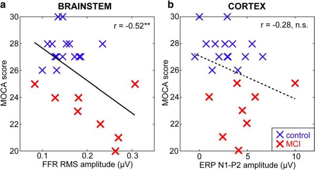
Mild cognitive impairment (MCI) is recognized as a transitional phase in the progression toward more severe forms of dementia and is an early precursor to Alzheimer's disease. Previous neuroimaging studies reveal that MCI is associated with aberrant sensory-perceptual processing in cortical brain regions subserving auditory and language function. However, whether the pathophysiology of MCI extends to speech processing before conscious awareness (brainstem) is unknown. Using a novel electrophysiological approach, we recorded both brainstem and cortical speech-evoked brain event-related potentials (ERPs) in older, hearing-matched human listeners who did and did not present with subtle cognitive impairment revealed through behavioral neuropsychological testing. We found that MCI was associated with changes in neural speech processing characterized as hypersensitivity (larger) brainstem and cortical speech encoding in MCI compared with controls in the absence of any perceptual speech deficits. Group differences also interacted with age differentially across the auditory pathway; brainstem responses became larger and cortical ERPs smaller with advancing age. Multivariate classification revealed that dual brainstem-cortical speech activity correctly identified MCI listeners with 80% accuracy, suggesting its application as a biomarker of early cognitive decline. Brainstem responses were also a more robust predictor of individuals' MCI severity than cortical activity. Our findings suggest that MCI is associated with poorer encoding and transfer of speech signals between functional levels of the auditory system and advance the pathophysiological understanding of cognitive aging by identifying subcortical deficits in auditory sensory processing mere milliseconds (<10 ms) after sound onset and before the emergence of perceptual speech deficits.SIGNIFICANCE STATEMENT Mild cognitive impairment (MCI) is a precursor to dementia marked by declines in communication skills. Whether MCI pathophysiology extends below cerebral cortex to affect speech processing before conscious awareness (brainstem) is unknown. By recording neuroelectric brain activity to speech from brainstem and cortex, we show that MCI hypersensitizes the normal encoding of speech information across the hearing brain. Deficient neural responses to speech (particularly those generated from the brainstem) predicted the presence of MCI with high accuracy and before behavioral deficits. Our findings advance the neurological understanding of MCI by identifying a subcortical biomarker in auditory-sensory processing before conscious awareness, which may be a precursor to declines in speech understanding.

Keywords: auditory evoked potentials; brainstem frequency-following response (FFR); cognitive aging; dementia biomarkers; event-related brain potential (ERPs); speech processing.


Figures

Figure 1.
Figure 1.
Behavioral speech listening skills are similar in MCI and control listeners. Shown are behavioral identification (a) and variability (CVs) in RTs (b) for speech identification in normal and MCI listeners. a, Psychometric identification functions illustrate the proportion of trials categorized as one of two phonetic classes (/u/ or /a/) across a vowel continuum. Inset shows the slope of psychometric functions estimated from the sigmodal fit. No group differences were observed in psychometric slopes. b, MCI and control listeners labeled speech tokens with similar average variability, as evident by the CV of the RTs. Error bars indicate ±1 SEM.
Figure 2.
Figure 2.
Dual brainstem and cortical neuroelectric recording paradigm. Schematic derivation of brainstem FFR (orange) and cortical ERP (green) responses from grand averaged speech-evoked activity via high- and low-pass filtering, respectively. Note that time is not to scale in the right traces. Gray trace, stimulus waveform. MRI anatomy illustrates the presumed source generators of brainstem and cortical potentials. In the current experiment, brainstem and cortical responses were recorded serially and isolated via this selective filtering technique.
Figure 3.
Figure 3.
Neuroelectric brain activity reveals deficient speech coding at subcortical and cortical levels in MCI. a, b, Brainstem FFR time waveforms (a) and spectra (b) illustrating responses to the two prototypical vowel tokens /u/ (vw1) and /a/ (vw5). Time waveforms show phase-locked neural activity from the brainstem. ▾ indicates the onset of the time-locking speech stimulus. MCI FFRs are characterized by an increase in amplitude that is largely attributed to hypersensitive coding of the vowel's fundamental frequency (H1 = 100 Hz) and integer-related harmonics (H2–H6). c, Cortical ERPs to speech. As with brainstem FFRs, MCI shows aberrant neural speech coding at the cortical level as indicated by overexaggerated N1–P2 magnitudes.
Figure 4.
Figure 4.
Brainstem and cortical speech coding is overexaggerated in the MCI brain. a, b, RMS amplitudes of brainstem FFRs (a) and cortical N1–P2 (b) across the speech vowel continuum. Relative to controls, MCI is marked by larger, more robust subcortical and cortical responsiveness to speech. c, d, Probability density functions for the distribution of neural measures (i.e., pooling across speech tokens) per group. Note that groups are linearly separable based on either their (passively evoked) brainstem or (actively evoked) cortical response to speech. e, f, Age dependence. Group differences diverge in brainstem (e), but converge in cortical responses (f) with advancing age. Error bars indicate ± 1 SEM.
Figure 5.
Figure 5.
Brainstem FFR and cortical ERP results with the control group split by median age (control analysis). Otherwise, as in Figure 4, c and d. In contrast to the substantial MCI versus control differences observed in FFRs, both brainstem (a) and cortical (b) responses were indistinguishable when comparing the oldest and youngest of the cognitively normal listeners. Absent effects in both brainstem and cortical ERPs confirms that the MCI-related changes that we find in speech processing are not due to aging alone.
Figure 6.
Figure 6.
MCI individuals are distinguished from controls based on the hierarchical neural processing of speech between brainstem and auditory cortex. Data points represent individual listeners' brainstem versus cortical response metric plotted in a 2D neural space (responses to all vowel stimuli are included). Dotted line indicates the decision boundary of the linear classifier. MCI and controls are linearly separable when considering their combined brainstem and cortical responses to speech. Data points marked with a cross (X) show misclassified listeners. Inset, Confusion matrix from group classification. Overall, MCI and control listeners are correctly classified with 80% accuracy (cross-validated) into their respective group based on their dual ERP measures alone.
Figure 7.
Figure 7.
Brainstem speech coding provides a stronger biomarker of MCI severity than cortex. a, Pooling across speech tokens, stronger neural activity at the level of the brainstem predicts higher severity of MCI (i.e., lower MOCA scores). b, MCI severity is not reliably predicted by cortical speech response amplitudes. Solid lines indicate significant correlation; dotted lines are insignificant correlation. **p < 0.01.

References

    1. Aiken SJ, Picton TW (2008) Envelope and spectral frequency-following responses to vowel sounds. Hear Res 245:35–47. 10.1016/j.heares.2008.08.004 - DOI - PubMed
    1. Alain C, Woods DL (1999) Age-related changes in processing auditory stimuli during visual attention: evidence for deficits in inhibitory control and sensory memory. Psychol Aging 14:507–519. 10.1037/0882-7974.14.3.507 - DOI - PubMed
    1. Alain C, McDonald K, Van Roon P (2012) Effects of age and background noise on processing a mistuned harmonic in an otherwise periodic complex sound. Hear Res 283:126–135. 10.1016/j.heares.2011.10.007 - DOI - PubMed
    1. Alain C, Roye A, Salloum C (2014) Effects of age-related hearing loss and background noise on neuromagnetic activity from auditory cortex. Front Syst Neurosci 8:8. 10.3389/fnsys.2014.00008 - DOI - PMC - PubMed
    1. Bajo R, Maestú F, Nevado A, Sancho M, Gutierrez R, Campo P, Castellanos NP, Gil P, Moratti S, Pereda E, Del-Pozo F (2010) Functional connectivity in mild cognitive impairment during a memory task: implications for the disconnection hypothesis. J Alzheimers Dis 22:183–193. 10.3233/JAD-2010-100177 - DOI - PubMed

Publication types

LinkOut - more resources