. 2017 Mar 21;114(12):E2357-E2364.
doi: 10.1073/pnas.1701219114. Epub 2017 Mar 7.

Genetic dissection of colorectal cancer progression by orthotopic transplantation of engineered cancer organoids

Affiliations

Genetic dissection of colorectal cancer progression by orthotopic transplantation of engineered cancer organoids

Arianna Fumagalli et al. Proc Natl Acad Sci U S A. .

Abstract

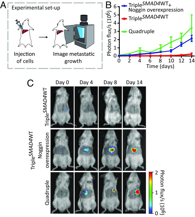
In the adenoma-carcinoma sequence, it is proposed that intestinal polyps evolve through a set of defined mutations toward metastatic colorectal cancer (CRC). Here, we dissect this adenoma-carcinoma sequence in vivo by using an orthotopic organoid transplantation model of human colon organoids engineered to harbor different CRC mutation combinations. We demonstrate that sequential accumulation of oncogenic mutations in Wnt, EGFR, P53, and TGF-β signaling pathways facilitates efficient tumor growth, migration, and metastatic colonization. We show that reconstitution of specific niche signals can restore metastatic growth potential of tumor cells lacking one of the oncogenic mutations. Our findings imply that the ability to metastasize-i.e., to colonize distant sites-is the direct consequence of the loss of dependency on specific niche signals.

Keywords: adenoma-carcinoma sequence; colorectal cancer; metastasis; niche independence; organoids.


Conflict of interest statement

The authors declare no conflict of interest.

Figures

Fig. 1.
Fig. 1.
Development of an orthotopic intestinal organoid transplantation model to study CRC progression. (A) Experimental setup of the orthotopic transplantation model. The day before transplantation, 250,000 cells were plated in type I collagen. The collagen drops with the organoids were subsequently transplanted into the caecal wall of immune-deficient mice. Approximately 6–8 wk later, mice were analyzed for tumor growth and presence of metastasis. (B) Representative merged tile scan image of a transplanted collagen drop with organoids (Left) and a cross-section of the graft 1 d after transplantation (Right). g, graft; m, mucosa; me, muscolaris externa; sm, submucosa. Dashed lines highlight the graft. (C) Representative H&E, β-catenin, and Ki-67 staining of a primary tumor, and liver and long metastases. The borders between tumors tissue (T) and healthy tissue (Ht) are indicated with a dotted line. (Scale bars: 100 μm.)
Fig. 2.
Fig. 2.
Orthotopic transplantation of different CRC driver mutation combinations reveals contribution of the separate mutations to progression of human CRC. (A) Human-specific cytokeratin, H&E, Ki-67, and cleaved caspase-3 immunostainings on tumors isolated from mice transplanted with the indicated mutant human colon organoids. Additionally, percentages of Ki-67 positive (average and SEMs) and cleaved caspase-3-positive cells are depicted. (Scale bars: 500 µm.) (B) List of engineered mutant human colon organoid lines transplanted into the caecal wall of immune-deficient mice with relative tumor take. (C) Graph representing average tumor growth. Tumor volume was measured weekly by palpation. Error bars indicate the SEM of biological replicates, TripleP53WT, TripleKRASWT, and TripleAPCWT n = 3, TripleSMAD4WT n = 8, quadruple n = 9.
Fig. 3.
Fig. 3.
Intravital imaging shows high migratory behavior of tumor cells in quadruple organoid-derived tumors. (A) Setup of the intravital imaging experiment. After a primary tumor has developed, an abdominal imaging window (AIW) (33) was implanted onto the tumor. Photoconversion was performed in randomly picked regions within the tumor. Mice recovered overnight in cage. Twenty-four hours after photoconversion, converted regions were retraced, and the photoswitched areas were analyzed for migration. (B) Representative intravital images of Dendra2-expressing TripleAPCWT, TripleSMAD4WT, and quadruple tumors in which green represents nonswitched Dendra2 and red the photoconverted Dendra2. Images of the photoswitched areas at time point 0 h (Left) and the same imaging area 24 h after photoconversion (Right). White dashed lines highlight the photoswitched areas at beginning of the experiment. Orange dashed lines mark the edges of the red-Dendra2 areas 24 h after photoconversion. (Scale bar: 100 µm.) (C) Displacement of the red-Dendra2 areas 24 h after photoconversion per each condition. Red lines show the median; each symbol represents an imaging field and different symbol shapes represent different animals. TripleAPCWT n = 2, TripleSMAD4WT and quadruple n = 6. Ns, not significant, *P < 0.05.
Fig. 4.
Fig. 4.
Quadruple mutant human colon organoids spontaneously metastasize and efficiently colonize and grow in the liver. (A) Liver and lungs of all transplanted mice were analyzed for presence of spontaneous metastases. The second column shows percentage and number of transplanted mice that spontaneously developed metastases. The third column represents the total number of metastases found, and the last column depicts the metastatic load in squared millimeters. (B) Representative confocal images of a Dendra2-positive liver metastasis in a TripleSMAD4WT-transplanted mouse (Left) and in a quadruple-transplanted mouse (Middle). At Right, representative pictures of a quadruple spontaneous lung metastasis revealed by human-specific cytokeratin immunostaining. (C) Mutant human colon organoids were dissociated, and subsequently 250,000 cells were injected into a mesenteric vein of immune-deficient mice. After 6 wk, livers were analyzed for the presence of metastases. (D) Quantification of the experiment described in C. Second column depicts percentages and numbers of mice that developed liver metastases. Total number of metastases and metastatic load are represented in columns 3 and 4, respectively. (E, Left) Representative picture of a liver containing quadruple mutant metastasis. Arrowheads indicate the metastatic foci. (E, Right) Ki-67 staining (red) of Dendra2-positive quadruple mutant metastasis (green). (Scale bars: B, 100 µm; E, 50 µm.)
Fig. 5.
Fig. 5.
Niche independence is a key contributor to metastatic growth. (A) Mutant human colon organoids were dissociated, and subsequently 50,000 cells were intrahepatically injected in mice. The niche was reconstituted by overexpression of Noggin. Tumor growth was monitored by bioluminescence. (B) The kinetics of metastatic growth. Points are averages, and error bars indicate SEM of biological replicates. Quadruple n = 3, TripleSMAD4WT and TripleSMAD4WT overexpressing Noggin n = 5. (C) Representative images at indicated timepoints.

References

    1. Fearon ER, Vogelstein B. A genetic model for colorectal tumorigenesis. Cell. 1990;61(5):759–767. - PubMed
    1. Fearon ER. Molecular genetics of colorectal cancer. Annu Rev Pathol. 2011;6:479–507. - PubMed
    1. Lengauer C, Kinzler KW, Vogelstein B. Genetic instability in colorectal cancers. Nature. 1997;386(6625):623–627. - PubMed
    1. Nguyen DX, Bos PD, Massagué J. Metastasis: From dissemination to organ-specific colonization. Nat Rev Cancer. 2009;9(4):274–284. - PubMed
    1. Sato T, et al. Long-term expansion of epithelial organoids from human colon, adenoma, adenocarcinoma, and Barrett’s epithelium. Gastroenterology. 2011;141(5):1762–1772. - PubMed

Publication types

MeSH terms