Review
. 2017 May;14(5):296-304.
doi: 10.1038/nrgastro.2017.12. Epub 2017 Mar 8.

Acinar cell plasticity and development of pancreatic ductal adenocarcinoma

Affiliations
Review

Acinar cell plasticity and development of pancreatic ductal adenocarcinoma

Peter Storz. Nat Rev Gastroenterol Hepatol. 2017 May.

Abstract

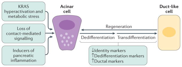
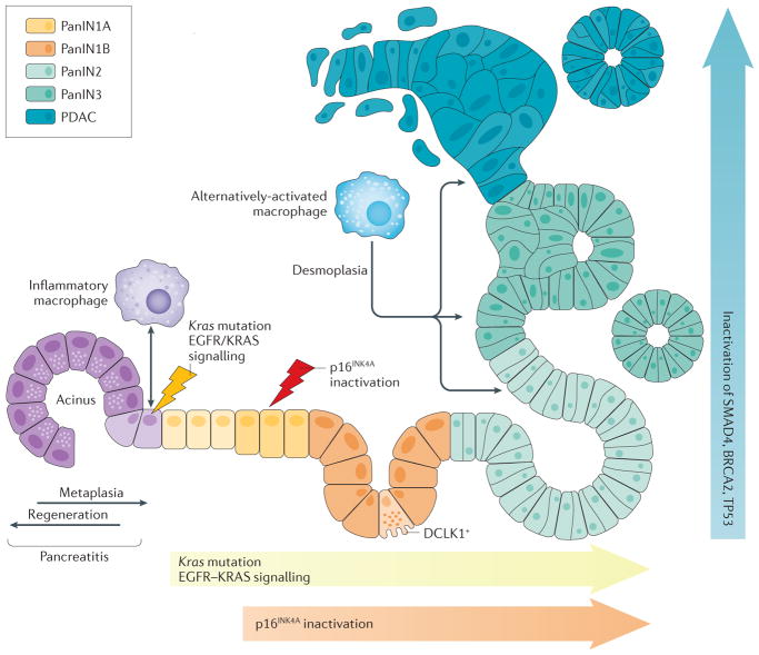
Acinar cells in the adult pancreas show high plasticity and can undergo transdifferentiation to a progenitor-like cell type with ductal characteristics. This process, termed acinar-to-ductal metaplasia (ADM), is an important feature facilitating pancreas regeneration after injury. Data from animal models show that cells that undergo ADM in response to oncogenic signalling are precursors for pancreatic intraepithelial neoplasia lesions, which can further progress to pancreatic ductal adenocarcinoma (PDAC). As human pancreatic adenocarcinoma is often diagnosed at a stage of metastatic disease, understanding the processes that lead to its initiation is important for the discovery of markers for early detection, as well as options that enable an early intervention. Here, the critical determinants of acinar cell plasticity are discussed, in addition to the intracellular and extracellular signalling events that drive acinar cell metaplasia and their contribution to development of PDAC.


Conflict of interest statement

Competing interests statement

The author declares no competing interests.

Figures

Figure 1
Figure 1. Acinar cell plasticity and metaplasia to duct-like cells in the adult pancreas
In response to pancreatic injury, the loss of cell–cell and cell–matrix contacts (contact-mediated signalling), loss of polarity, KRAS hyperactivity and increased inflammatory signalling can drive acinar cells to undergo dedifferentiation and transdifferentiation to a duct-like phenotype that is needed for pancreatic regeneration. Acinar-to-ductal metaplasia becomes irreversible in the presence of an oncogenic Kras mutation and persistent growth factor signalling, leading to metabolic and signalling changes that lock the duct-like cells in their transdifferentiated state and initiate further progression to low-grade precancerous lesions.
Figure 2
Figure 2. Inflammatory macrophage-driven signalling leading to acinar-to-ductal metaplasia
Inflammatory macrophages can initiate acinar-to-ductal metaplasia (ADM) through NF-κB activation as caused by secreted inflammatory cytokines such as TNF and the chemokine CCL5. Macrophage-secreted IL-6 contributes to ADM and the development of PDAC through JAK–STAT3 signalling. In addition, macrophage-secreted matrix metalloproteinases (MMPs) contribute to extracellular matrix (ECM) degradation and activate Notch signalling (NICD, Notch intracellular domain). Other transcription factors activated in acinar cells after inflammation and contributing to ADM are NFATC1 and NFATC4.
Figure 3
Figure 3. KRAS-driven intrinsic signalling pathways leading to irreversible acinar-to-ductal metaplasia in mice
To initiate irreversible acinar-to-ductal metaplasia (ADM), both oncogenic KRAS and wild-type KRAS activities need to be increased. This KRAS signalling mediates induction of similar transcription factors to inflammatory macrophages, but facilitates persistent signalling leading to irreversibility of the ADM process. Signalling hubs downstream of wild-type and mutant KRAS that relay signals to activate transcription factors driving ADM are PRKD1 and phosphatidylinositol 3-kinase (PI3K). PRKD1 can be activated by mutant KRAS-initiated metabolic changes and increases in mitochondrial reactive oxygen species (mROS). PRKD1 then initiates NF-κB and Notch (NICD; Notch intracellular domain) signalling and upregulated expression of matrix metalloproteinases (MMPs), epidermal growth factor (EGF), EGF receptor (EGFR) and transforming growth factor (TGF)-α (via NF-κB), and SOX9 and PDX1 (via NCID). Increased intrinsic EGFR signalling leads to further activation of wild-type KRAS and signal amplification. MMPs can contribute to extracellular matrix degradation as well as activation of Notch. PI3K induces cytoskeletal reorganization by activating small GTPases such as RAC1 and RHOA, but also activates ERK1/2 and AKT. STAT3 and NFATC1 or NFATC4 are activated via PI3K or AKT signalling. NFATC1 or NFATC4 mediate upregulation of SOX2 and SOX9. Mutant KRAS also upregulates the expression of intercellular adhesion molecule 1 (ICAM1), a surface molecule that initiates chemoattraction of inflammatory macrophages into the ADM region. Red arrows indicate signalling mediated by oncogenic KRAS. Blue arrows indicate signalling mediated by EGFR or wild-type KRAS. Grey arrows indicate signalling downstream of both mutant and wild-type KRAS. Green arrows indicate feedback signalling that potentiates KRAS-initiated signalling.
Figure 4
Figure 4. Oncogenic KRAS and inflammation as drivers of acinar-to-ductal metaplasia and clonal expansion
Schematic showing how macrophage subtypes and genetic mutations contribute to acinar-to-ductal metaplasia (ADM), clonal expansion and progression to pancreatic cancer. During pancreatitis ADM is a reversible process, but becomes irreversible when an oncogenic Kras mutation is present. The accumulation of KRAS activity as caused by oncogenic Kras mutations and epidermal growth factor receptor (EGFR)–wild-type KRAS signalling, as well as loss of senescence due to an additional inactivation of cyclin-dependent kinase inhibitor 2A (CDKN2A, also known as p16INK1A), is needed for progression. Further progression to pancreatic intraepithelial neoplasia (PanIN)-2, carcinoma in situ (PanIN3) and pancreatic ductal adenocarcinoma (PDAC) occurs after acquisition of additional gene mutations inTp53 (p53), Brca2 and Smad4. The progression to cancerous lesions occurs with an increase in desmoplasia. Cells positive for the serine/threonine-protein kinase DCLK1 are of acinar origin, are formed mainly in low-grade PanIN lesions (PanIN1A, PanIN1B and PanIN2) and have cancer stem cell functions.

References

    1. Stanger BZ, Hebrok M. Control of cell identity in pancreas development and regeneration. Gastroenterology. 2013;144:1170–1179. - PMC - PubMed
    1. Pinho AV, et al. Adult pancreatic acinar cells dedifferentiate to an embryonic progenitor phenotype with concomitant activation of a senescence programme that is present in chronic pancreatitis. Gut. 2011;60:958–966. - PubMed
    1. Rooman I, Real FX. Pancreatic ductal adenocarcinoma and acinar cells: a matter of differentiation and development? Gut. 2012;61:449–458. - PubMed
    1. Liou GY, et al. Macrophage-secreted cytokines drive pancreatic acinar-to-ductal metaplasia through NF-kappaB and MMPs. J Cell Biol. 2013;202:563–577. - PMC - PubMed
    1. Logsdon CD, Ji B. Ras activity in acinar cells links chronic pancreatitis and pancreatic cancer. Clin Gastroenterol Hepatol. 2009;7:S40–S43. - PMC - PubMed

Publication types