. 2017:2017:6848430.
doi: 10.1155/2017/6848430. Epub 2017 Feb 8.

Growth Arrest-Specific 6 Enhances the Suppressive Function of CD4+CD25+ Regulatory T Cells Mainly through Axl Receptor

Affiliations

Growth Arrest-Specific 6 Enhances the Suppressive Function of CD4+CD25+ Regulatory T Cells Mainly through Axl Receptor

Guang-Ju Zhao et al. Mediators Inflamm. 2017.

Abstract

Background. Growth arrest-specific (Gas) 6 is one of the endogenous ligands of TAM receptors (Tyro3, Axl, and Mertk), and its role as an immune modulator has been recently emphasized. Naturally occurring CD4+CD25+ regulatory T cells (Tregs) are essential for the active suppression of autoimmunity. The present study was designed to investigate whether Tregs express TAM receptors and the potential role of Gas6-TAM signal in regulating the suppressive function of Tregs. Methods. The protein and mRNA levels of TAM receptors were determined by using Western blot, immunofluorescence, flow cytometry, and RT-PCR. Then, TAM receptors were silenced using targeted siRNA or blocked with specific antibody. The suppressive function of Tregs was assessed by using a CFSE-based T cell proliferation assay. Flow cytometry was used to determine the expression of Foxp3 and CTLA4 whereas cytokines secretion levels were measured by ELISA assay. Results. Tregs express both Axl and Mertk receptors. Gas6 increases the suppressive function of Tregs in vitro and in mice. Both Foxp3 and CTLA-4 expression on Tregs are enhanced after Gas6 stimulation. Gas6 enhances the suppressive activity of Tregs mainly through Axl receptor. Conclusion. Gas6 has a direct effect on the functions of CD4+CD25+Tregs mainly through its interaction with Axl receptor.


Conflict of interest statement

The authors declare that there is no conflict of interests regarding the publication of this paper.

Figures

Figure 1
Figure 1
TAM receptors were expressed in mouse CD4+CD25+Tregs. (a) Protein expression from CD4+CD25+Tregs, cardiac tissue, and kidney tissue was analyzed by Western blot using specific anti-Axl (2 μg/ml) and anti-Mertk (2 μg/ml) antibodies. (b) Axl and Mertk mRNA expressions in CD4+CD25+Tregs and marcrophages were determined by semiquantitative RT-PCR. Expression of β-actin was shown as an internal control. (c) CD4+CD25+Tregs were incubated with anti-Axl (10 μg/ml) and anti-Mertk (10 μg/ml) monoclonal Ab, respectively, followed by FITC-labeled goat anti-rat IgG as the secondary Ab (1 : 30). The cells were analyzed by flow cytometry.
Figure 2
Figure 2
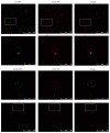
Immunofluorescence detection of TAM receptors. CD4+CD25+Tregs were incubated with anti-Axl (10 μg/ml) and anti-Mertk (10 μg/ml) monoclonal Ab, respectively, followed by FITC-labeled goat anti-rat IgG as the secondary Ab (1 : 30). The cells were analyzed by fluorescent confocal microscopy. Representative photomicrographs show that FITC-positive cells (green) were detected among PE-positive CD4+CD25+Tregs (red). Those double-stained cells are shown in yellow.
Figure 3
Figure 3
CD4+CD25+Tregs from rmGas6-treated mice exhibited increased Foxp3 levels and suppressive activity. BALB/c mice were administrated with 1, 3, or 6 μg/mouse of rmGas6 via tail vein. After 24 hours, CD4+CD25+T cells were purified from spleens. (a-b) MFI analyses showing expression levels of Foxp3 protein in CD4+CD25+T cells from rmGas6-treated mice and control mice (two mice each in two independent experiments). P < 0.05 compared with the value for the control group. (c) CD4+CD25T cells from normal mice were cocultured with CD4+CD25+T cells from control or Gas6-treated mice for 3 days with or without anti-CD3 and anti-CD28 stimulation (three mice each in two independent experiments). Histogram of CFSE-labeled CD4+CD25T cells are displayed, and the percentage of cells that have proliferated as shown in gates. One representative example is shown. (d) Mean percentage of suppression was calculated (n = 6). CD4+CD25+T cells from Gas6-treated mice had increased suppressive activity when compared with those from control mice. P < 0.05 compared with the value for anti-CD3/CD28 group.
Figure 4
Figure 4
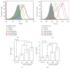
Effects of Gas6 on Foxp3 and CTLA-4 expression in CD4+CD25+Tregs in vitro. Representative examples of Foxp3 (a) and CTLA-4 (b) expressions in CD4+CD25+Tregs after 20, 100, and 500 ng/ml rmGas6 stimulation for 24 hours. The average levels of Foxp3 (c) and CTLA-4 (d) were increased after Gas6 stimulation (n = 4/group). P < 0.05.
Figure 5
Figure 5
Effects of Gas6 on suppressive activity of CD4+CD25+Tregs in vitro. CD4+CD25+Tregs were pretreated with PBS or rmGas6, respectively, for 24 h. Then, prestimualted CD4+CD25+Tregs were mixed with CD4+CD25T cells in the presence of anti-CD3/CD28 Abs. After 72 h, the proliferation of CD4+CD25T cells and the levels of cytokine in the conditioned media were determined. (a) Suppressive activity of CD4+CD25+Tregs was measured by a CFSE assay (n = 4/group). The levels of IL-2 (b) as well as IL-4 and IFN-γ (c) in the conditioned media were determined by ELISA (n = 4/group). P < 0.05 compared with the value for the control Tregs group, and #P < 0.05 compared with the value for anti-CD3/CD28 group.
Figure 6
Figure 6
Gas6 modulates the Foxp3 and CTLA4 expression mainly through Axl receptor. (a–d) CD4+CD25+Tregs were treated with anti-Axl or anti-Mertk Abs or PBS in the presence of 100 ng/ml rmGas6. After 24 h of incubation, the expression of CTLA-4 and Foxp3 was determined by flow cytometry (n = 4/group). P < 0.05 compared with the value for the Gas6 group, and #P < 0.05 compared with the value for rmGas6+anti-Mertk group. (e–h) The expression of CTLA-4 and Foxp3 in Tregs with or without Axl knockout was determined by flow cytometry. P < 0.05 compared with the value for the Gas6 group.
Figure 7
Figure 7
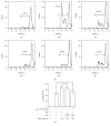
Gas6 modulates the suppressive function of Tregs mainly through Axl receptor. Axl knockdown Tregs were pretreated with 100 ng/ml rmGas6 for 24 h and then mixed with CD4+CD25T cells in presence of anti-CD3/CD28 Abs. After 72 h, the suppressive function of CD4+CD25+T cells was determined by CFSE assay (a and b) (n = 4/group). (A) CD4+CD25T cells alone. (B) CD4+CD25T cells were stimulated with a combination of soluble anti-CD3 and anti-CD28 antibodies. (C) CD4+CD25+Tregs and (D) rmGas6-pretreated Tregs were mixed with CFSE-labeled CD4+CD25T cells at a ratio of 1 : 1 in the presence of anti-CD3 and anti-CD28 antibodies. (E) siRNA-control and (F) siRNA-Axl transfected CD4+CD25+Tregs pretreated with Gas6 were cocultured with CFSE-labeled CD4+CD25T cells. P < 0.05.

References

    1. Belkaid Y. Regulatory T cells and infection: a dangerous necessity. Nature Reviews Immunology. 2007;7(11):875–888. doi: 10.1038/nri2189. - DOI - PubMed
    1. Sakaguchi S., Yamaguchi T., Nomura T., Ono M. Regulatory T cells and immune tolerance. Cell. 2008;133(5):775–787. doi: 10.1016/j.cell.2008.05.009. - DOI - PubMed
    1. Luan Y.-Y., Yin C.-F., Qin Q.-H., et al. Effect of regulatory T cells on promoting apoptosis of t lymphocyte and its regulatory mechanism in sepsis. Journal of Interferon and Cytokine Research. 2015;35(12):969–980. doi: 10.1089/jir.2014.0235. - DOI - PMC - PubMed
    1. Alunno A., Manetti M., Caterbi S., et al. Altered immunoregulation in rheumatoid arthritis: the role of regulatory T cells and proinflammatory Th17 cells and therapeutic implications. Mediators of Inflammation. 2015;2015:12. doi: 10.1155/2015/751793.751793 - DOI - PMC - PubMed
    1. Bluestone J. A., Buckner J. H., Fitch M., et al. Type 1 diabetes immunotherapy using polyclonal regulatory T cells. Science Translational Medicine. 2015;7(315) doi: 10.1126/scitranslmed.aad4134.315ra189 - DOI - PMC - PubMed

MeSH terms

Substances