. 2017 Feb;24(1):e55-e60.
doi: 10.3747/co.24.3299. Epub 2017 Feb 27.

Nasopharyngeal non-intestinal-type adenocarcinoma: a case report and updated review of the literature

Affiliations

Nasopharyngeal non-intestinal-type adenocarcinoma: a case report and updated review of the literature

C Jain et al. Curr Oncol. 2017 Feb.

Abstract

Background: Non-intestinal-type adenocarcinoma is a malignancy traditionally found in the sinonasal cavity. To our knowledge, this case is the first reported of this rare condition originating in the nasopharynx.

Case presentation: A 67-year-old woman with nasopharyngeal non-intestinal-type adenocarcinoma, with an accompanying parapharyngeal mass received primary radiation treatment for both lesions. Her tumour subsequently persisted, with a concomitant conversion in pathology from a low- to a high-grade malignancy.

Results: Non-intestinal-type and intestinal-type adenocarcinomas of the nasopharynx are extremely rare tumours and do not appear in the World Health Organization classification system. We review the pathophysiologic features of these malignancies and propose modifications to the current classification system.

Conclusions: Non-intestinal-type adenocarcinoma should be included in the differential diagnosis of nasopharyngeal masses. In our experience, this tumour in this location showed a partial response to primary radiation but later converted from a low- to a high-grade adenocarcinoma.

Keywords: Non-intestinal adenocarcinoma; disease classifications; intestinal adenocarcinoma; nasopharynx; sinonasal cavity.


Figures
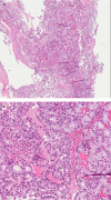
FIGURE 1
FIGURE 1
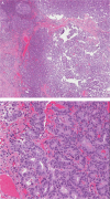
Endoscopic biopsy of the pedunculated nasopharynx mass. (A) The low-power view demonstrates a non-intestinal-type adenocarcinoma having numerous uniform glandular structures arranged in a back-to-back pattern with little intervening stroma and invading the submucosa. Hematoxylin and eosin stain, 20× original magnification. (B) The higher power view confirms that the glands are lined by a single layer of columnar cells with uniform round nuclei showing mild-to-moderate nuclear atypia and with ample foamy and eosinophilic cytoplasm. Hematoxylin and eosin stain, 200× original magnification.
FIGURE 2
FIGURE 2
Intraoperative photograph of the nasopharyngeal non-intestinal-type adenocarcinoma before resection. The septum has been resected for adequate exposure of the tumour.
FIGURE 3
FIGURE 3
Subsequent post-radiotherapy excision of the nasopharyngeal mass. (A,B) Although the histopathologic features are similar to those in the original biopsy (Figure 1), the excised specimen showed significantly less mucinous differentiation, a greater degree of cytologic atypia (increased nuclear size, increased nuclear-to-cytoplasmic ratio, and conspicuous nucleoli), nuclear stratification, and numerous mitotic figures, all indicating transformation to a high-grade non-intestinal-type adenocarcinoma. Hematoxylin and eosin stain, 20× and 200× magnification respectively.
FIGURE 4
FIGURE 4
Immunohistochemical staining profiles for both the primary tumour biopsy and the resected specimen were identical, with the exception of the Ki-67 labelling index, (A) which was markedly increased (36.8%) in the resected specimen, compared with (B) the biopsy specimen (6.1%). The Ki-67 labelling indices were manually counted by image analysis software in “hot spots.” As in the biopsy specimen, (C) neoplastic cells in the resected specimen were positive for cytokeratin 7 and (D) negative for markers of intestinal differentiation, including cytokeratin 20. All images 200× original magnification.
FIGURE 5
FIGURE 5
Modification of the 2005 World Health Organization classification of nasopharyngeal carcinoma. This figure expands the current classification system to include adenocarcinoma as a subtype of nasopharyngeal carcinoma, as highlighted in blue. It further incorporates non-ITAC and ITAC as possible subsets of nasopharyngeal adenocarcinoma.

References

    1. Guo ZM, Liu WW, He JH. A retrospective cohort study of nasopharyngeal adenocarcinoma: a rare histological type of nasopharyngeal cancer. Clin Otolaryngol. 2009;34:322–7. doi: 10.1111/j.1749-4486.2009.01952.x. - DOI - PubMed
    1. Chan JKC, Bray F, McCarron P, et al. Tumours of the nasopharynx: nasopharyngeal carcinoma. In: Barnes EL, Eveson JW, Reichart P, et al., editors. Pathology and Genetics of Head and Neck Tumours: World Health Organization Classification of Tumours. Lyon, France: iarc Press; 2005. pp. 85–97.
    1. Barnes L, Tse LLY, Hunt JL, Brandwein-Gensler M, Curtin HD, Boffetta P. Chapter 1 Tumours of the nasal cavity and paranasal sinuses. In: Barnes EL, Eveson JW, Reichart P, et al., editors. Pathology and Genetics of Head and Neck Tumours: World Health Organization Classification of Tumours. Lyon, France: iarc Press; 2005. pp. 9–80.
    1. Bayrak R, Haltas H, Yenidunya S. The value of cdx2 and cytokeratins 7 and 20 expression in differentiating colorectal adenocarcinomas from extraintestinal gastrointestinal adenocarcinomas: cytokeratin 7−/20+ phenotype is more specific than cdx2 antibody. Diagn Pathol. 2012;7:9. doi: 10.1186/1746-1596-7-9. - DOI - PMC - PubMed
    1. Bhaijee F, Carron J, Bell D. Low-grade nonintestinal sinonasal adenocarcinoma: a diagnosis of exclusion. Ann Diagn Pathol. 2011;15:181–4. doi: 10.1016/j.anndiagpath.2010.10.002. - DOI - PubMed

LinkOut - more resources