. 2017 Jan 10;8(2):764-775.
doi: 10.1364/BOE.8.000764. eCollection 2017 Feb 1.

Clinically compatible flexible wide-field multi-color fluorescence endoscopy with a porcine colon model

Affiliations

Clinically compatible flexible wide-field multi-color fluorescence endoscopy with a porcine colon model

Gyugnseok Oh et al. Biomed Opt Express. .

Abstract

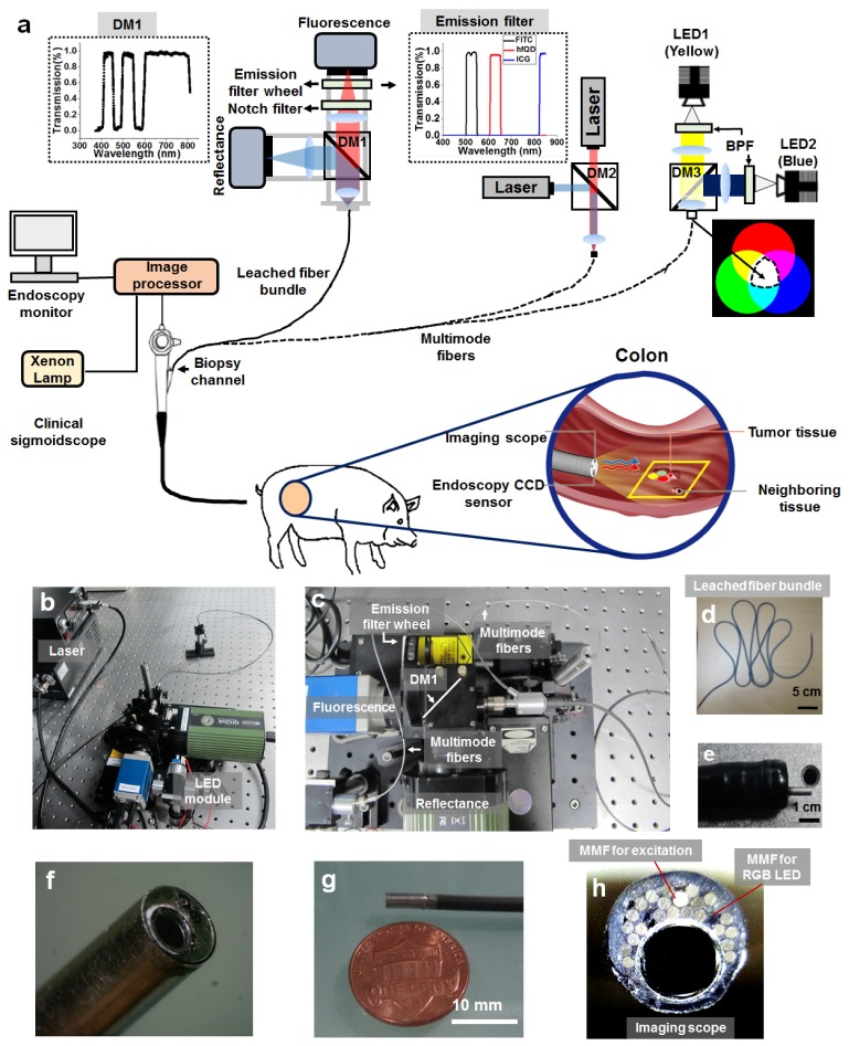
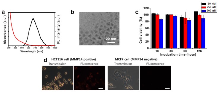
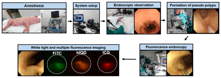
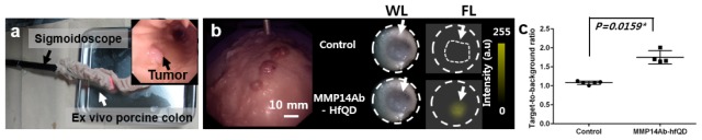
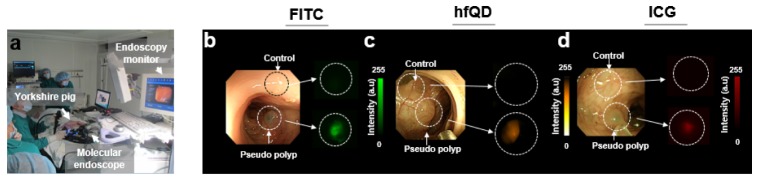
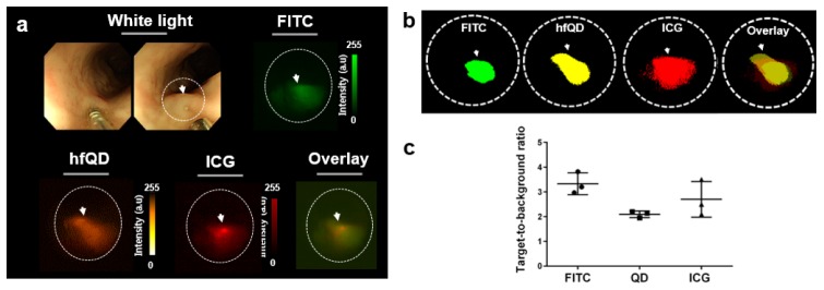
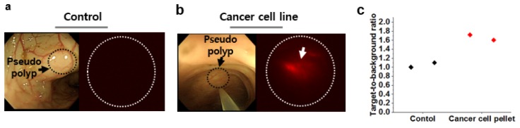
Early detection of structural or molecular changes in dysplastic epithelial tissues is crucial for cancer screening and surveillance. Multi-targeting molecular endoscopic fluorescence imaging may improve noninvasive detection of precancerous lesions in the colon. Here, we report the first clinically compatible, wide-field-of-view, multi-color fluorescence endoscopy with a leached fiber bundle scope using a porcine model. A porcine colon model that resembles the human colon is used for the detection of surrogate tumors composed of multiple biocompatible fluorophores (FITC, ICG, and heavy metal-free quantum dots (hfQDs)). With an ex vivo porcine colon tumor model, molecular imaging with hfQDs conjugated with MMP14 antibody was achieved by spraying molecular probes on a mucosa layer that contains xenograft tumors. With an in vivo porcine colon embedded with surrogate tumors, target-to-background ratios of 3.36 ± 0.43, 2.70 ± 0.72, and 2.10 ± 0.13 were achieved for FITC, ICG, and hfQD probes, respectively. This promising endoscopic technology with molecular contrast shows the capacity to reveal hidden tumors and guide treatment strategy decisions.

Keywords: (110.0110) Imaging systems; (170.2150) Endoscopic imaging; (170.4580) Optical diagnostics for medicine.


Figures

Fig. 1
Fig. 1
Configuration of the clinically-compatible flexible wide-field multi-color fluorescence endoscope system. (a) Schematic of multi-color fluorescence endoscope designed to be inserted through the biopsy channel (diameter: 3.2 mm) of a clinical sigmoidoscope. The system is comprised of two detection channels: reflectance and fluorescence. Multiple-channel fluorescence imaging is obtained by the rotating filter wheel in front of the fluorescence camera detector. (b–c) Photographs of the imaging device and system. (c) An enlarged view of the detection part in the endoscopic system. (d) Photograph of highly flexible leached fiber bundle used for home-made probes. Note that the leached fiber bundle in (a) is not visible in (c). (e) Photograph of combined imaging scope and sigmoidoscope. (f–g) Close-up photograph of the distal end of the imaging scope. The scope was enclosed and sealed within an aluminum sheath. The magnified imaging scope shows a leached fiber bundle and multimode fibers for delivering excitation light and white light (from LEDs). (h) Frontal view of the distal end of the imaging scope. Acronyms are defined as follows: (LED: Light-emitting diode, DM: Dichroic mirror, MMF: Multi-mode fiber, BPF: Band-pass filter, RGB: Red green blue).
Fig. 2
Fig. 2
(a) Absorption and fluorescence profiles of the hfQDs. (b) Transmission electron microscopic image of the hfQDs. (c) Cytotoxicity measurements of HCT116 cells with hfQDs. For QDs toxicity, cells were treated with 50, 250, and 500 nM of quantum dots for 1–12 h at 37 °C. After the incubation, cell viability was measured by CCK-8 assay. (d) Fluorescence imaging with MMP14-QDs in HCT116 and MCF7 cells. Scale bar = 50 µm.
Fig. 3
Fig. 3
In vivo fluorescence endoscopic imaging protocol in porcine colon. Prior to the imaging, a 30 kg Yorkshire pig was anesthetized with isoflurane (1.5%–2.0%) through endotracheal intubation and the multi-color fluorescence endoscope was set. Next, the observation of the porcine colon using the clinical sigmoidoscope was performed to remove any existing stool with PBS solution. Prior to fluorescence imaging, a mixture of fluorophores was injected into the colonic wall to form surrogate tumors using the clinical injector.
Fig. 4
Fig. 4
Ex vivo tumor-targeted fluorescence image with MMP14Ab-hfQD probe in the porcine colon. (a) Experimental configuration. (b) Tumor-targeted fluorescence imaging using MMP14Ab-hfQD and PBS solution as control with grafted mouse tumors in the porcine colon. (c) Quantified TBR using PBS solution and MMP14ab-hfQD ex vivo with 1.08 ± 0.05 and 1.74 ± 0.06, respectively. p < 0.05 by unpaired t-test.
Fig. 5
Fig. 5
In vivo fluorescence endoscopic imaging of surrogate tumors in the porcine colon with different fluorophores. (a) Experimental configuration. (b) Fluorescence imaging of surrogate tumors with FITC, ICG, and hfQDs in the porcine colon.
Fig. 6
Fig. 6
In vivo multi-color fluorescence endoscopic imaging in the porcine colon. (a) (upper) Generation of a surrogate tumor with mixtures of multiple fluorophores. Fluorescence images of each dye were obtained with corresponding emission filters. (b) Threshold images of multiple fluorophores with the endoscope showing the distributions of each fluorophore and a merged image. (c) TBR of fluorophores in the surrogate tumor at the same concentration of 1 mg/ml.
Fig. 7
Fig. 7
Intra-vital in vivo fluorescence endoscopic imaging of SL4-DsRed cancer cell pellet in porcine colon. (a) Surrogate tumor imaging of PBS injection (control experiment). (b) Surrogate tumor fluorescence imaging of SL4-DsRed cancer cells. (c) Mean TBR value at the injection site of the colon cancer cells and control.

References

    1. Pasha S. F., Leighton J. A., Das A., Harrison M. E., Gurudu S. R., Ramirez F. C., Fleischer D. E., Sharma V. K., “Comparison of the yield and miss rate of narrow band imaging and white light endoscopy in patients undergoing screening or surveillance colonoscopy: a meta-analysis,” Am. J. Gastroenterol. 107(3), 363–371 (2012). 10.1038/ajg.2011.436 - DOI - PubMed
    1. Sturm, M.B. and T.D. Wang,” Emerging optical methods for surveillance of Barrett's oesophagus,” Gut, gutjnl-2013–306706 (2015). - PMC - PubMed
    1. Oh G., Chung E., Yun S. H., “Optical fibers for high-resolution in vivo microendoscopic fluorescence imaging,” Opt. Fiber Technol. 19(6), 760–771 (2013). 10.1016/j.yofte.2013.07.008 - DOI
    1. Oh G., Yoo S. W., Jung Y., Ryu Y.-M., Park Y., Kim S.-Y., Kim K. H., Kim S., Myung S.-J., Chung E., “Intravital imaging of mouse colonic adenoma using MMP-based molecular probes with multi-channel fluorescence endoscopy,” Biomed. Opt. Express 5(5), 1677–1689 (2014). 10.1364/BOE.5.001677 - DOI - PMC - PubMed
    1. Weissleder R., “Molecular imaging in cancer,” Science 312(5777), 1168–1171 (2006). 10.1126/science.1125949 - DOI - PubMed

LinkOut - more resources