. 2017 Jan 30;8(2):1221-1239.
doi: 10.1364/BOE.8.001221. eCollection 2017 Feb 1.

Ultrahigh-resolution OCT imaging of the human cornea

Affiliations

Ultrahigh-resolution OCT imaging of the human cornea

René M Werkmeister et al. Biomed Opt Express. .

Abstract

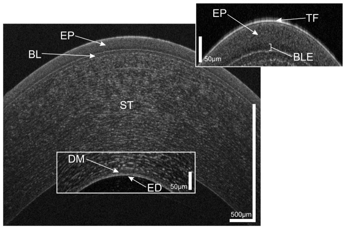
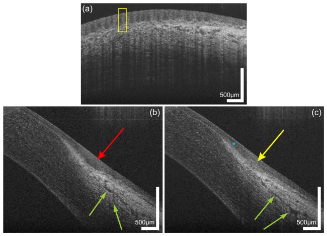
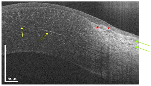
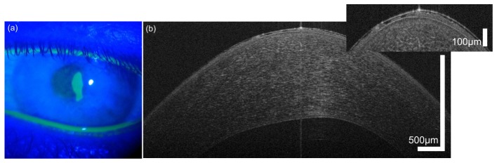
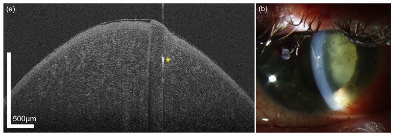
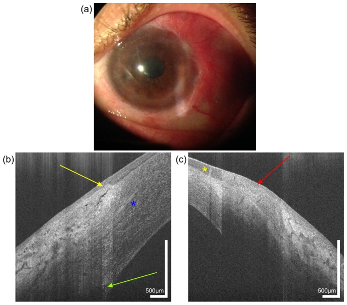
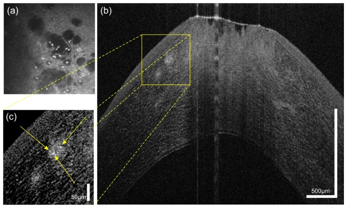
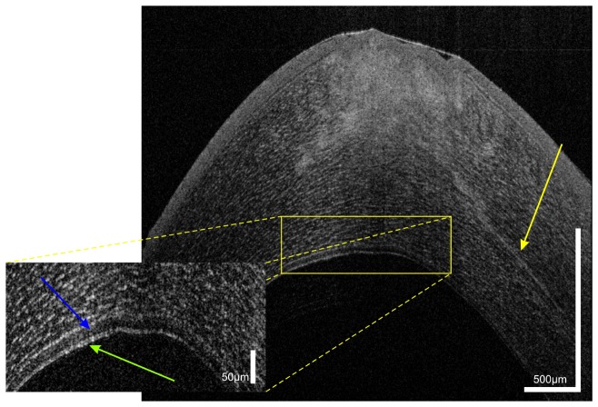
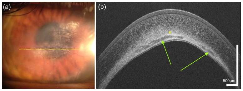
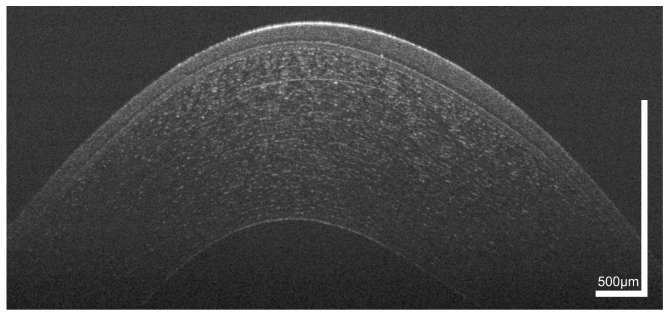
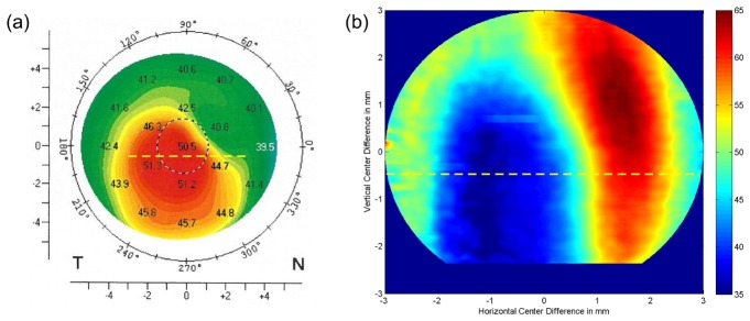
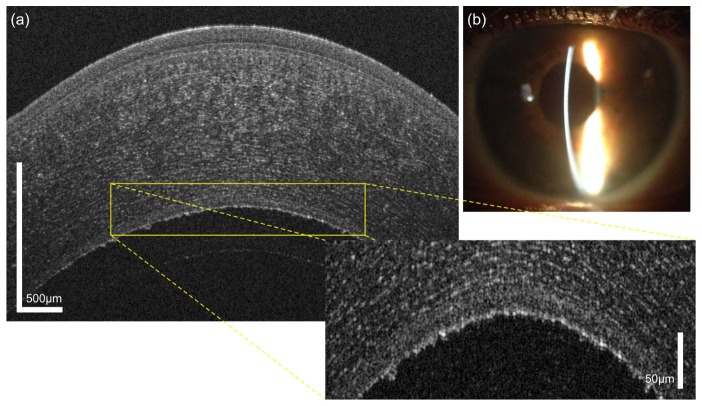
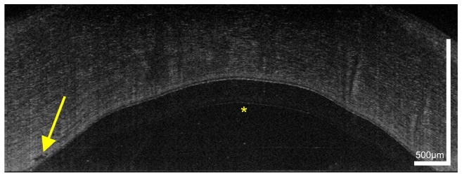
We present imaging of corneal pathologies using optical coherence tomography (OCT) with high resolution. To this end, an ultrahigh-resolution spectral domain OCT (UHR-OCT) system based on a broad bandwidth Ti:sapphire laser is employed. With a central wavelength of 800 nm, the imaging device allows to acquire OCT data at the central, paracentral and peripheral cornea as well as the limbal region with 1.2 µm x 20 µm (axial x lateral) resolution at a rate of 140 000 A-scans/s. Structures of the anterior segment of the eye, not accessible with commercial OCT systems, are visualized. These include corneal nerves, limbal palisades of Vogt as well as several corneal pathologies. Cases such as keratoconus and Fuchs's endothelial dystrophy as well as infectious changes caused by diseases like Acanthamoeba keratitis and scarring after herpetic keratitis are presented. We also demonstrate the applicability of our system to visualize epithelial erosion and intracorneal foreign body after corneal trauma as well as chemical burns. Finally, results after Descemet's membrane endothelial keratoplasty (DMEK) are imaged. These clinical cases show the potential of UHR-OCT to help in clinical decision-making and follow-up. Our results and experience indicate that UHR-OCT of the cornea is a promising technique for the use in clinical practice, but can also help to gain novel insight in the physiology and pathophysiology of the human cornea.

Keywords: (170.0110) Imaging systems; (170.4460) Ophthalmic optics and devices; (170.4500) Optical coherence tomography.


Figures

Fig. 1
Fig. 1
UHR-OCT tomogram of the central and paracentral zone of the cornea of a 38 year old healthy male. TF, tear film; EP, epithelium; BLE, basal layer of epithelium; BL, Bowman’s layer; ST, corneal stroma; DM, Descemet’s membrane; ED, endothelium.
Fig. 2
Fig. 2
Limbal palisades of Vogt of a 38 year old male subject. (a) Transversal scan at the inferior limbus revealing the fence-like structure of the palisades with highly reflective collagen ridges. (b,c) Sagital scans at (b) inter-palisade and (c) palisade positions yielding the difference in reflectivity at the putative location of the stem cell niches. The blue asterisk indicates the hyperreflective extension of the corneo-scleral junction. Green arrows indicate parts of the conventional aqueous humor drainage pathway.
Fig. 3
Fig. 3
UHR-OCT tomogram of the paracentral and peripheral cornea and limbus of the same subject as in Fig. 1. In the mid stroma, a highly reflective corneal nerve (yellow arrows) with a thickness between 5µm (central) and 10 µm (paracentral) is visible. In the limbus, blood vessels (red asterisk) as well as conjunctival and episclearal plexus (green arrows) are shown.
Fig. 4
Fig. 4
Corneal epithelial abrasion in a 25 year old male with progressive keratoconus, image taken 4 days after epi-off corneal collagen cross-linking procedure. (a) slit lamp photograph with Fluorescein staining showing the defect, (b) corresponding OCT image.
Fig. 5
Fig. 5
Intrastromal foreign body in a 43 year old male patient who wore glasses during an accident. The injury was accompanied by a globe perforation. During the slit lamp examination after surgery, a corneal foreign body was noted; yet, it was not clear whether it needed immediate removal through a second surgical intervention or not. (a) OCT image, (b) slit lamp photograph.
Fig. 6
Fig. 6
Image of a 44 year old female patient with a chemical burn in her left eye, caused by a bathroom cleaner 10 years ago. Slit lamp photography (a) of the left eye reveals corneal neovacularization. UHR-OCT cross-sectional images of the temporal limbus of the (b) right and (c) left eye. Yellow arrow, limbal palisade of Vogt. Red arrow, scar tissue with absence of palisades. Yellow asterisk indicates the position of a newly formed vessel in the conjunctiva. Green arrow, Schlemm’s canal. Blue asterisk, corneal nerve.
Fig. 7
Fig. 7
45 year old male patient with Acanthamoeba keratitis, (a) Confocal microscopy depicting Acanthamoeba cysts. (b) and (c) UHR-OCT scans of the same patient revealing at least three cysts (yellow arrows) and defects in epithelium and anterior stroma.
Fig. 8
Fig. 8
UHR-OCT of Acanthamoeba keratitis in the same patient as shown in Fig. 7. The cross-sectional image reveals radial keratoneuritis with a thickened corneal nerve and a hyporeflective space (blue arrow) above the double banded structure of the epithelium.
Fig. 9
Fig. 9
Sequelae after herpetic keratitis in a 34 year old male who was first diagnosed 3 years ago. The slit lamp photography (a) as well as the OCT image (b) reveal neovascularization. Yellow dotted line in (a) indicates the location of the UHR-OCT scan; Green arrows, corneal neovascularization; yellow asterisk, calcifications/lipid.
Fig. 10
Fig. 10
Cross sectional image of the paracentral zone of the cornea of a 38 year old male patient with progressive keratoconus. Epithelial thinning in the paracentral cornea is visualized.
Fig. 11
Fig. 11
Corneal topography of the same 38 year old patient as shown in Fig. 10. (a) Corneal refractive power as measured via Scheimpflug tomography. (b) Epithelial thickness map retrieved from a three-dimensional UHR-OCT data set. Yellow dashed lines in both figures indicate the position of the cross-sectional scan depicted in Fig. 11. Due to reduced signal-to-noise-ratio in the periphery, the evaluated region was cropped to a circular pattern with a diameter of 6 mm. The thinnest region of the cornea is present in the temporal inferior part and corresponds well with the thinnest zone of the epithelium as measured by UHR-OCT.
Fig. 12
Fig. 12
Fuchs’ endothelial dystrophy that was newly diagnosed in a 60 year old female patient, (a) OCT image, (b) corresponding slit lamp photograph.
Fig. 13
Fig. 13
UHR-OCT image of the cornea of a 74 year old female patient who underwent DMEK for Fuchs’ dystrophy (24 hours postoperatively). Yellow arrow marks an irregularity in the recipient stroma. Yellow asterisk indicates an image artifact due to the internal fixation target of the OCT.

References

    1. Whitcher J. P., Srinivasan M., Upadhyay M. P., “Corneal blindness: a global perspective,” Bull. World Health Organ. 79(3), 214–221 (2001). - PMC - PubMed
    1. Klintworth G. K., “Corneal dystrophies,” Orphanet J. Rare Dis. 4(1), 7 (2009).10.1186/1750-1172-4-7 - DOI - PMC - PubMed
    1. Belin M. W., Khachikian S. S., “An introduction to understanding elevation-based topography: how elevation data are displayed - a review,” Clin. Experiment. Ophthalmol. 37(1), 14–29 (2009).10.1111/j.1442-9071.2008.01821.x - DOI - PubMed
    1. Cairns G., McGhee C. N., “Orbscan computerized topography: attributes, applications, and limitations,” J. Cataract Refract. Surg. 31(1), 205–220 (2005).10.1016/j.jcrs.2004.09.047 - DOI - PubMed
    1. Rosales P., Marcos S., “Pentacam Scheimpflug quantitative imaging of the crystalline lens and intraocular lens,” J. Refract. Surg. 25(5), 421–428 (2009).10.3928/1081597X-20090422-04 - DOI - PubMed

LinkOut - more resources