Case Reports
. 2017 Jul;46(7):967-974.
doi: 10.1007/s00256-017-2610-0. Epub 2017 Mar 7.

Metal wear-induced pseudotumour following an endoprosthetic knee replacement for Ewing sarcoma

Affiliations
Case Reports

Metal wear-induced pseudotumour following an endoprosthetic knee replacement for Ewing sarcoma

Richard Craig et al. Skeletal Radiol. 2017 Jul.

Abstract

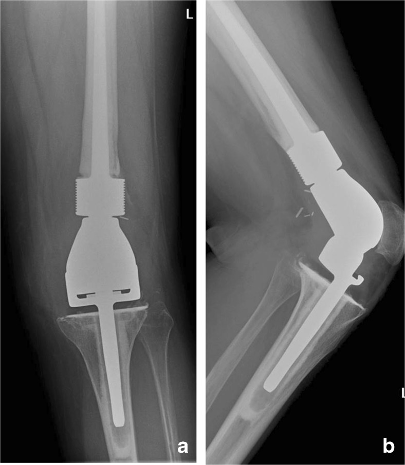
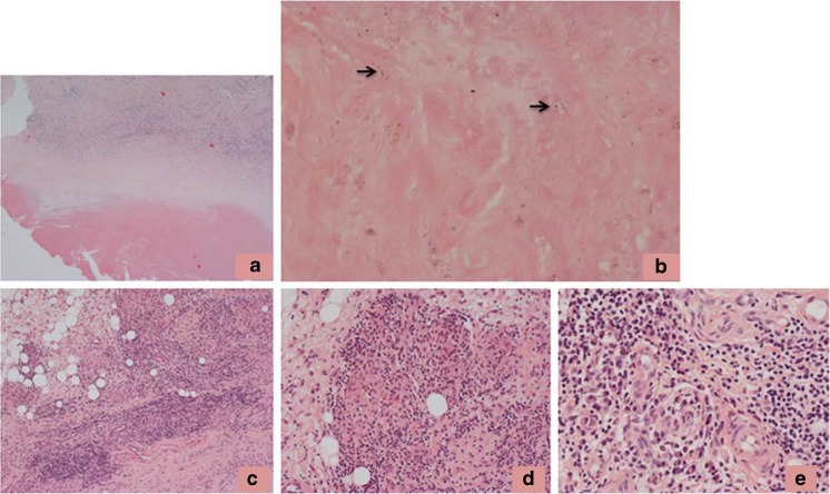
Pseudotumours are well recognised as a complication of metal-on-metal hip arthroplasties and are thought to develop on the basis of an innate and adaptive immune response to cobalt-chrome (Co-Cr) wear particles. We report a case of a large pseudotumour that developed following a knee endoprosthetic replacement (EPR) undertaken for Ewing sarcoma. The lesion contained necrotic and degenerate connective tissue in which there were numerous scattered metal wear-containing macrophages, eosinophil polymorphs, lymphocytes, plasma cells and aseptic lymphocyte-dominated vascular-associated lesion-like lymphoid aggregates. Metal ion levels were elevated. No evidence of infection or tumour was noted and it was concluded that the lesion was most likely an inflammatory pseudotumour developing on the basis of an innate and adaptive immune response to components of Co-Cr metal wear derived from the knee EPR.

Keywords: Endoprosthesis; Knee; Metal wear; Pseudotumour.


Conflict of interest statement

Funding

MV was an EU-funded visitor on the Erasmus + program

Conflicts of interest

The authors declare that they have no conflicts of interest

Figures

Fig. 1
Fig. 1
Imaging of the original tumour. a Coronal short tau inversion recovery (STIR) magnetic resonance (MR) image demonstrates a high signal lesion in the distal femoral metaphysis contacting the intercondylar notch and physis with adjacent bone marrow and soft-tissue oedema. b The lateral plain radiograph does not demonstrate the original lesion well, apart from posterior periosteal thickening (arrow). Histology was consistent with a Ewing sarcoma
Fig. 2
Fig. 2
a Anteroposterior and b lateral plain radiographs post-resection of the distal femur and proximal tibia, with insertion of a distal femoral endoprosthesis, coupled to a rotating hinged tibial component
Fig. 3
Fig. 3
Histology of the capsule/synovium 42 months post-index surgery showing: a necrosis on the tissue surface with underlying diffuse inflammatory infiltrate; b focally heavy inflammatory infiltrate around small vessels lined by plump endothelial cells (arrows); c inflammatory infiltrate containing lymphocytes and macrophages, some of which contain black wear particles (arrowed); d prominent eosinophil polymorph infiltrate (arrows) with e evidence of vasculitis
Fig. 4
Fig. 4
a Coronal STIR, b coronal T1, c axial T1-weighted MRI and d axial ultrasound image 6 years post-index surgery show a complex, predominantly thin-walled cystic soft-tissue mass intimately related to the antero-lateral aspect of the femoral component of the prosthesis (arrows). The lesion is well-defined, returns largely low STIR and is isointense to high T1-weighted signal relative to skeletal muscle, consistent with a pseudotumour. The diagnosis is supported by the homogeneous, hypoechoic appearance on ultrasound, with no Doppler activity
Fig. 5
Fig. 5
Histology of the pseudotumour 64 months post-index surgery showing: a characteristic zonal arrangement of the pseudotumour with a band of necrosis on the surface, an underlying zone of partly necrotic fibrous tissue containing macrophages, and deeper tissue containing a macrophage and lymphoid infiltrate; b necrotic macrophages and metal wear particles (some with arrows) in the superficial zone of the pseudotumour; c perivascular lymphoid infiltrate and scattered macrophages in deeper tissue; d granuloma-like collections of foreign body macrophages; e aggregates of lymphocytes and plasma cells around small vessels in the deeper tissue

References

    1. Pandit H, Glyn-Jones S, McLardy-Smith P, et al. Pseudotumours associated with metal-on-metal hip resurfacing. J Bone Joint Surg (Br) 2008;90:847–851. doi: 10.1302/0301-620X.90B7.20213. - DOI - PubMed
    1. Daniel J, Holland J, Quigley L, Sprague S, Bhandari M. Pseudotumours associated with total hip arthroplasty. J Bone Joint Surg Am. 2012;94-A:86–93. doi: 10.2106/JBJS.J.01612. - DOI - PubMed
    1. Hallab NJ, Jacobs JJ. Biological effects of implant debris. Bull NYU Hosp Jt Dis. 2009;67:182–188. - PubMed
    1. Athanasou NA. The pathobiology and pathology of aseptic implant failure. Bone Joint Res. 2016;5:162–168. doi: 10.1302/2046-3758.55.BJR-2016-0086. - DOI - PMC - PubMed
    1. Kenan S, Kahn L, Haramati N, Kenan S. A rare case of pseudotumour formation associated with methyl methacrylate hypersensitivity in a patient following cemented total knee arthroplasty. Skeletal Radiol. 2016;45:1115–1122. doi: 10.1007/s00256-016-2372-0. - DOI - PubMed

Publication types

LinkOut - more resources