. 2017 Jun;143(6):981-990.
doi: 10.1007/s00432-017-2370-1. Epub 2017 Mar 8.

Long non-coding RNA UCA1 regulates the expression of Snail2 by miR-203 to promote hepatocellular carcinoma progression

Affiliations

Long non-coding RNA UCA1 regulates the expression of Snail2 by miR-203 to promote hepatocellular carcinoma progression

Ji-Nan Xiao et al. J Cancer Res Clin Oncol. 2017 Jun.

Abstract

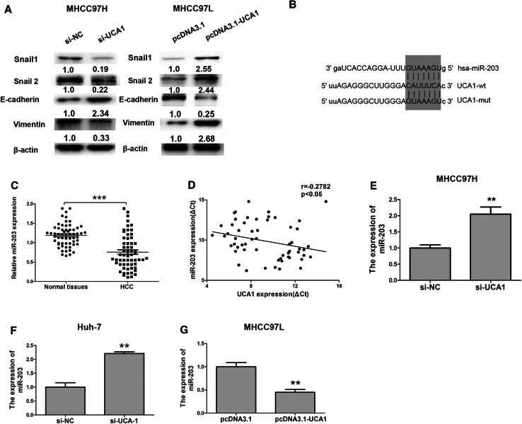
Purpose: Long non-coding RNA (LncRNA) urothelial carcinoma-associated 1 (UCA1) is reported to be dysregulated in hepatocellular carcinoma (HCC) progression. However, the functions of UCA1 in HCC still need further study. The aim is to detect the role of UCA1 involving in HCC cells proliferation and invasion, and epithelial-mesenchymal transition (EMT).

Methods: The quantitative real-time PCR was used to detect the UCA1 and miR-203 expression levels in 60 cases' HCC tissues and adjacent normal tissues. Western blotting analysis was performed to detect the EMT markers E-cadherin, Vimentin and transcription factor Snail1, Snail2 expression. Luciferase reporter assay, RNA immunoprecipitation (RIP) and pull-down assays were used to evaluate whether miR-203 was a target of UCA1.

Results: Our results showed that UCA1 was markedly upregulated in HCC tissues and higher UCA1 expression in HCC was positively associated with tumor size, vascular invasion and American Joint Committee on Cancer (AJCC) stage (P < 0.05). Furthermore, gain-of-function and loss-of-function analysis showed that UCA1 knockdown inhibited HCC cells proliferation and invasion in vitro and xenograft tumour growth in vivo. Moreover, UCA1 overexpression promoted cell epithelial-mesenchymal transition (EMT) in HCC via effectively sponging to miR-203 and thereby activating the expression of transcription factor Snail2.

Conclusions: Our results identified that UCA1/miR-203/Snail2 pathway might involve in HCC progression. Inhibition of UCA1 acted as a promising therapeutic target for HCC patients.

Keywords: Epithelial–mesenchymal transition; Snail2; Urothelial carcinoma-associated 1; miR-203.


Conflict of interest statement

The authors declare no conflict of interest.

Figures

Fig. 1
Fig. 1
UCA1 was upregulated in HCC tissues and cell lines. a Relative UCA1 expression in HCC tissues and adjacent normal tissues was determined by qRT-PCR (n = 60). GAPDH was used as internal control. b The correlation between the UCA1 expression and tumor size. c The correlation between the UCA1 expression and vascular invasion. d The correlation between the UCA1 expression and AJCC stage. e The expression levels of UCA1 in four HCC cell lines (Huh7, Sk-hep-1, MHCC97H and MHCC97L) were significantly increased compared to that in the LO2 cells. GAPDH was used as internal control. f, g The expression of UCA was examined by qRT-PCR after transfecting two siRNA-UCA1 oligos in MHCC97H or Huh7 cell lines. GAPDH was used as internal control. h The expression of UCA was examined by qRT-PCR after tranfecting pcDNA3.1-UCA1 plasmid in MHCC97L and Sk-hep-1 cell lines. GAPDH was used as internal control. **P < 0.05, error bars represent the mean ± SD of triplicate experiments
Fig. 2
Fig. 2
Knockdown of UCA inhibited the HCC cell proliferation and invasion in vitro and tumor growth in vivo. a Cell proliferation is shown after transfection with si-NC or si-UCA1 into MHCC97H cell and b ias shown after transfection with pcDNA3.1 or pcDNA3.1-UCA1 into Sk-hep-1 cell. The growth index was assessed at 0, 24, 48 and 72 h using CCK8 proliferation assay, then the absorbance of the plate was measured at 450 nm ,**P < 0.05. c Cell invasion abilities and d cell invasion number analysis was evaluated after transfection with si-NC or si-UCA1 into MHCC97H cell and was evaluated after transfection with pcDNA3.1 or pcDNA3.1-UCA1into Sk-hep-1 cell **P < 0.05. e In vivo, MHCC97H cells infected with Lentivirus control or Lentivirus-shRNA-UCA1 were subcutaneously injected into the flanks of nude mice, and f tumor formation was monitored and the volume was evaluated every weeks, **P < 0.05, error bars represent the mean ± SD of triplicate experiments
Fig. 3
Fig. 3
UCA1 promoted the EMT in HCC cells and was negatively correlated with miR-203. a Knockdown of UCA1 downregulated the expression of Snail1, Snai2 and Vimentin and upregulated the expression of E-cadherin in MHCC97H (left panel), and overexpression of UCA1 upregulated the expression of Snail1, Snail2 and Vimentin and downregulated the expression of E-cadherin in MHCC97L. b The miR-203 target site in the sequence of UCA1, as predicted by miRanda (http://mircrorna.org). c Relative miR-203 expression in 60 cases HCC tissues and adjacent normal tissues were determined by qRT-PCR; GAPDH was used as internal control. d The correlation analyses were performed between the levels of UCA1 expression and miR-203 expression in HCC tissues. e, f Knockdown of UCA1 in MHCC97H and Huh7 cells upregulated miR-203 level. g Overexpression of UCA1 in MHCC97L cell downregulated the level of miR-203, **P < 0.05, error bars represent the mean ± SD of triplicate experiments
Fig. 4
Fig. 4
MiR-203 was identified as a target of UCA1 in HCC cell. a, b Dual luciferase assays showed a decrease in reporter activity following cotransfection of pmirGLO-wt-UCA1 and miR-203 mimic in MHCC97H cells and human HEK293T, whereas the cotransfection of pmirGLO-mut-UCA1 and miR-203 had no effect on reporter activity. c RNA immunoprecipitation (RIP) results with anti-IgG, anti-Ago2 and 10% input were presented. Relative RNA levels of U1 in anti-SNRNP70 group versus anti-IgG immunoprecipitates were used as positive control. IgG antibody was used as negative control group in MHCC97H cells. d Biotinylated UCA1 or antisense RNA was incubated with cell extracts of MHCC97H cells, qRT-PCR assays was used to detect the miR-203 expression. **P < 0.05, ***P < 0.01, error bars represent the mean ± SD of triplicate experiments, n.s. not significant
Fig. 5
Fig. 5
UCA1 enhanced the expression of Snail2 by negatively regulating the miR-203 a, b After transfection at 48 h with si-NC, si-UCA1, miR-203 inhibitor miR-203 inhibitor + si-UCA1, the mRNA and protein level of Snail2 in MHCC97H cells was examined by qRT-PCR or western blot assays. f After transfected at 48 h with pcDNA3.1, pcDNA3.1-UCA1, miR-203 mimic, miR-203 mimic + pcDNA3.1-UCA1, the mRNA and protein level of Snail2 in MHCC97L cells were examined by qRT-PCR or western blot assays. **P < 0.05, error bars represent the mean ± SD of triplicate experiments, n.s. not significant

References

    1. Bian Z, Jin L, Zhang J et al (2016) LncRNA-UCA1 enhances cell proliferation and 5-fluorouracil resistance in colorectal cancer by inhibiting miR-204-5p. Sci Rep 6:23892 - PMC - PubMed
    1. Cheng N, Cai W, Ren S et al (2015) Long non-coding RNA UCA1 induces non-T790M acquired resistance to EGFR-TKIs by activating the AKT/mTOR pathway in EGFR-mutant non-small cell lung cancer. Oncotarget 6:23582–23593 - PMC - PubMed
    1. Dhanasekaran R, Limaye A, Cabrera R (2012) Hepatocellular carcinoma: current trends in worldwide epidemiology, risk factors, diagnosis, and therapeutics. Hepat Med 4:19–37 - PMC - PubMed
    1. Ding X, Park SI, McCauley LK et al (2013) Signaling between transforming growth factor beta (TGF-beta) and transcription factor SNAI2 represses expression of microRNA miR-203 to promote epithelial-mesenchymal transition and tumor metastasis. J Biol Chem 288:10241–10253 - PMC - PubMed
    1. Ergun S, Oztuzcu S (2015) Oncocers: ceRNA-mediated cross-talk by sponging miRNAs in oncogenic pathways. Tumour Biol 36:3129–3136 - PubMed

MeSH terms

LinkOut - more resources