. 2017 Jun;104(5):700-711.
doi: 10.1111/mmi.13663. Epub 2017 Mar 27.

Multi-layered inhibition of Streptomyces development: BldO is a dedicated repressor of whiB

Affiliations

Multi-layered inhibition of Streptomyces development: BldO is a dedicated repressor of whiB

Matthew J Bush et al. Mol Microbiol. 2017 Jun.

Abstract

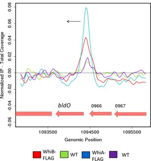
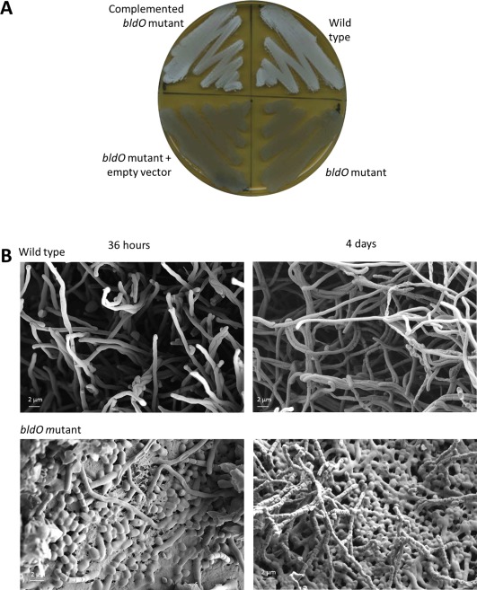
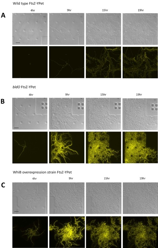
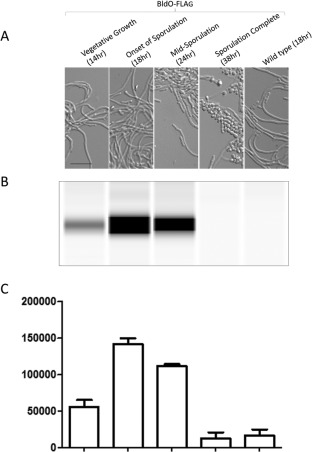
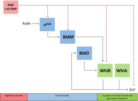
BldD-(c-di-GMP) sits on top of the regulatory network that controls differentiation in Streptomyces, repressing a large regulon of developmental genes when the bacteria are growing vegetatively. In this way, BldD functions as an inhibitor that blocks the initiation of sporulation. Here, we report the identification and characterisation of BldO, an additional developmental repressor that acts to sustain vegetative growth and prevent entry into sporulation. However, unlike the pleiotropic regulator BldD, we show that BldO functions as the dedicated repressor of a single key target gene, whiB, and that deletion of bldO or constitutive expression of whiB is sufficient to induce precocious hypersporulation.


Figures

Figure 1
Figure 1
ChIP‐seq data showing that bldO is a WhiAB target. Colour‐coding of the ChIP samples is as follows: 3xFLAG‐[Gly4Ser]3‐WhiB strain (WhiB‐FLAG, red), corresponding S. venezuelae wild‐type anti‐FLAG negative control (WT, green), 3xFLAG‐[Gly4Ser]3‐WhiA strain (WhiA‐FLAG, blue) and corresponding S. venezuelae wild‐type anti‐FLAG negative control (WT, purple). The plot spans approximately 3 kb of DNA sequence. Genes running right to left are shown in red. The black arrow indicates the gene subject to WhiA and WhiB regulation (bldO). These data are derived from the ChIP‐seq datasets described in Bush et al., mBio, 2016, 7, e00523‐16 (with permission), but sven0965 (bldO) was not specifically discussed in that paper.
Figure 2
Figure 2
Deletion of bldO causes precocious hypersporulation on solid medium. A. The phenotypes of wild‐type S.venezuelae, the bldO mutant, the bldO mutant carrying the empty vector, and the complemented bldO mutant, photographed after four days of growth on DNA medium. B. Scanning electron micrographs showing the precocious hypersporulation phenotype of the bldO mutant after 36 h and 4 days of growth on DNA medium.
Figure 3
Figure 3
Deletion of bldO or overexpression of WhiB causes precocious hypersporulation in liquid medium. Time‐lapse images (4, 9, 15 and 19 h) of (A) wild‐type S. venezuelae, (B) the bldO mutant and (C) wild‐type S. venezuelae constitutively expressing whiB from the ermE* promoter, grown in DNB, in the microfluidic system. All three strains carry the same FtsZ‐YPet translational fusion expressed from the native ftsZ promoter, and both the DIC (upper) and fluorescence (lower) images are shown. For the corresponding movies, please see Supporting Information Movies S1A/B, S2A/B and S6A/B. Scale bars = 10 µm.
Figure 4
Figure 4
Automated Western blot analysis of the C‐terminally FLAG‐tagged version of BldO, expressed in trans from the native promoter in the bldO null mutant background. Equal amounts (2.5 µg) of total protein were loaded for each sample and BldO‐FLAG was detected with anti‐FLAG antibody using the quantitative ‘Wes’ capillary electrophoresis and blotting system (ProteinSimple – San Jose, CA; see Supplementary Material). Wild‐type S. venezuelae expressing non‐FLAG‐tagged BldO was used as a negative control. Both the BldO FLAG‐tagged strain and wild type were grown in DNB medium. Top: DIC images of the culture at each time point. Middle: virtual Western blot. Bottom: quantitation of BldO levels (area under each peak; arbitrary units). All experimental samples were analysed in triplicate and the mean value and its Standard Error are shown for each sample.
Figure 5
Figure 5
Spatial localisation of bldO transcription. Fluorescence images of wild‐type S. venezuelae carrying the bldOp‐ypet transcriptional fusion or the empty ypet reporter vector. Strains were imaged on coverslips after 18 h of growth in DNB, following the addition of FM4‐64 membrane dye. For the corresponding movies of the same strains, grown in the microfluidic system, please see Supporting Information Movies S4A/B and S5A/B.
Figure 6
Figure 6
BldO has a single target, whiB. A. Genome‐wide distribution of BldO binding sites identified by ChIP‐seq analysis using M2 anti‐FLAG antibody, conducted at the onset of sporulation on the bldO null mutant complemented in trans with a functional bldO‐3xFLAG allele expressed under the control of the native promoter from the ΦBT1 integration site. Strains were grown in DNB medium. The peak upstream of whiB is marked by the red arrow. Peaks also seen in the negative control (wild‐type S. venezuelae), such as those indicated by asterisks, were excluded from further analysis. B. Close‐up of a ∼5‐kb region around whiB. Colour‐coding of the ChIP samples is as follows: the onset of sporulation (bldO‐3xFLAG, green), S. venezuelae wild‐type anti‐FLAG negative control (WT, purple). Genes running left to right are shown in green, and genes running right to left are shown in red. The black arrow indicates the gene (whiB) subject to BldO regulation.
Figure 7
Figure 7
BldO binds to a sequence overlapping the transcriptional start site and the −10 region of the developmentally induced whiBp2 promoter. A. DNase I footprinting analysis of BldO bound to radiolabelled probes derived from the (forward) and (reverse) sequence upstream of whiB. 5′ end‐labelled probes were incubated with increasing concentrations of BldO (indicated in µM above the lanes) and subjected to DNase I footprinting analysis. The footprints are flanked by Maxam and Gilbert sequence ladders (GA) and the black brackets indicate the positions of the BldO‐protected regions. Asterisks mark phosphodiester bonds that are not protected from cleavage. B. Summary of the DNase I footprinting results, showing the protected sequences on the forward and reverse strands (black brackets) relative to the transcriptional start site (+1) and the −10 and −35 sequences (underlined). The sequence of the protected region is also shown above with the hyphenated inverted repeat highlighted in red. Asterisks mark phosphodiester bonds that are not protected from cleavage.
Figure 8
Figure 8
BldO functions to repress whiB transcription. whiB mRNA abundance determined by qRT‐PCR in the wild type (black bars) and the bldO mutant (white bars) throughout development. Strains were grown in DNB medium. Expression values were calculated relative to the accumulation of the constitutively expressed hrdB reference mRNA and normalised to the wild‐type value at 10 h.
Figure 9
Figure 9
Simplified representation of the developmental regulatory network, highlighting the involvement of BldO and the importance of whiB as a node (adapted from Nat Rev Microbiol 2015, 13, 749–760, with permission). Both BldM (Al‐Bassam et al., 2014) and σBldN (of which RsbN is the cognate anti‐sigma factor; Bibb et al., 2012) are required for the formation of aerial hyphae. During vegetative growth, whiB expression is repressed both by BldD‐(c‐di‐GMP) and by BldO. Relief from repression by BldD (presumably due to a drop in c‐di‐GMP levels) and BldO (by an unknown mechanism) and activation by BldM leads to expression of whiB. Once produced, WhiB combines with WhiA to initiate developmental cell division.

References

    1. Aínsa, J.A. , Ryding, N.J. , Hartley, N. , Findlay, K.C. , Bruton, C.J. , and Chater, K.F. (2000) WhiA, a protein of unknown function conserved among Gram‐positive bacteria, is essential for sporulation in Streptomyces coelicolor A3(2). J Bacteriol 182: 5470–5478. - PMC - PubMed
    1. Al‐Bassam, M.M. , Bibb, M.J. , Bush, M.J. , Chandra, G. , and Buttner, M.J. (2014) Response regulator heterodimer formation controls a key stage in Streptomyces development. PLoS Genet 10: e1004554. - PMC - PubMed
    1. Bibb, M.J. , Domonkos, A. , Chandra, G. , and Buttner, M.J. (2012) Expression of the chaplin and rodlin hydrophobic sheath proteins in Streptomyces venezuelae is controlled by σBldN and a cognate anti‐sigma factor, RsbN. Mol Microbiol 84: 1033–1049. - PubMed
    1. Brown, N.L. , Stoyanov, J.V. , Kidd, S.P. , and Hobman, J.L. (2003) The MerR family of transcriptional regulators. FEMS Microbiol Rev 27: 145–163. - PubMed
    1. Bush, M.J. , Bibb, M.J. , Chandra, G. , Findlay, K.C. , and Buttner, M.J. (2013) Genes required for aerial growth, cell division, and chromosome segregation are targets of WhiA before sporulation in Streptomyces venezuelae . mBio 4: e00684‐13. - PMC - PubMed

MeSH terms