. 2017 Jan;21(1):27-37.
doi: 10.1089/omi.2016.0158.

Understanding Epstein-Barr Virus Life Cycle with Proteomics: A Temporal Analysis of Ubiquitination During Virus Reactivation

Affiliations

Understanding Epstein-Barr Virus Life Cycle with Proteomics: A Temporal Analysis of Ubiquitination During Virus Reactivation

Dong-Wen Lv et al. OMICS. 2017 Jan.

Abstract

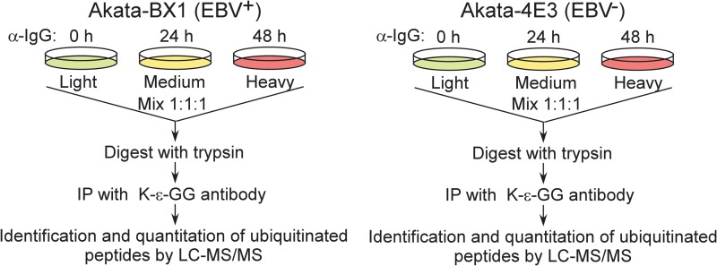
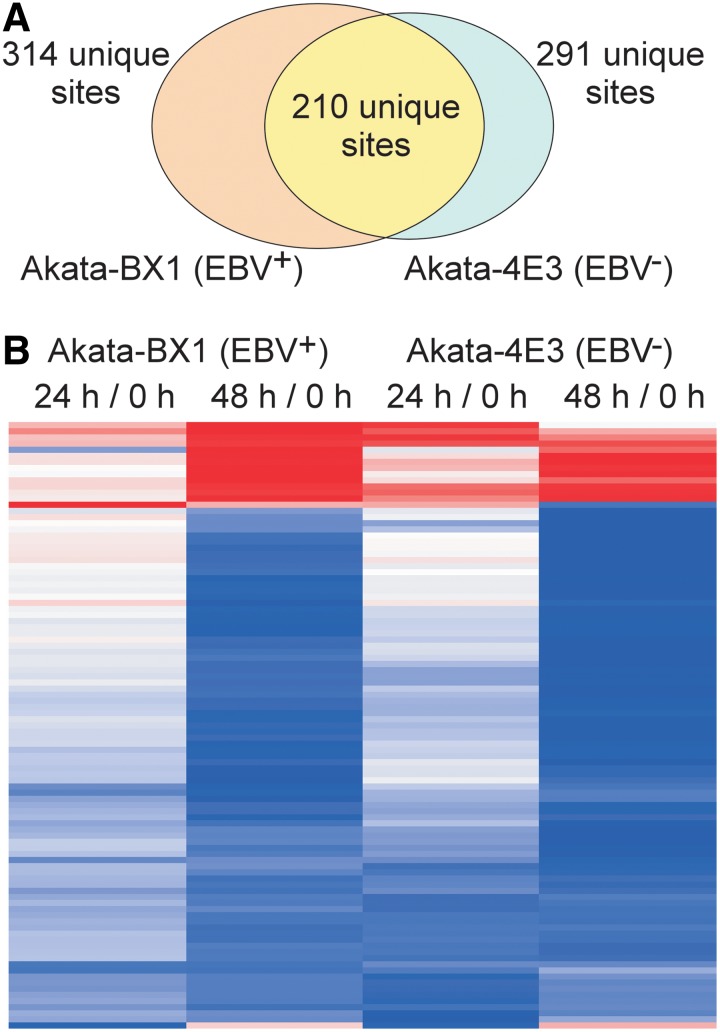
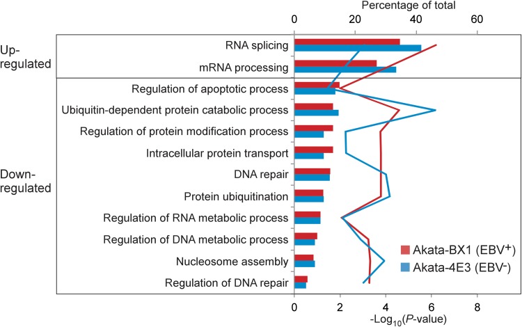
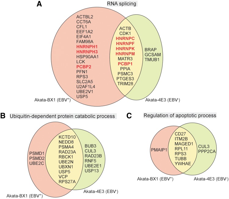
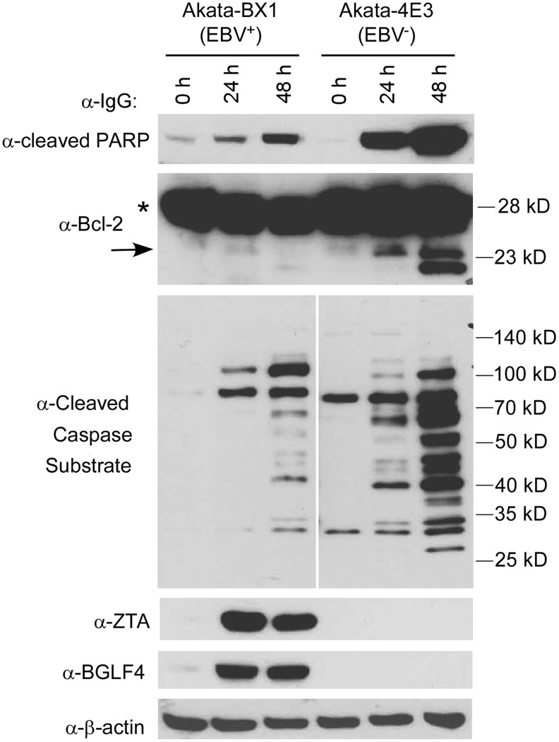
Epstein-Barr virus (EBV) is a human γ-herpesvirus associated with cancer, including Burkitt lymphoma, nasopharyngeal, and gastric carcinoma. EBV reactivation in latently infected B cells is essential for persistent infection whereby B cell receptor (BCR) activation is a physiologically relevant stimulus. Yet, a global view of BCR activation-regulated protein ubiquitination is lacking when EBV is actively replicating. We report here, for the first time, the long-term effects of IgG cross-linking-regulated protein ubiquitination and offer a basis for dissecting the cellular environment during the course of EBV lytic replication. Using the Akata-BX1 (EBV+) and Akata-4E3 (EBV-) Burkitt lymphoma cells, we monitored the dynamic changes in protein ubiquitination using quantitative proteomics. We observed temporal alterations in the level of ubiquitination at ∼150 sites in both EBV+ and EBV- B cells post-IgG cross-linking, compared with controls with no cross-linking. The majority of protein ubiquitination was downregulated. The upregulated ubiquitination events were associated with proteins involved in RNA processing. Among the downregulated ubiquitination events were proteins involved in apoptosis, ubiquitination, and DNA repair. These comparative and quantitative proteomic observations represent the first analysis on the effects of IgG cross-linking at later time points when the majority of EBV genes are expressed and the viral genome is actively being replicated. In all, these data enhance our understanding of mechanistic linkages connecting protein ubiquitination, RNA processing, apoptosis, and the EBV life cycle.

Keywords: Association Study; big data; proteomics.


Conflict of interest statement

The authors declare that no conflicting financial interests exist.

Figures

<b>FIG. 1.</b>
FIG. 1.
Schematic illustration of the SILAC-based quantitative proteomic approach. Akata-BX1 (EBV+) and Akata-4E3 (EBV) cells were cultured in light, medium, and heavy media, as indicated. EBV, Epstein-Barr virus; SILAC, stable isotope labeling by amino acid in cell culture.
<b>FIG. 2.</b>
FIG. 2.
Widespread changes in protein ubiquitination upon IgG cross-linking. (A) IgG cross-linking triggers widespread changes of protein ubiquitination. Scatter plot showing the SILAC ratio (24 h/0 h and 48 h/0 h) and signal intensity of ubiquitinated peptides identified from EBV+ and EBV B cells. Red dots: peptides with ubiquitination downregulated or upregulated; Blue dots: peptides not regulated by IgG cross-linking. (B) IgG cross-linking triggers minimal changes of nonubiquitinated peptides. Scatter plot showing the SILAC ratio (24 h/0 h and 48 h/0 h) and signal intensity of nonubiquitinated peptides identified from EBV+ and EBV B cells. Red dots: downregulated or upregulated peptides; Blue dots: peptides not regulated by IgG cross-linking. See also Supplementary Tables S1 and S2 and Supplementary Figure S1.
<b>FIG. 3.</b>
FIG. 3.
Analysis of IgG-triggered protein ubiquitination in EBV+ and EBV B cells. (A) Overlap of identified ubiquitinated sites in EBV+ and EBV B cells. (B) Heat map analysis of ubiquitinated sites with significant changes in EBV+ B cells and the corresponding changes of these sites in EBV B cells. Red: highest abundance; Blue: lowest abundance. See also Supplementary Table S3.
<b>FIG. 4.</b>
FIG. 4.
GO enrichment of hyper- and hypoubiquitinated proteins from EBV+ and EBV B cells. Significantly over-represented (p < 0.05) biological processes are shown by line chart. p values were determined by hypergeometric test after FDR correction. The percentage of each GO item is shown by bar chart. FDR, false discovery rate; GO, gene ontology.
<b>FIG. 5.</b>
FIG. 5.
The overlap of proteins involved in three major GO items between EBV+ and EBV B cells. (A) The overlap of proteins with upregulated ubiquitination sites between EBV+ and EBV B cells. Proteins highlighted by red and bold belong to the enriched GO item of RNA splicing. (B) The overlap of proteins (containing downregulated ubiquitination sites) involved in ubiquitin-dependent protein catabolic process between EBV+ and EBV B cells. (C) The overlap of proteins (containing downregulated ubiquitination sites) involved in regulation of apoptotic process between EBV+ and EBV B cells.
<b>FIG. 6.</b>
FIG. 6.
IgG cross-linking reduces ubiquitin chain formation. (A) MS spectra showing the relative abundance of ubiquitin K63 containing ubiquitinated peptide at 24 and 48 h after IgG cross-linking of both EBV+ and EBV Akata cells. (B) Validation of SILAC results. EBV+ and EBV B cells were treated with IgG cross-linking as indicated. Western blot analysis of K63 ubiquitination on ubiquitin (RPS27A) was performed by using K63-linkage-specific antibody. β-Actin was included as loading control.
<b>FIG. 7.</b>
FIG. 7.
IgG cross-linking triggers apoptosis induction. EBV+ and EBV B cells were treated with IgG cross-linking as indicated. Western blot analysis showing the cleavage of PARP, Bcl-2, and general caspase substrates. Asterisk denotes uncleaved Bcl-2 and arrow denotes cleaved Bcl-2. EBV lytic transactivator ZTA and protein kinase BGLF4 blots were included to indicate lytic induction efficiency. β-Actin was included as loading control.

Similar articles

Cited by

References

    1. Adhikari A, and Chen ZJ. (2009). Diversity of polyubiquitin chains. Dev Cell 16, 485–486 - PubMed
    1. Bentz GL, Bheda-Malge A, Wang L, Shackelford J, Damania B, and Pagano JS. (2014). KSHV LANA and EBV LMP1 induce the expression of UCH-L1 following viral transformation. Virology 448, 293–302 - PMC - PubMed
    1. Berard M, Casamayor-Palleja M, Billian G, Bella C, Mondiere P, and Defrance T. (1999). Activation sensitizes human memory B cells to B-cell receptor-induced apoptosis. Immunology 98, 47–54 - PMC - PubMed
    1. Bryant H, and Farrell PJ. (2002). Signal transduction and transcription factor modification during reactivation of Epstein-Barr virus from latency. J Virol 76, 10290–10298 - PMC - PubMed
    1. Cai Q, Guo Y, Xiao B, et al. (2011). Epstein-Barr virus nuclear antigen 3C stabilizes Gemin3 to block p53-mediated apoptosis. PLoS Pathog 7, e1002418. - PMC - PubMed

Publication types