. 2017 Apr 28;28(17):174003.
doi: 10.1088/1361-6528/aa655e. Epub 2017 Mar 8.

Multidomain proteins under force

Affiliations

Multidomain proteins under force

Jessica Valle-Orero et al. Nanotechnology. .

Abstract

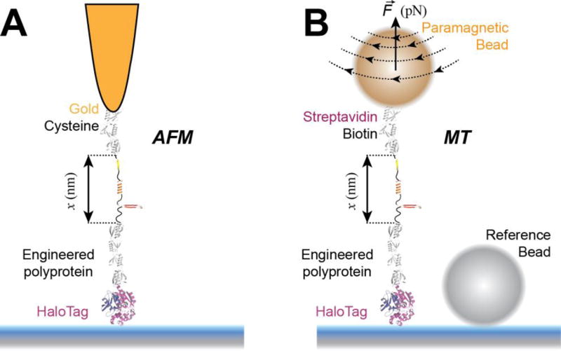
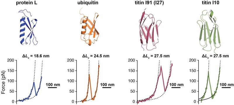
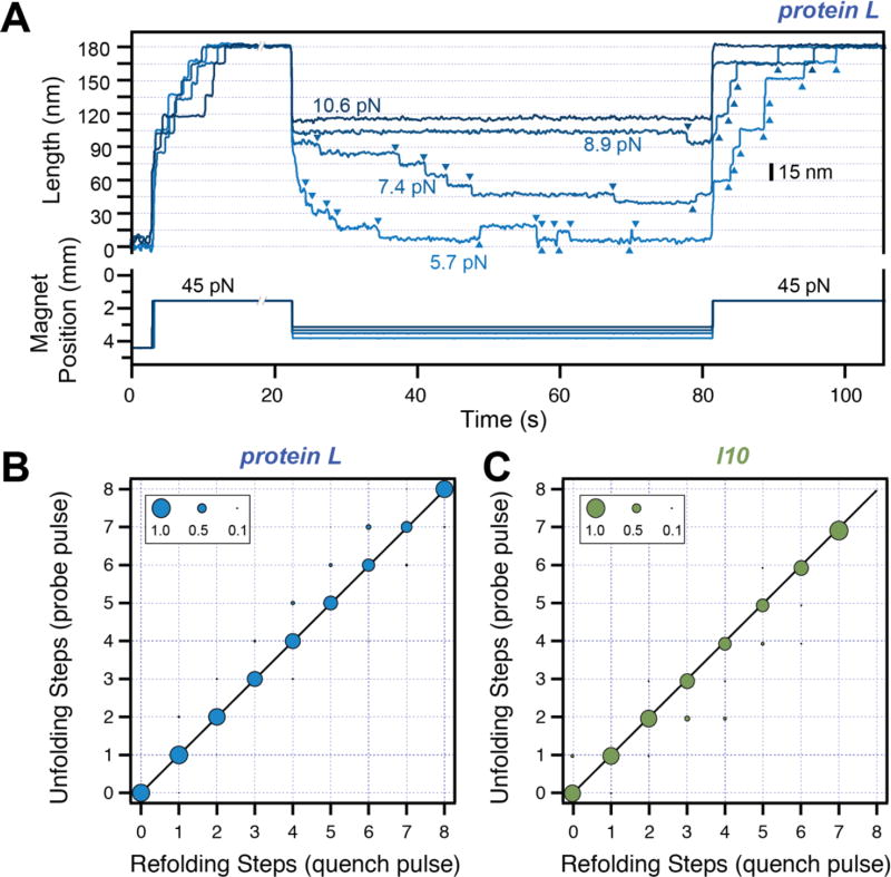
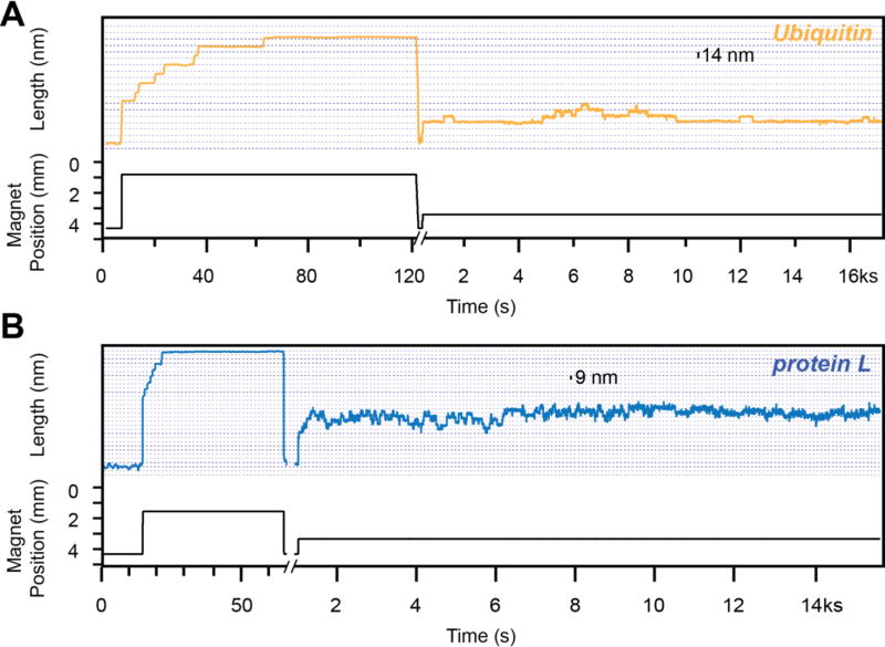
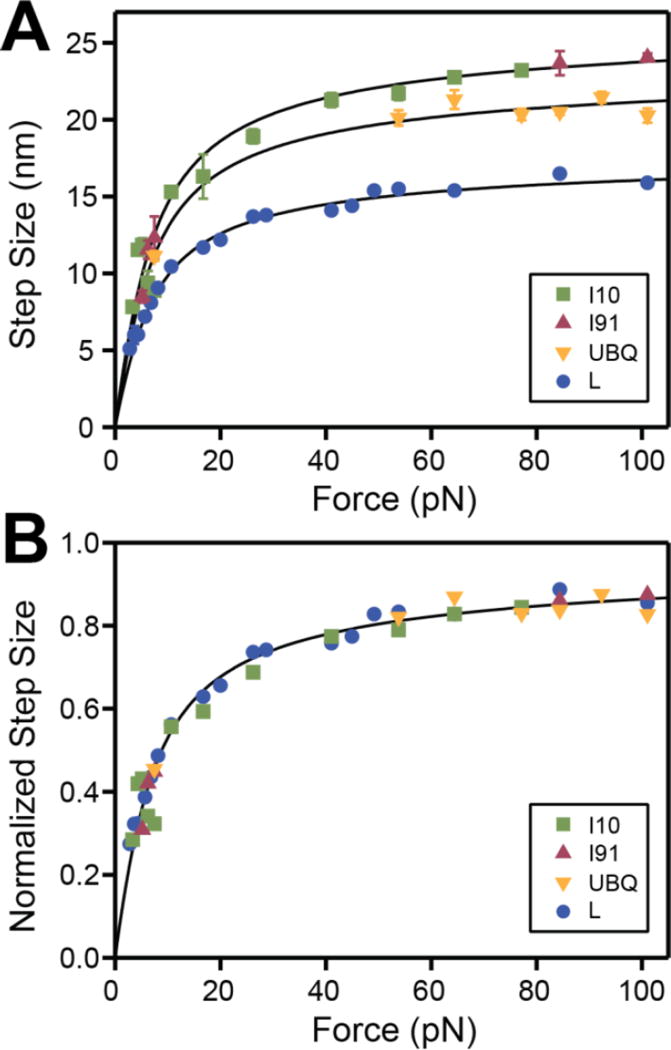
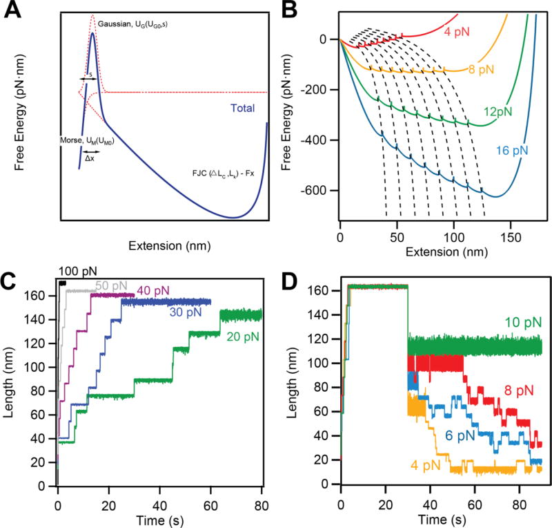
Advancements in single-molecule force spectroscopy techniques such as atomic force microscopy and magnetic tweezers allow investigation of how domain folding under force can play a physiological role. Combining these techniques with protein engineering and HaloTag covalent attachment, we investigate similarities and differences between four model proteins: I10 and I91-two immunoglobulin-like domains from the muscle protein titin, and two α + β fold proteins-ubiquitin and protein L. These proteins show a different mechanical response and have unique extensions under force. Remarkably, when normalized to their contour length, the size of the unfolding and refolding steps as a function of force reduces to a single master curve. This curve can be described using standard models of polymer elasticity, explaining the entropic nature of the measured steps. We further validate our measurements with a simple energy landscape model, which combines protein folding with polymer physics and accounts for the complex nature of tandem domains under force. This model can become a useful tool to help in deciphering the complexity of multidomain proteins operating under force.


Figures

Figure 1
Figure 1. Single molecule force spectroscopy techniques used to study protein folding
A) Schematics of an atomic force microscopy (AFM) experiment. A multidomain protein construct is covalently attached to the surface using HaloTag chemistry. A gold-coated cantilever with a tip having a radius of ~10 nm is used to pull the construct from the opposite end using gold-thiol attachment. Denaturation of protein domains leads to unfolding steps in the measured extension (in force-clamp mode) or to peaks in the measured force (in force-extension mode). B) Schematics of a magnetic tweezers (MT) experiment. A multidomain protein construct is covalently attached to the surface using HaloTag chemistry and tethered to a paramagnetic bead using the biotin-streptavidin bond. A reference non-magnetic bead glued to the glass surface is used to correct for drift. Denaturation of protein domains leads to unfolding steps in the measured extension. AFM is ideal for measuring fast occurring processes, such as unfolding of proteins at high forces, while MT excels on measuring slow-occurring events, such as unfolding and refolding of protein domains at low forces, taking place on a minute-to-hour time scale.
Figure 2
Figure 2. Protein domains investigated with force spectroscopy
Top: Cartoon representation of the four proteins considered for this study: protein L (PDB code: 1HZ5), ubiquitin (PDB code: 1UBQ), titin I91 domain (former I27, PDB code 1TIT), and titin I10 domain (PDB code: 1PGA). Our constructs contain nine repeats for ubiquitin and eight repeats for the other proteins. Bottom: Characteristic AFM force-extension traces showing the unfolding and extension of a protein domain at a loading rate of 400 nm/s (~6 nN/s). Protein L unfolds with a specific contour length of 18.6 nm at an average force of ~130 pN, ubiquitin unfolds with a contour length of 24.5 nm at an average force of ~200 pN, I91 and I10 unfold with a contour length of 27.5 nm at a force of ~210 pN, and ~140 pN, respectively.
Figure 3
Figure 3. Refolding under force
A) Typical traces measured with magnetic tweezers. Protein L is exposed to a high-force pulse (45 pN –fingerprint pulse), where tethering of a single protein construct is confirmed by unfolding all its domains. The force is then quenched to different low values, where the protein domains refold with a characteristic step size and force-dependent kinetics. A final high-force probe pulse is used to determine the number of refolded domains at low force. B) and C) Number of unfolding steps in the probe pulse as a function of refolding steps in the quench pulse for protein L (B) and titin I10 (C). The one-to-one correspondence indicates that the folded tertiary structure forms immediately after the collapse steps for these two proteins.
Figure 4
Figure 4. Protein unfolding at low force
Traces obtained with magnetic tweezers using a ubiquitin construct (A) and a protein L construct (B). Initially, a high-force pulse protocol (fingerprint pulse) is used to confirm the tethering of a single protein construct. Following a force quench at ~4 pN, when the protein is allowed to refold, we expose the construct to a constant low force (8 pN). At this force ubiquitin shows unfolding and refolding steps of 14 nm, while protein L shows similar steps of 9 nm. The kinetics of these steps differs as well for the two proteins: the unfolding and refolding transition of protein L take place on a much faster kinetics than ubiquitin.
Figure 5
Figure 5. Size dependency of the folding transitions as a function of force for different proteins
A) Measured size of the unfolding and refolding steps as a function of force for I10 (green squares), I91 (red triangles), ubiquitin (orange triangles) and protein L (blue circles). The lines represent the behavior predicted by the worm-like chain (WLC) model, using a persistence of 0.58 nm and the contour length increments from Figure 2. B) The same data-points as in A, normalized for each protein to the specific contour length increment. The points collapse on a master curve that can be described by a WLC model (black line).
Figure 6
Figure 6. Interpretation of the measured data using a simple energy landscape model
A) Schematics depicting the construction of our energy landscape by combining the FJC energy at a given force with a Morse potential and a Gaussian barrier, which separate the folded and unfolded states. B) The effect of force on the energy landscape. Force affects both the height of the barrier between the folded and unfolded states of each domain, as well as the final extension. C) and D) Langevin dynamics simulations reproducing the unfolding (C) and refolding (D) behaviors for protein L in different force regimes.

References

    1. Vogel V, Sheetz M. Local Force and Geometry Sensing Regulate Cell Functions. Nature Reviews Molecular Cell Biology. 2006;7:265–75. - PubMed
    1. Liu BY, Chen W, Evavold BD, Zhu C. Accumulation of Dynamic Catch Bonds between TCR and Agonist Peptide-MHC Triggers T Cell Signaling. Cell. 2014;157:357–68. - PMC - PubMed
    1. Wong VW, Rustad KC, Akaishi S, Sorkin M, Glotzbach JP, Januszyk M, Nelson ER, Levi K, Paterno J, Vial IN, Kuang AA, Longaker MT, Gurtner GC. Focal adhesion kinase links mechanical force to skin fibrosis via inflammatory signaling. Nat Med. 2012;18:148–52. - PMC - PubMed
    1. Engler AJ, Sen S, Sweeney HL, Discher DE. Matrix elasticity directs stem cell lineage specification. Cell. 2006;126:677–89. - PubMed
    1. Alegre-Cebollada J, Kosuri P, Giganti D, Eckels E, Rivas-Pardo JA, Hamdani N, Warren CM, Solaro RJ, Linke WA, Fernandez JM. S-glutathionylation of cryptic cysteines enhances titin elasticity by blocking protein folding. Cell. 2014;156:1235–46. - PMC - PubMed

LinkOut - more resources