

Dissection of the interventricular septum: Echocardiographic features
- PMID: 28272209
- PMCID: PMC5348157
- DOI: 10.1097/MD.0000000000006191
Dissection of the interventricular septum: Echocardiographic features
Abstract
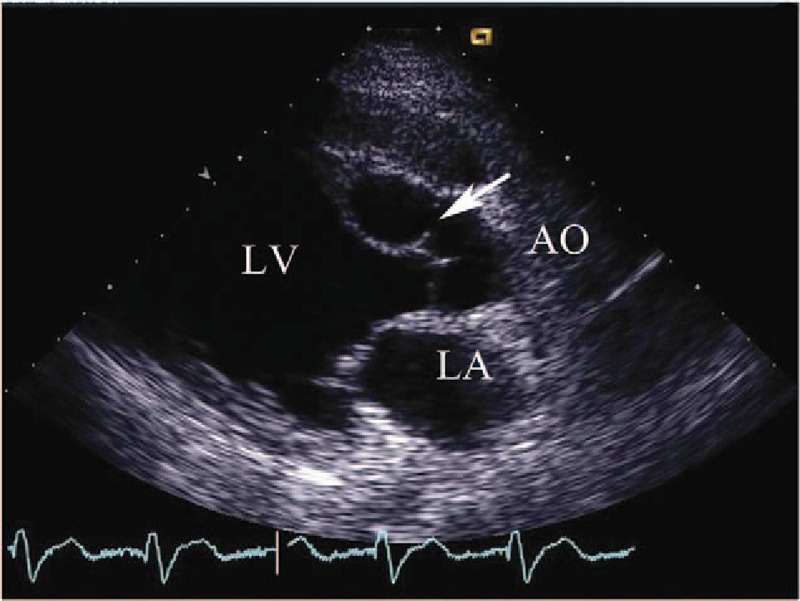
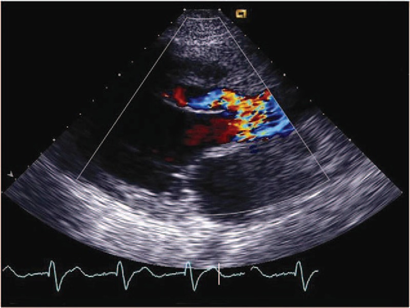
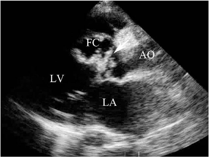
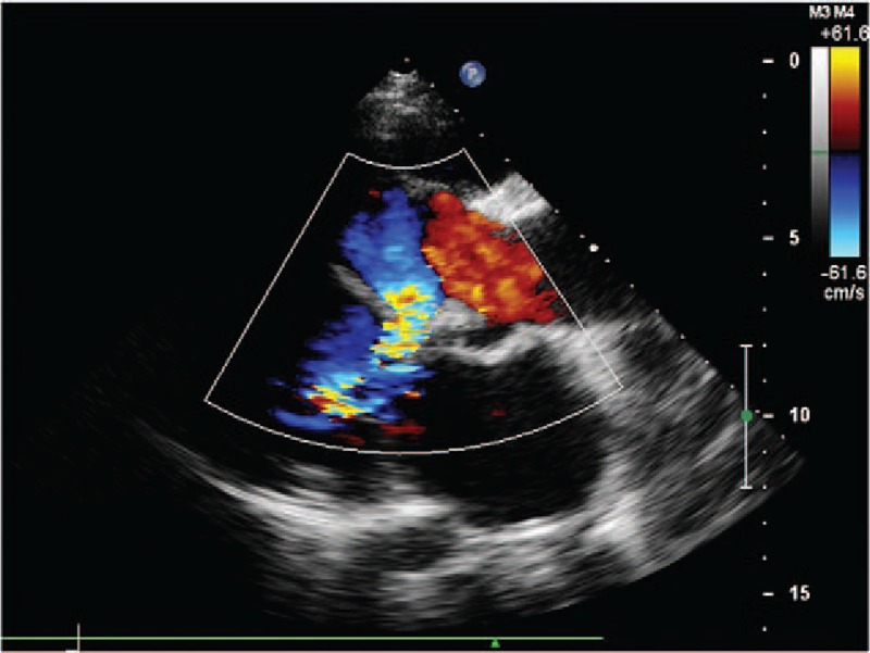
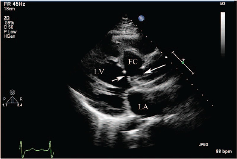
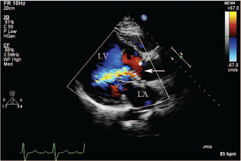
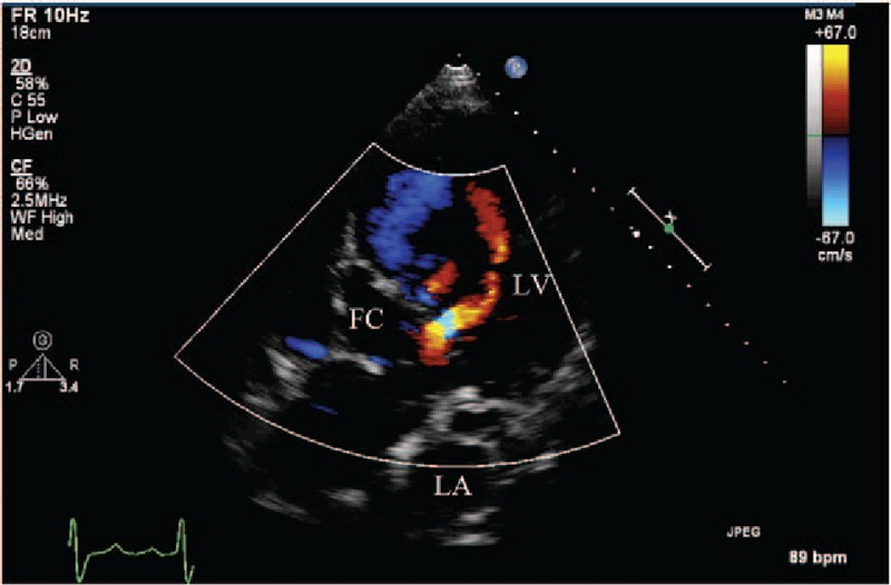
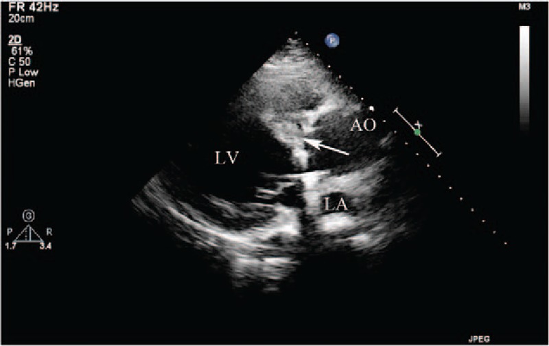
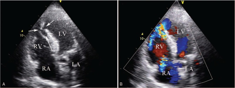
Dissection of the interventricular septum (IVS) is an extremely rare entity. An institutional echocardiographic database was retrospectively reviewed; 13 patients with a diagnosis of IVS dissection were found and confirmed by cardiac surgery. The purposes of the study were: to determine the value of transthoracic echocardiography (TTE) in establishing the diagnosis of IVS dissection, and to detail the TTE features of IVS dissection.Thirteen patients with IVS dissection diagnosed by TTE, 8 males and 5 females were taken from 789,114 TTE studies performed between 1985 and 2014. All underwent cardiac surgery during which their diagnosis was confirmed. The etiology, location, 2-dimensional morphology, and color Doppler findings of IVS dissection were noted.The right sinus of Valsalva (SOV) was involved in 11 of the 13 patients. In 5 patients, a single aneurysm of the right SOV was seen dissecting into the IVS. One patient with a combination of a bicuspid aortic valve and a right SOV aneurysm dissected into the IVS. In 4 patients, aortic valve infective endocarditis resulted in IVS dissection. In 1 patient, mechanical aortic valve prosthetic replacement was complicated by annular detachment and a severe paravalvular leak causing IVS dissection. In all 11 patients, TTE showed a dissecting cystic-like mass in the IVS from the base to the mid-septum or confined to the septal base. The path of the dissection in these 11 patients was traced to the right SOV and communications between the IVS dissection and the aortic root were identified. In the remaining 2 patients, IVS dissection followed septal rupture due to a myocardial infarction, and communication was seen between the IVS dissection and the right ventricle.The study showed that most of the dissections of the IVS commence in the right SOV, due to either congenital anomalies or infective endocarditis, or following aortic valve replacement or myocardial infarction. The TTE characteristic of IVS dissection is a cystic-like mass seen in the IVS.
Conflict of interest statement
The authors have no conflicts of interest to disclose.
Figures
References
-
- Engel PJ, Held JS, van der Bel-Kahn J, et al. Echocardiographic diagnosis of congenital sinus of Valsalva aneurysm with dissection of the interventricular septum. Circulation 1981;63:705–11. - PubMed
-
- Raffa H, Mosieri J, Sorefan AA, et al. Sinus of Valsalva aneurysm eroding into the interventricular septum. Ann Thorac Surg 1991;51:996–8. - PubMed
-
- Fasoli G, Della Valentina P, Scognamiglio R. Echocardiographic findings in left ventricular septal aneurysm. Int J Cardiol 1988;18:441–3. - PubMed
-
- Branco LM, Feliciano J, Cacela D, et al. Giant septal cavity due to coronary artery fistula and ventricular septal dissection after cardiac surgery. Eur J Echocardiogr 2008;9:163–6. - PubMed
-
- Dubel HP, Romaniuk P, Warnke H. Coronary ventricular fistulas in patients with heart transplants. Herz 1991;16:55–9. - PubMed
MeSH terms
LinkOut - more resources
Full Text Sources
Other Literature Sources
