. 2017 Mar 6;18(3):569.
doi: 10.3390/ijms18030569.

Effects of Silver Nanoparticles on Multiple Drug-Resistant Strains of Staphylococcus aureus and Pseudomonas aeruginosa from Mastitis-Infected Goats: An Alternative Approach for Antimicrobial Therapy

Affiliations

Effects of Silver Nanoparticles on Multiple Drug-Resistant Strains of Staphylococcus aureus and Pseudomonas aeruginosa from Mastitis-Infected Goats: An Alternative Approach for Antimicrobial Therapy

Yu-Guo Yuan et al. Int J Mol Sci. .

Abstract

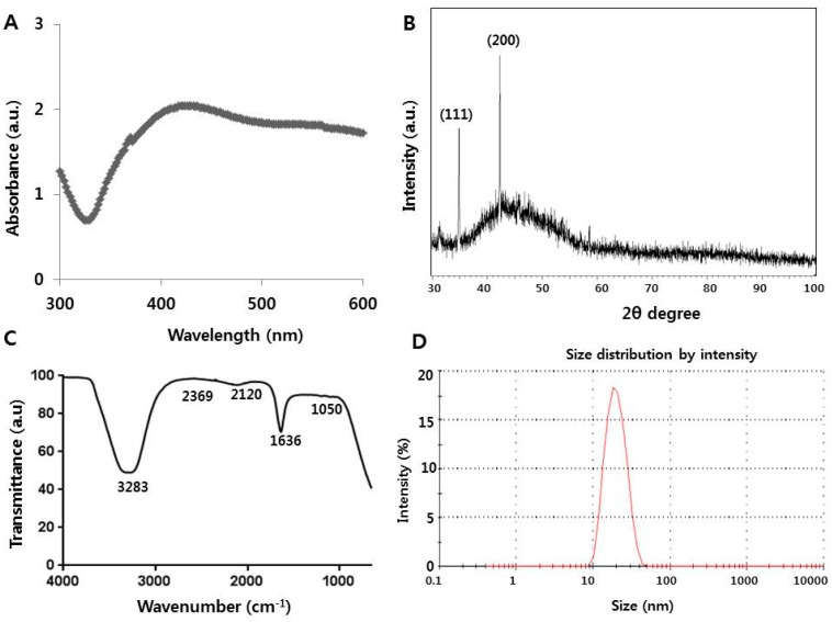
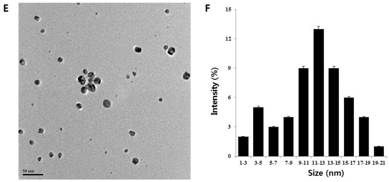
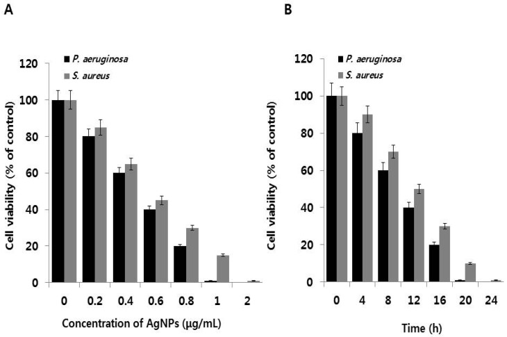
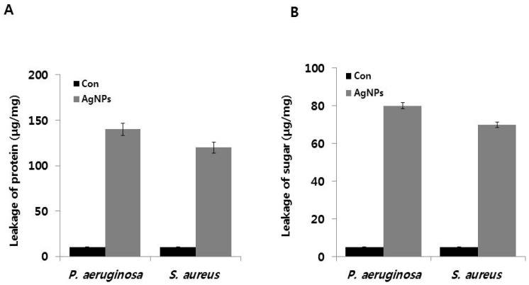
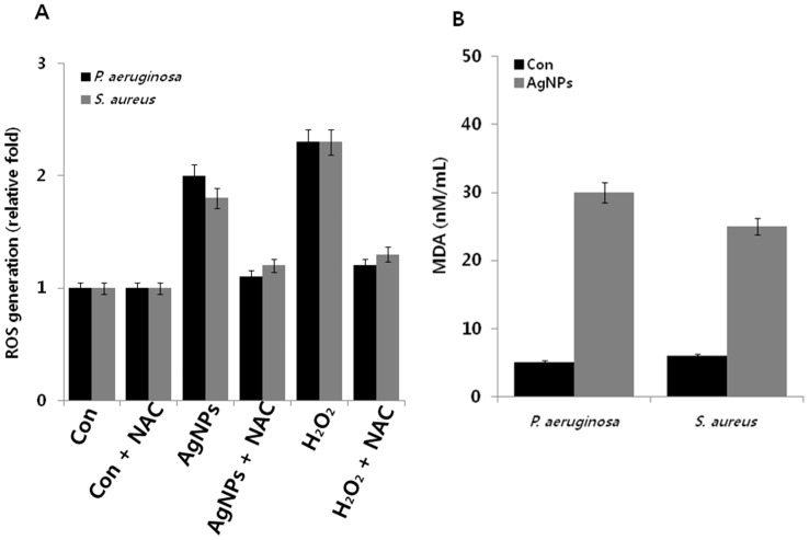
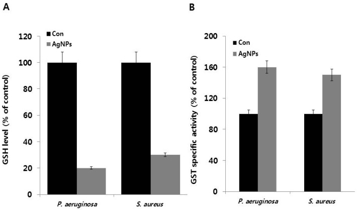
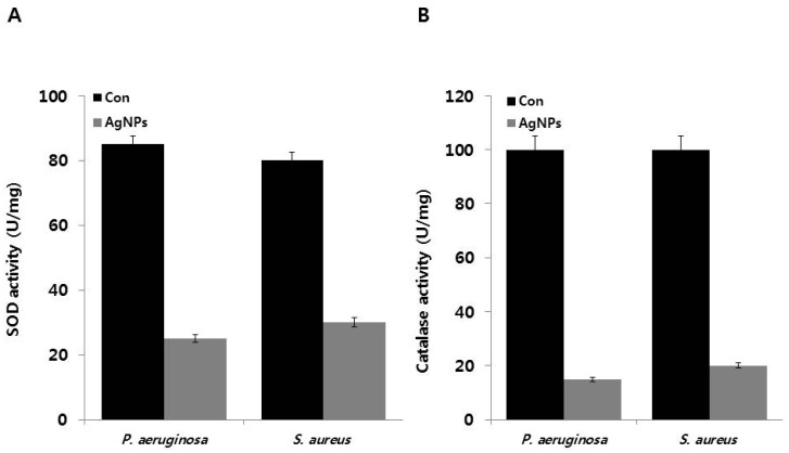
Recently, silver nanoparticles (AgNPs) have been widely used in various applications as antimicrobial agents, anticancer, diagnostics, biomarkers, cell labels, and drug delivery systems for the treatment of various diseases. Microorganisms generally acquire resistance to antibiotics through the course of antibacterial therapy. Multi-drug resistance (MDR) has become a growing problem in the treatment of infectious diseases, and the widespread use of broad-spectrum antibiotics has resulted in the development of antibiotic resistance by numerous human and animal bacterial pathogens. As a result, an increasing number of microorganisms are resistant to multiple antibiotics causing continuing economic losses in dairy farming. Therefore, there is an urgent need for the development of alternative, cost-effective, and efficient antimicrobial agents that overcome antimicrobial resistance. Here, AgNPs synthesized using the bio-molecule quercetin were characterized using various analytical techniques. The synthesized AgNPs were highly spherical in shape and had an average size of 11 nm. We evaluated the efficacy of synthesized AgNPs against two MDR pathogenic bacteria, namely, Pseudomonas aeruginosa and Staphylococcus aureus, which were isolated from milk samples produced by mastitis-infected goats. The minimum inhibitory concentrations (MICs) of AgNPs against P. aeruginosa and S. aureus were found to be 1 and 2 μg/mL, respectively. Our findings suggest that AgNPs exert antibacterial effects in a dose- and time-dependent manner. Results from the present study demonstrate that the antibacterial activity of AgNPs is due to the generation of reactive oxygen species (ROS), malondialdehyde (MDA), and leakage of proteins and sugars in bacterial cells. Results of the present study showed that AgNP-treated bacteria had significantly lower lactate dehydrogenase activity (LDH) and lower adenosine triphosphate (ATP) levels compared to the control. Furthermore, AgNP-treated bacteria showed downregulated expression of glutathione (GSH), upregulation of glutathione S-transferase (GST), and downregulation of both superoxide dismutase (SOD) and catalase (CAT). These physiological and biochemical measurements were consistently observed in AgNP-treated bacteria, thereby suggesting that AgNPs can induce bacterial cell death. Thus, the above results represent conclusive findings on the mechanism of action of AgNPs against different types of bacteria. This study also demonstrates the promising use of nanoparticles as antibacterial agents for use in the biotechnology and biomedical industry. Furthermore, this study is the first to propose the mode of action of AgNPs against MDR pathogens isolated from goats infected with subclinical mastitis.

Keywords: Pseudomonas aeruginosa; Staphylococcus aureus; metabolic activity; multiple drug resistance; oxidative stress; silver nanoparticles.


Conflict of interest statement

The authors declare no conflict of interest.

Figures

Figure 1
Figure 1
Synthesis and characterization of silver nanoparticles (AgNPs) using quercetin: (A) absorption spectrum of AgNPs synthesized using quercetin (B) X-ray diffraction spectra of AgNPs; (C) Fourier transform infrared spectra of AgNPs; (D) size distribution of AgNPs based on dynamic light scattering (DLS); (E) TEM images of AgNPs; and (F) several fields were used to measure AgNPs particle size; micrograph shows AgNPs with sizes ranging from 1 to 21 nm based on TEM images.
Figure 1
Figure 1
Synthesis and characterization of silver nanoparticles (AgNPs) using quercetin: (A) absorption spectrum of AgNPs synthesized using quercetin (B) X-ray diffraction spectra of AgNPs; (C) Fourier transform infrared spectra of AgNPs; (D) size distribution of AgNPs based on dynamic light scattering (DLS); (E) TEM images of AgNPs; and (F) several fields were used to measure AgNPs particle size; micrograph shows AgNPs with sizes ranging from 1 to 21 nm based on TEM images.
Figure 2
Figure 2
Effect of AgNPs on cell viability: (A) P. aeruginosa and S. aureus cells were incubated with various concentrations of AgNPs. Bacterial survival was determined at 24 h based on a CFU count assay; (B) Time dependent effect of AgNPs on P. aeruginosa and S. aureus. The experiment was performed with various controls, including a positive control (AgNPs and MHB, without inoculum) and a negative control (MHB and inoculum, without AgNPs). Results are expressed as the means ± SD of three separate experiments, with three replicates per experiment. Statistically significant differences between treatment and control groups were determined using student’s t-test (p < 0.05).
Figure 3
Figure 3
Effects of AgNPs on metabolic activity: (A) P. aeruginosa and S. aureus cells were incubated with respective MIC of AgNPs for 12 h. LDH activity was determined by measuring the reduction of NAD+ to NADH and H+ during the oxidation of lactate to pyruvate; (B) ATP levels were determined by measuring luminescence levels and comparing against an ATP standard curve. Results are expressed as the means ± SD of three separate experiments, with three replicates per experiment. Statistically significant differences between treatment and control groups were determined using student’s t-test (p < 0.05).
Figure 4
Figure 4
Effect of AgNPs on leakage of protein and sugars: (A) for protein leakage analysis, P. aeruginosa and S. aureus cells were treated with the respective MIC of AgNPs for 12 h, and protein levels were measured using the Bradford assay; and (B) for sugar analysis, all test strains were treated with respective MIC of AgNPs for 12 h, and sugar concentrations were measured. Results are expressed as the means ± SD of three separate experiments, with three replicates per experiment. Statistically significant differences between treatment and control groups were determined using student’s t-test (p < 0.05).
Figure 5
Figure 5
Effect of AgNPs on ROS generation and MDA: (A) P. aeruginosa and S. aureus cells were treated with respective MIC of AgNPs for 12 h. ROS generation was measured using DCFDA; and (B) MDA levels were measured using TBARS assay. Results are expressed as the means ± SD of three separate experiments, with three replicates per experiment. Statistically significant differences between treatment and control groups were determined using student’s t-test (p < 0.05).
Figure 6
Figure 6
Effect of AgNPs on GSH and GST activity: (A) P. aeruginosa and S. aureus cells were treated with respective MIC of AgNPs for 12 h. GSH levels were measured enzymatically in the clear supernatant based on the reduction of 5,5′-dithiobis-(2-nitrobenzoic acid) by the GSH reductase system; (B) GST activity was determined as described in the Materials and Methods Section. Results are expressed as the means ± SD of three separate experiments, with three replicates per experiment. Statistically significant differences between treatment and control groups were determined using student’s t-test (p < 0.05).
Figure 7
Figure 7
Effect of AgNPs on SOD and CAT activity: (A) P. aeruginosa and S. aureus cells were treated with respective MIC of AgNPs for 12 h. SOD (A) and CAT activities (B) were measured as described in the Materials and Methods Section. Results are expressed as the means ± SD of three separate experiments, with three replicates per experiment. Statistically significant differences between treatment and control groups were determined using student’s t-test (p < 0.05).

Similar articles

Cited by

References

    1. Harmon R.J. Physiology of mastitis and factors affecting somatic cell counts. J. Dairy Sci. 1994;77:2103–2112. doi: 10.3168/jds.S0022-0302(94)77153-8. - DOI - PubMed
    1. Oliszewski R., Núñez D.K.M., González S., Oliver G. β-Glucuronidase method to determine mastitis levels in goat milk. Methods Mol. Biol. 2004;268:475–479. - PubMed
    1. Ezzat A.M., Quintela-Baluja M., Böhme K., Fernández-No I., Caamaño-Antelo S., Calo-Mata P., Barros-Velázquez J. The Immunology of mammary gland of dairy ruminants between healthy and inflammatory conditions. J. Vet. Med. 2014;2014:659801. - PMC - PubMed
    1. Bannerman D.D., Chockalingam A., Paape M.J., Hope J.C. The bovine innate immune response during experimentally-induced Pseudomonas aeruginosa mastitis. Vet. Immunol. Immunopathol. 2005;107:201–215. doi: 10.1016/j.vetimm.2005.04.012. - DOI - PubMed
    1. Akira S., Uematsu S., Takeuchi O. Pathogen recognition and innate immunity. Cell. 2006;124:783–801. doi: 10.1016/j.cell.2006.02.015. - DOI - PubMed

MeSH terms