. 2017 Mar 8:7:43567.
doi: 10.1038/srep43567.

Amigo2-upregulation in Tumour Cells Facilitates Their Attachment to Liver Endothelial Cells Resulting in Liver Metastases

Affiliations

Amigo2-upregulation in Tumour Cells Facilitates Their Attachment to Liver Endothelial Cells Resulting in Liver Metastases

Yusuke Kanda et al. Sci Rep. .

Abstract

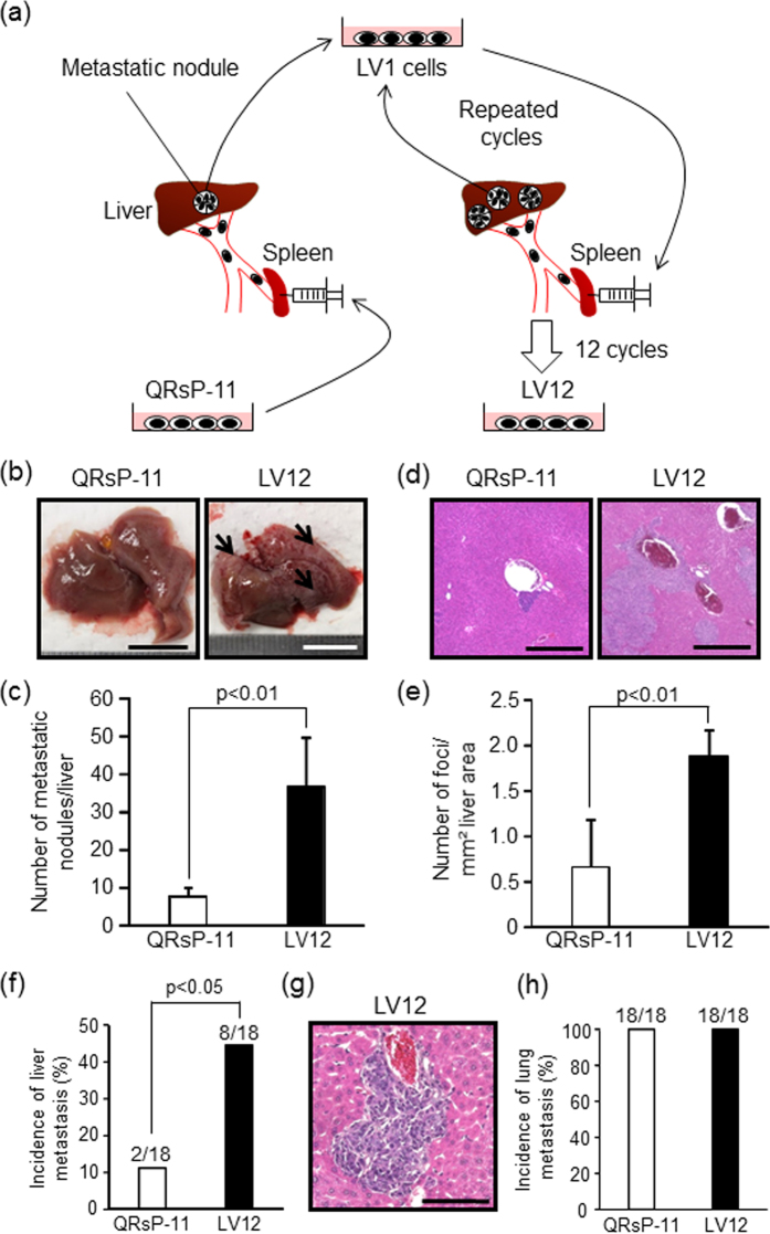
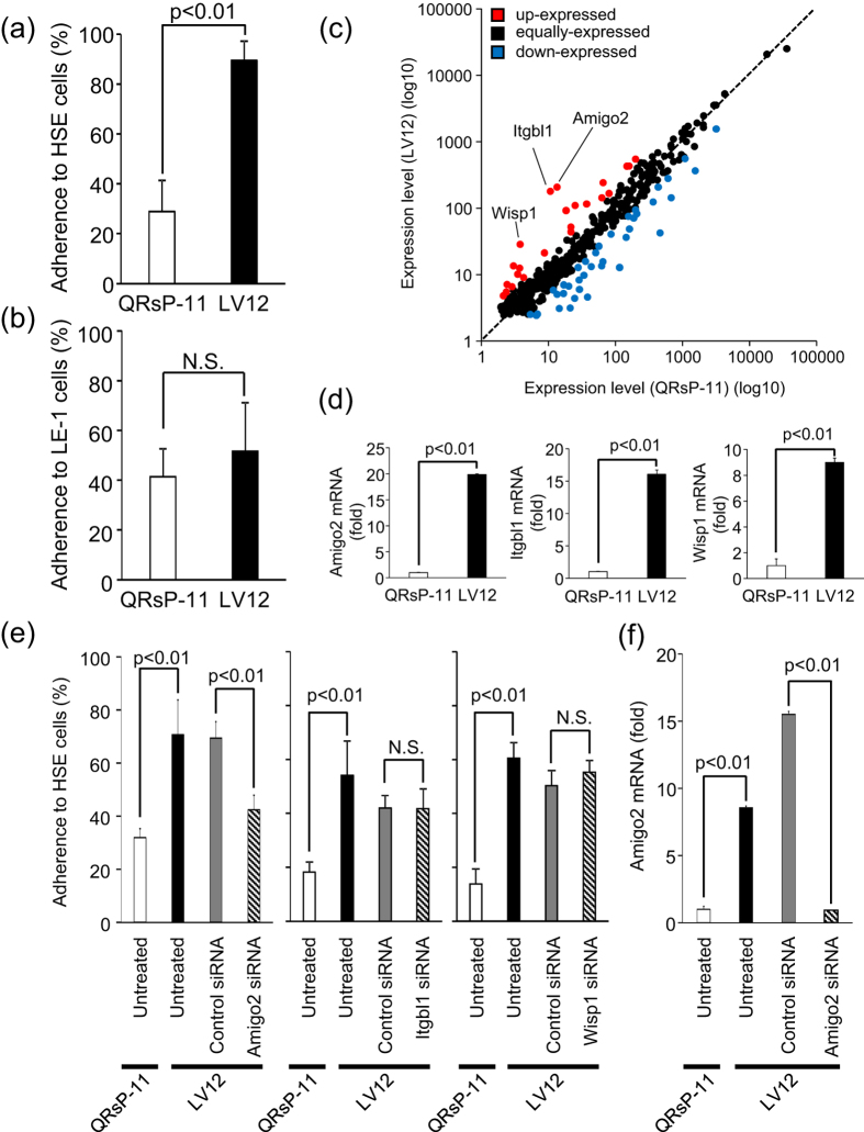
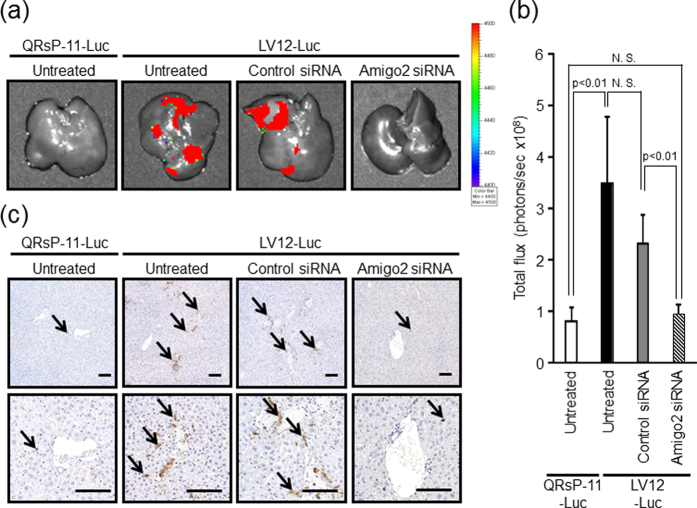
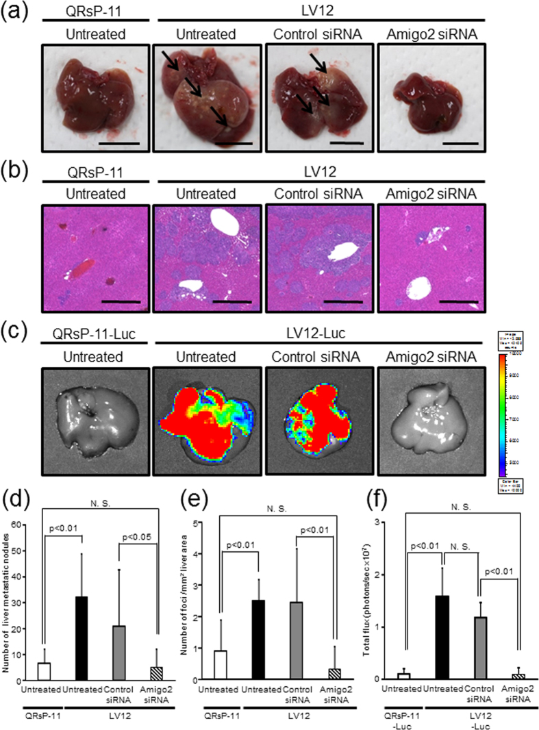
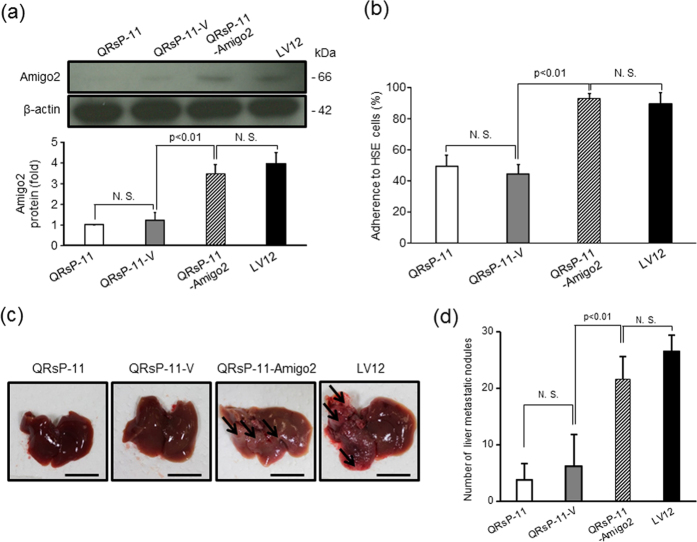
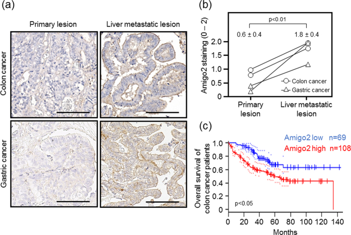
Since liver metastasis is the main cause of death in cancer patients, we attempted to identify the driver gene involved. QRsP-11 fibrosarcoma cells were injected into the spleens of syngeneic mice to isolate tumour sub-populations that colonize the liver. Cells from liver metastatic nodules were established and subsequently injected intrasplenically for selection. After 12 cycles, the cell subline LV12 was obtained. Intravenous injection of LV12 cells produced more liver metastases than QRsP-11 cells, whereas the incidence of lung metastases was similar to that of QRsP-11 cells. LV12 cells adhered to liver-derived but not to lung-derived endothelial cells. DNA chip analysis showed that amphoterin-induced gene and open reading frame 2 (Amigo2) was overexpressed in LV12 cells. siRNA-mediated knockdown of Amigo2 expression in LV12 cells attenuated liver endothelial cell adhesion. Ex vivo imaging showed that suppression of Amigo2 in luciferase-expressing LV12 cells reduced attachment/metastasis to liver to the same level as that observed with QRsP-11 cells. Forced expression of Amigo2 in QRsP-11 cells increased liver endothelial cell adhesion and liver metastasis. Additionally, Amigo2 expression in human cancers was higher in liver metastatic lesions than in primary lesions. Thus, Amigo2 regulated tumour cell adhesion to liver endothelial cells and formation of liver metastases.


Conflict of interest statement

The authors declare no competing financial interests.

Figures

Figure 1
Figure 1. LV12 cells possess a high liver-metastatic potential and give rise to multiple liver colonies by intrasplenic and intravenous injections.
(a) Schematic representation of in vivo sequential selection of a liver-metastatic variant subline (LV12) from QRsP-11 mouse fibrosarcoma cells. Metastatic nodules in the livers of C57BL/6 mice, which formed from 1 × 106 QRsP-11 cells previously injected into the spleen, were harvested and a cell culture subline was established. These cells were then injected into the spleens of other mice, and liver metastases subsequently formed. The metastatic foci were excised and expanded as a new subline. This procedure was repeated 12 times, yielding a cell subline designated as LV12. (b) Macroscopic views of liver metastasis formation 7 days after intrasplenic injection of 1 × 106 LV12 or QRsP-11 cells. Arrows indicate representative metastatic nodules. Scale bar: 10 mm. (c) The numbers of metastases on the liver surface were determined. Bar graphs show means ± SD (n = 5 in each group). (d) Representative H&E staining of the livers from Fig. 1b. Scale bar: 500 μm. (e)The number of metastatic foci per mm2 of liver area was determined using image analysis software. Bar graphs show means ± SD (n = 5 in each group). (f) The incidence of liver metastatic colonization 7 days after tail vein injection of 1 × 106 LV12 or QRsP-11 cells was evaluated using histology. Bar graphs show means ± SD from three independent experiments with similar results (n = 18 in each group). (g) Microscopic appearance of the liver metastatic foci formed by LV12 cells. Scale bar: 100 μm. (h) The incidence of lung metastasis was determined by the presence of metastatic nodules on the lung surface on day 7 post-injection. The incidence of metastatic colonization is indicated above each bar (number of mice with metastasis/number of mice examined).
Figure 2
Figure 2. Amigo2 functions in the adhesion of LV12 cells to liver endothelial cells.
(a,b) LV12 and QRsP-11 cells were labelled with fluorescent dye and were placed onto endothelial cells derived from liver (HSE) or lung (LE-1) for 30 min. The percentages of adherent cells were determined by measuring fluorescence intensity. Bar graphs show means ± SD (n = 5 in each group). (c) Scatter plot of adhesion-related mRNA expression profiles of LV12 cells and QRsP-11 cells. Amigo2, Itgbl1 and Wisp1 were included in the upregulated genes in LV12 cells. (d)The relative levels of Amigo2, Itgbl1 and Wisp1 mRNA in LV12 cells compared to QRsP-11 cells as measured using qRT-PCR. Bar graphs show means ± SD (n = 5 in each group). (e) LV12 cells were transfected with siRNA targeting Amigo2, Itgbl1, Wisp1 or with control siRNA. After 24 h, the cells were harvested and their adhesion to HSE cells was assayed. Bar graphs show means ± SD (n = 5 in each group). (f) Downregulation of Amigo2 mRNA expression by Amigo2 siRNA. Transfection of LV12 cells was confirmed using qRT-PCR. Bar graphs show means ± SD (n = 4 in each group).
Figure 3
Figure 3. RNA interference-mediated Amigo2 suppression reduces intrahepatic attachment of LV12 cells.
(a) An expression vector for firefly luciferase was introduced into LV12 and QRsP-11 cells (LV12-Luc and QRsP-11-Luc, respectively). Representative bioluminescent images of livers 4 hours after intrasplenic injection of 5 × 106 QRsP-11-Luc or LV12-Luc cells transfected with either Amigo2 or control siRNA are shown. (b) Quantification of total flux (photons per sec) from the livers shown in Fig. 3a. Bar graphs show means ± SD (n = 4 in each group). (c) Tumour cells in the liver tissues were detected using immunohistochemistry with an anti-luciferase antibody. LV12-Luc or QRsP-11-Luc cells are indicated by arrows. Top, ×10 low-power field; bottom, ×20 high-power field. Scale bar: 100 μm.
Figure 4
Figure 4. Downregulation of Amigo2 mRNA in LV12 cells decreased liver metastasis.
(a) Representative pictures show the macroscopic metastatic nodules (indicated by arrows) on the liver surface 7 days after the mice were injected intrasplenically with LV12 cells transfected with Amigo2 or control siRNA. Scale bar: 10 mm. (b) H&E staining of the liver metastatic foci. Scale bar: 500 μm. (c) Representative bioluminescent images of livers 7 days after intrasplenic injection of QRsP-11-Luc or LV12-Luc cells transfected with either Amigo2 or control siRNA are shown. (d) Metastatic nodules on the liver surface in individual mice were counted. Bar graphs show means ± SD from two independent experiments with similar results. QRsP-11 and untreated LV12 cells, n = 12; control siRNA or Amigo2 siRNA-transfected LV12 cells, n = 11. (e) The number of metastatic foci per mm2 of liver area was determined. Bar graphs show means ± SD from two independent experiments with similar results. QRsP-11 and untreated LV12 cells, n = 12; control siRNA or Amigo2 siRNA-transfected LV12 cells, n = 11. (f) Quantification of total flux (photons per sec) from the livers shown in Fig. 4c. Bar graphs show means ± SD (n = 9 in each group).
Figure 5
Figure 5. Amigo2 overexpression in QRsP-11 cells promotes their adhesion to liver endothelial (HSE) cells and liver metastasis.
(a) QRsP-11 cells were stably transfected with an empty (QRsP-11-V) or an Amigo2 expression (QRsP-11-Amigo2) vector. Amigo2 overexpression was confirmed by western blot analysis. β-actin served as the internal control. Bar graphs show means ± SD (n = 3 in each group). Full-length blots were presented in Supplementary Fig. S5. (b)The adhesion of the transfectants to HSE liver endothelial cells was quantified. Bar graphs show means ± SD (n = 5 in each group). (c) Macroscopic observation of the livers of mice 7 days after intrasplenic injection of control or Amigo2-overexpressing QRsP-11 cells are shown. The arrows highlight metastatic nodules. Scale bar: 10 mm. (d) The liver surface nodules in each mouse were counted. Bar graphs show means ± SD (n = 5 in each group).
Figure 6
Figure 6. Amigo2 is overexpressed in liver metastases in human colon and gastric carcinomas.
(a) Immunohistochemical staining of Amigo2 in matched primary and liver metastatic lesions of human colon and gastric cancers. Scale bar: 100 μm. (b) Amigo2 staining intensity in paired specimens of primary colon and gastric carcinomas and their metastases. Results are shown as means ± SD (n = 4 in each group). (c) Kaplan-Meier analysis of overall survival in colon cancer patients with high versus low Amigo2 mRNA expression. Dotted lines indicate 95% confidence intervals for each group. These data were obtained from PrognoScan (http://www.abren.net/PrognoScan/).

References

    1. Li F., Tiede B., Massague J. & Kang Y. Beyond tumorigenesis: cancer stem cells in metastasis. Cell Res 17, 3–14, doi: 10.1038/sj.cr.7310118 (2007). - DOI - PubMed
    1. Talmadge J. E. & Fidler I. J. AACR centennial series: the biology of cancer metastasis: historical perspective. Cancer Res 70, 5649–5669, doi: 10.1158/0008-5472.CAN-10-1040 (2010). - DOI - PMC - PubMed
    1. Budczies J. et al.. The landscape of metastatic progression patterns across major human cancers. Oncotarget 6, 570–583, doi: 10.18632/oncotarget.2677 (2015). - DOI - PMC - PubMed
    1. Fidler I. J. & Kripke M. L. Metastasis results from preexisting variant cells within a malignant tumor. Science 197, 893–895 (1977). - PubMed
    1. Chiang A. C. & Massague J. Molecular basis of metastasis. N Engl J Med 359, 2814–2823, doi: 10.1056/NEJMra0805239 (2008). - DOI - PMC - PubMed

Publication types

MeSH terms