Review
. 2017 Apr 1;74(4):407-415.
doi: 10.1001/jamapsychiatry.2016.3325.

An Integrated Neuroscience Perspective on Formulation and Treatment Planning for Posttraumatic Stress Disorder: An Educational Review

Affiliations
Review

An Integrated Neuroscience Perspective on Formulation and Treatment Planning for Posttraumatic Stress Disorder: An Educational Review

David A Ross et al. JAMA Psychiatry. .

Abstract

Importance: Posttraumatic stress disorder (PTSD) is a common psychiatric illness, increasingly in the public spotlight in the United States due its prevalence in the soldiers returning from combat in Iraq and Afghanistan. This educational review presents a contemporary approach for how to incorporate a modern neuroscience perspective into an integrative case formulation. The article is organized around key neuroscience "themes" most relevant for PTSD. Within each theme, the article highlights how seemingly diverse biological, psychological, and social perspectives all intersect with our current understanding of neuroscience.

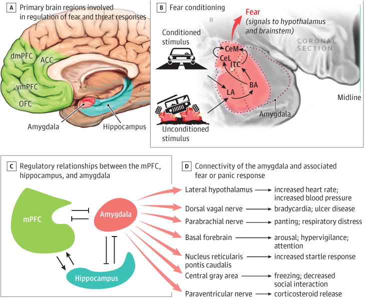
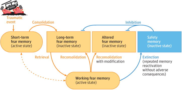
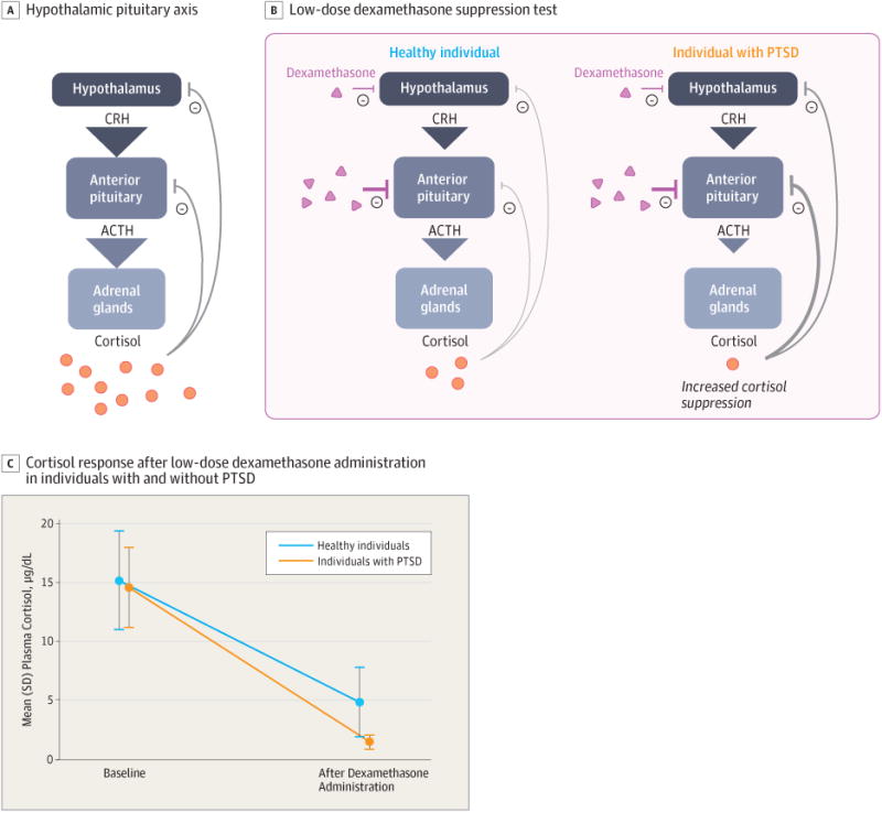
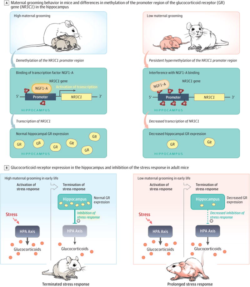
Observations: Any contemporary neuroscience formulation of PTSD should include an understanding of fear conditioning, dysregulated circuits, memory reconsolidation, epigenetics, and genetic factors. Fear conditioning and other elements of basic learning theory offer a framework for understanding how traumatic events can lead to a range of behaviors associated with PTSD. A circuit dysregulation framework focuses more broadly on aberrant network connectivity, including between the prefrontal cortex and limbic structures. In the process of memory reconsolidation, it is now clear that every time a memory is reactivated it becomes momentarily labile-with implications for the genesis, maintenance, and treatment of PTSD. Epigenetic changes secondary to various experiences, especially early in life, can have long-term effects, including on the regulation of the hypothalamic-pituitary-adrenal axis, thereby affecting an individual's ability to regulate the stress response. Genetic factors are surprisingly relevant: PTSD has been shown to be highly heritable despite being definitionally linked to specific experiences. The relevance of each of these themes to current clinical practice and its potential to transform future care are discussed.

Conclusions and relevance: Together, these perspectives contribute to an integrative, neuroscience-informed approach to case formulation and treatment planning. This may help to bridge the gap between the traditionally distinct viewpoints of clinicians and researchers.


Conflict of interest statement

Conflict of Interest Disclosures: Drs Ross, Arbuckle, and Travis, ascochairs of the National Neuroscience Curriculum Initiative, receive support from the National Institutes of Health (R25 MH10107602S1 and R25 MH086466 07S1).

Figures

Figure 1
Figure 1. Schematic Diagram of Neural Circuitry Involved in Fear Conditioning and Posttraumatic Stress Disorder
A, Primary brain regions involved in regulating fear and threat responses are theamygdala, the hippocampus, and the medial prefrontal cortex, which iscomprised of dorsal (dmPFC) and ventral (vmPFC) subdivisions, theorbitofrontal cortex (OFC), and the anterior cingulate cortex (ACC). B, Amygdala-specific circuits that are involved in fear conditioning. The sensory information representing the conditioned stimulus (eg, previously neutralstimulus such as driving a car) is integrated within the amygdala with the unconditioned stimulus information (eg, a traumatic event such as an explosionin a car). The amygdala is central in the neural circuit involved in regulating fearconditioning. In general, input to the lateral nucleus (LA) of the amygdala leadsto learning about fear, whereas the central amygdala (lateral [CeL] and medial [CeM] subdivisions) is responsible for sending output signals about fear to the hypothalamus and brainstem structures. The intercalated cell masses (ITC) are thought to regulate inhibition of information flow between the basal nucleus (BA) and central amygdala.C and D, Interactions between components of the mPFC and the hippocampus constantly regulate the amygdala's output to subcortical brain regions activating the fear reflex. The mPFC (in particular, the vmPFC) is classically thought to inhibit amygdala activity and reduce subjective distress, while the hippocampus plays a role both in the coding of fear memories and also in the regulation of the amygdala. The hippocampus and mPFC also interact in regulating context and fear modulation. Panels C and D adapted from Parsons RG and Ressler KJ.
Figure 2
Figure 2. Process of Memory Reconsolidation
Schematic diagram of the primary phases of memory consolidation, reconsolidation, and extinction after a traumatic event. Shortly after the fear conditioning process (conditioned stimulus–unconditioned stimulus pairing illustrated in Figure 1), a memory is in an active state in short-term memory until it gets consolidated and stabilized into long-term memory. As short-term memories are immediately available to conscious awareness, these memories are temporarily available to working memory as they are also being consolidated. At later time points, the retrieval of a consolidated memory returns the memory from an inactive state in long-term memory to an active state in working memory from which it needs to be stabilized anew. The process during which reactivated memories are stabilized again is called reconsolidation. Reconsolidation occurs most readily after brief reactivation, which strengthens the long-term memory. During reconsolidation, the active memory traces are potentially vulnerable to modification. Repeated reactivation of a memory without adverse consequences creates an extinction, or safety, memory which inhibits the original fear memory. Reconsolidation and extinction are opposing processes that act to strengthen or inhibit, respectively, fear memory expression over time.
Figure 3
Figure 3. Regulation of the Hypothalamic-Pituitary-Adrenal (HPA) Axis in Posttraumatic Stress Disorder (PTSD)
Evidence suggests that the sensitivity to glucocorticoid receptor activation in the HPA feedback system is increased in individuals with PTSD compared with healthy individuals. In studies that compared the response of individuals with and without PTSD to the low-dose dexamethasone suppression test, individuals with PTSD demonstrated increased suppression of the HPA axis as measured by plasma cortisol levels. When dexamethasone, a cortisol agonist, activates glucocorticoid receptors in individuals with PTSD, increased negative feedback on the HPA axis results in more rapid and robust decreases in ACTH release and in adrenal activation and cortisol release. Data in the graph (C) are from Yehuda et al.
Figure 4
Figure 4. Early Life Experience and Epigenetic Modulation of the Stress Response in Mice
A, left: Mice reared in a high licking and grooming (enriched or supportive) environment have less stress during the early part of their lives. In these mice, the glucocorticoid receptor (GR) promotor region is demethylated, which allows binding of transcription factors like NGFI-A and normal hippocampal GR expression. B, left: Normal GR expression results in more efficient feedback inhibition of the hypothalamic-pituitary-adrenal (HPA) axis and lower overall stress reactivity as adults. A, right: Mice reared in a low licking and grooming (neglectful) environment have persistent hypermethylation of the promoter region of the GR gene in the hippocampus, resulting in decreased GR expression. B, right: Reduced activity of hippocampal GRs results in decreased inhibition of the HPA axis with prolonged activation of the stress response. Consistent with rodent studies, hypermethylation of hippocampal GRs has been demonstrated in postmortem brains of patients with histories of childhood abuse.

References

    1. Ross DA, Travis MJ, Arbuckle MR. Posttraumatic stress disorder in a young adult military veteran. JAMA Psychiatry. doi: 10.1001/jamapsychiatry.2016.3334. published online March 8, 2017. - DOI - PMC - PubMed
    1. Daly RJ. Samuel Pepys and post-traumatic stress disorder. Br J Psychiatry. 1983;143:64–68. - PubMed
    1. Shay J. Odysseus in America: Combat Trauma and the Trials of Homecoming. New York, NY: Scribner; 2002.
    1. American Psychiatric Association. Diagnostic and Statistical Manual of Mental Disorders. 5th. Washington, DC: American Psychiatric Association; 2013.
    1. Friedman MJ, Kilpatrick DG, Schnurr PP, Weathers FW. Correcting misconceptions about the diagnostic criteria for posttraumatic stress disorder in DSM-5. JAMA Psychiatry. 2016;73(7):753–754. - PubMed

MeSH terms