. 2017 Mar 3;18(3):547.
doi: 10.3390/ijms18030547.

Role of uL3 in Multidrug Resistance in p53-Mutated Lung Cancer Cells

Affiliations

Role of uL3 in Multidrug Resistance in p53-Mutated Lung Cancer Cells

Annapina Russo et al. Int J Mol Sci. .

Abstract

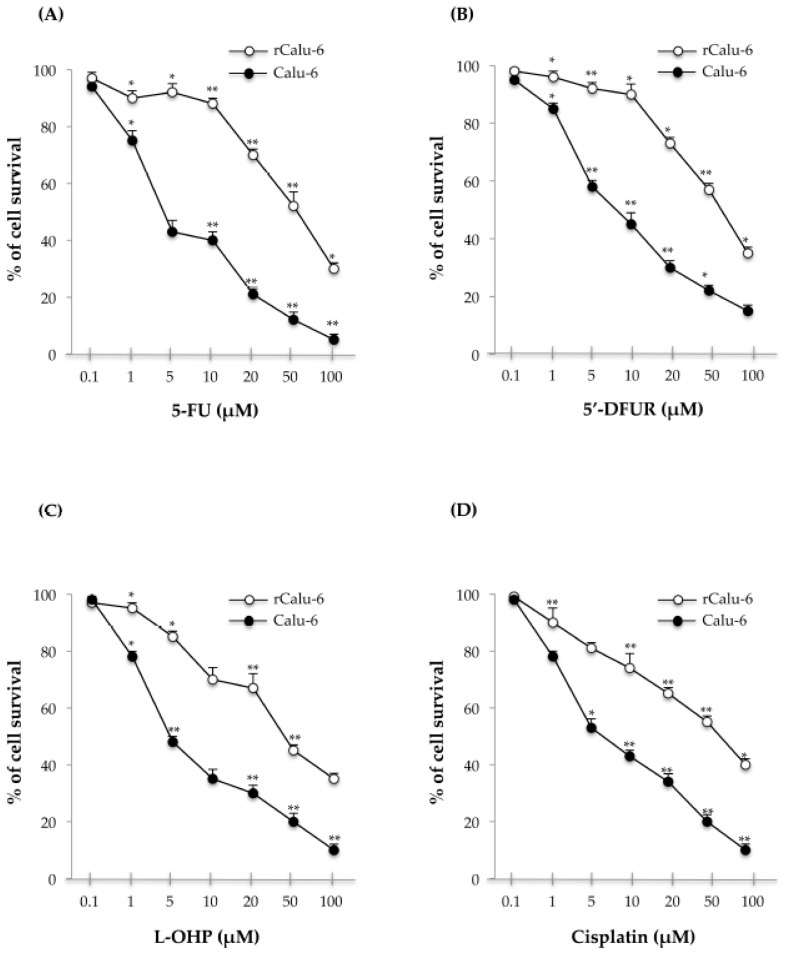
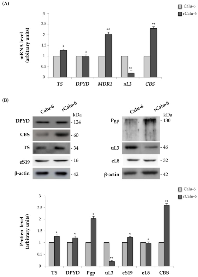
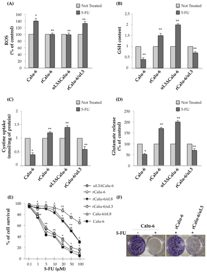
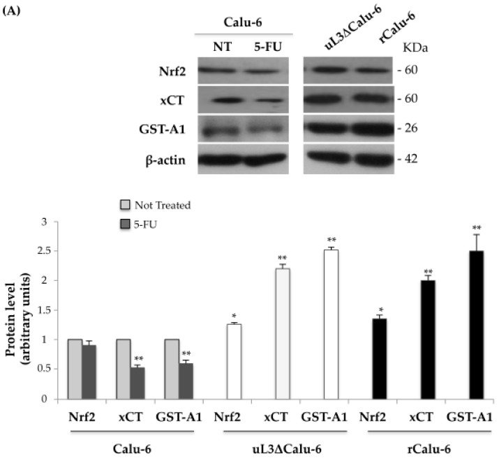
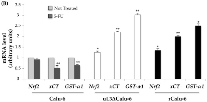
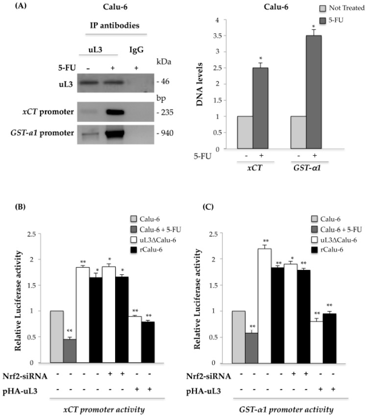
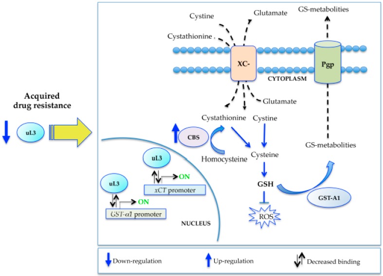
Cancer is one of the most common causes of death among adults. Chemotherapy is crucial in determining patient survival and quality of life. However, the development of multidrug resistance (MDR) continues to pose a significant challenge in the management of cancer. In this study, we analyzed the role of human ribosomal protein uL3 (formerly rpL3) in multidrug resistance. Our studies revealed that uL3 is a key determinant of multidrug resistance in p53-mutated lung cancer cells by controlling the cell redox status. We established and characterized a multidrug resistant Calu-6 cell line. We found that uL3 down-regulation correlates positively with multidrug resistance. Restoration of the uL3 protein level re-sensitized the resistant cells to the drug by regulating the reactive oxygen species (ROS) levels, glutathione content, glutamate release, and cystine uptake. Chromatin immunoprecipitation experiments and luciferase assays demonstrated that uL3 coordinated the expression of stress-response genes acting as transcriptional repressors of solute carrier family 7 member 11 (xCT) and glutathione S-transferase α1 (GST-α1), independently of Nuclear factor erythroid 2-related factor 2 (Nrf2). Altogether our results describe a new function of uL3 as a regulator of oxidative stress response genes and advance our understanding of the molecular mechanisms underlying multidrug resistance in cancers.

Keywords: GST-α1; MDR1; Nrf2; chemoresistance; lung cancer; multidrug resistance; nucleolar stress; ribosomal protein; uL3; xCT.


Conflict of interest statement

The authors declare no conflict of interest.

Figures

Figure 1
Figure 1
In vitro sensitivity of Calu-6 and rCalu-6 cells to 5-Fluorouracil (5-FU), 5′-Deoxy-5-fluorouridine (5′-DFUR), Oxaliplatin (L-OHP), and Cisplatin. Cells were cultured with increasing concentrations of 5-FU for 48 h and cell growth was assessed by sulforhodamine B (SRB) colorimetric assay. Cell growth was expressed as the percentage of control for each time point. ** p < 0.01, * p < 0.05 vs. untreated cells. The results illustrated in Figure 1, Figure 2, Figure 3, Figure 4 and Figure 5 are representative of three independently performed experiments; error bars represent the standard deviation.
Figure 2
Figure 2
Analysis of mRNAs and proteins related to chemoresistance. (A) Total RNA from Calu-6 and rCalu-6 cells was subjected to Reverse Transcription quantitative Polymerase Chain Reaction (RT-qPCR) with primers specific for the indicated mRNAs. The quantification of signals is shown. ** p < 0.01, * p < 0.05 vs. mRNA levels in Calu 6 cells set at 1; (B) Protein extracts from Calu-6 and rCalu-6 cells were analyzed by Western blotting with antibodies against the indicated proteins. β-actin was used as the loading control. The quantification of signals is shown. ** p < 0.01, * p < 0.05 vs. protein levels in Calu 6 cells set at 1.
Figure 3
Figure 3
uL3 contributes to Reactive Oxygen Species (ROS) defense in rCalu-6 cells. (A) Histogram quantifying ROS production in indicated cell lines after treatment with 5-FU. ** p < 0.01, * p < 0.05 vs. respective untreated cells set at 1; (B) Glutathione (GSH) content in Calu-6 cells and derivative sublines after treatment with 5-FU. The quantification of signals is shown. ** p < 0.01, * p < 0.05 vs. respective untreated cells set at 1; (C) Changes in cystine uptake after incubation of indicated cells with 5-FU. ** p < 0.01, * p < 0.05 vs. respective untreated cells set at 1; (D) Evaluation of glutamate release in indicated cell lines after treatment with 5-FU. ** p < 0.01, * p < 0.05 vs. respective untreated cells set at 1; (E) uL3ΔCalu-6, rCalu-6, rCalu-6/uL3, rCalu-6/eL8, Calu-6/eL8 and Calu-6 cells were cultured with increasing concentrations of 5-FU for 48 h and cell growth was assessed by SRB colorimetric assay. Cell growth was expressed as the percentage of control for each time point. Ectopic expression of uL3 (rCalu-6/uL3 cells) abolished resistance to 5-FU. ** p < 0.01, * p < 0.05 vs. Calu 6 cells; (F) Representative image of the clonogenic analysis for cell proliferation of Calu-6, rCalu-6, and rCalu-6/uL3 after treatment with 10 μM 5-FU for 48 h.
Figure 4
Figure 4
Influence of uL3 on Nrf2, xCT, and GST-A1 expression levels in Calu-6 cells and resistant sublines. (A) Protein extracts from Calu-6 cells treated or not with 10 μM 5-FU for 48 h, uL3ΔCalu-6 and rCalu-6 cells were analysed by Western blotting with the indicated antibodies. Anti-β-actin was used as the loading control. The quantification of signals is shown. ** p < 0.01, * p < 0.05 vs. protein levels in untreated Calu-6 cells set at 1; (B) Total RNA from Calu-6 treated or not with 10 μM 5-FU for 48 h, uL3ΔCalu-6 and rCalu-6 cells were subjected to qPCR with primers specific for the indicated genes. The quantification of signals is shown. ** p < 0.01,* p < 0.05 vs. mRNA levels in untreated Calu-6 cells set at 1.
Figure 4
Figure 4
Influence of uL3 on Nrf2, xCT, and GST-A1 expression levels in Calu-6 cells and resistant sublines. (A) Protein extracts from Calu-6 cells treated or not with 10 μM 5-FU for 48 h, uL3ΔCalu-6 and rCalu-6 cells were analysed by Western blotting with the indicated antibodies. Anti-β-actin was used as the loading control. The quantification of signals is shown. ** p < 0.01, * p < 0.05 vs. protein levels in untreated Calu-6 cells set at 1; (B) Total RNA from Calu-6 treated or not with 10 μM 5-FU for 48 h, uL3ΔCalu-6 and rCalu-6 cells were subjected to qPCR with primers specific for the indicated genes. The quantification of signals is shown. ** p < 0.01,* p < 0.05 vs. mRNA levels in untreated Calu-6 cells set at 1.
Figure 5
Figure 5
Analysis of the interaction between uL3 and xCT and GST-α1 gene promoters. (A) Protein samples of DNA-uL3 or DNA-IgG immunocomplexes from Calu-6 cells untreated or treated with 10 μM 5-FU for 48 h were analysed by Western blotting with antibodies against uL3. Note the absence of signal in the DNA-IgG immunocomplex. The same DNA-immunoprecipitates were subjected to qPCR with primers specific for xCT or GST-α1 gene promoters. The quantification of signals is shown. *p < 0.05 vs. DNA levels in untreated cells set at 1. Calu-6 cells treated or not treated with 10 μM 5-FU for 48 h, uL3ΔCalu-6 and rCalu-6 cells were transiently transfected with (B) xCT promoter luciferase reporter plasmid or (C) GST-α1 promoter luciferase reporter plasmid in presence or absence of Nrf2 siRNA or pHA-uL3 plasmid. Analysis of the relative luciferase activity, normalized against Renilla Luciferase (pRL) activity, of the samples is shown. ** p < 0.01, * p < 0.05 vs. untreated Calu-6 cells set at 1.
Figure 6
Figure 6
Proposed model of acquired multidrug resistance based on uL3 status.

References

    1. DeSantis C.E., Lin C.C., Mariotto A.B., Siegel R.L., Stein K.D., Kramer J.L., Alteri R., Robbins A.S., Jemal A. Cancer treatment and survivorship statistics, 2014. CA Cancer J. Clin. 2014;64:252–271. doi: 10.3322/caac.21235. - DOI - PubMed
    1. Maione P., Perrone F., Gallo C., Manzione L., Piantedosi F., Barbera S., Cigolari S., Rosetti F., Piazza E., Robbiati S.F., et al. Pretreatment quality of life and functional status assessment significantly predict survival of elderly patients with advanced non-small-cell lung cancer receiving chemotherapy: A prognostic analysis of the multicenter italian lung cancer in the elderly study. J. Clin. Oncol. 2005;23:6865–6872. - PubMed
    1. Chang A. Chemotherapy, chemoresistance and the changing treatment landscape for NSCLC. Lung Cancer. 2011;71:3–10. doi: 10.1016/j.lungcan.2010.08.022. - DOI - PubMed
    1. Chunga Y.M., Parka S., Parka J.K., Kimb Y., Kanga Y., Yooa Y.D. Establishment and characterization of 5-fuorouracil-resistant gastric cancer cells. Cancer Lett. 2000;159:95–101. doi: 10.1016/S0304-3835(00)00535-8. - DOI - PubMed
    1. Gu W., Fang F.-F., Li B., Cheng B.-B., Ling C.-Q. Characterization and resistance mechanisms of a 5-fluorouracil-resistant hepatocellular carcinoma cell line. Asian Pac. J. Cancer Prev. 2012;13:4807–4814. doi: 10.7314/APJCP.2012.13.9.4807. - DOI - PubMed

MeSH terms