. 2017 Mar 7;7(1):101.
doi: 10.1038/s41598-017-00109-8.

YidC and SecYEG form a heterotetrameric protein translocation channel

Affiliations

YidC and SecYEG form a heterotetrameric protein translocation channel

Ilie Sachelaru et al. Sci Rep. .

Abstract

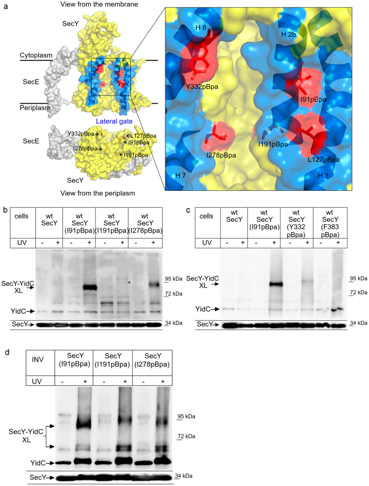
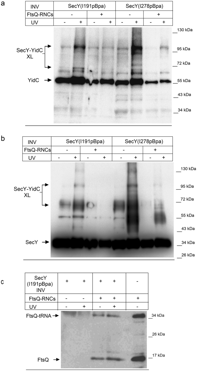
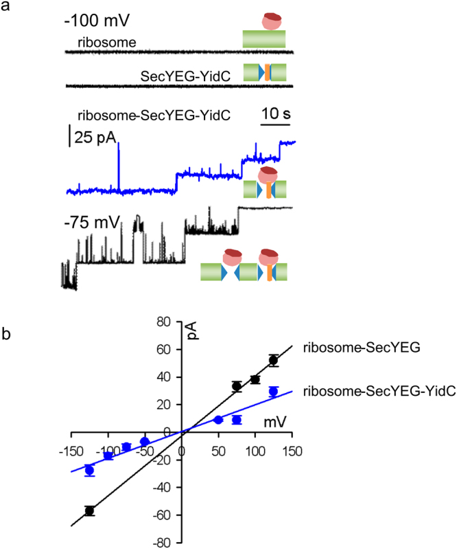
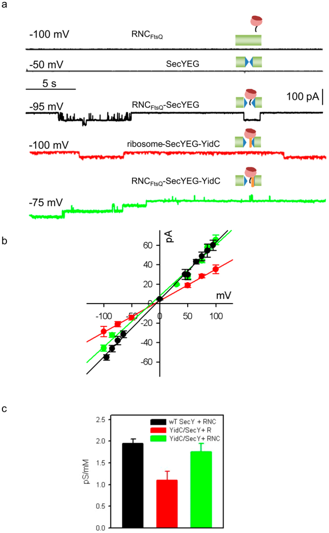
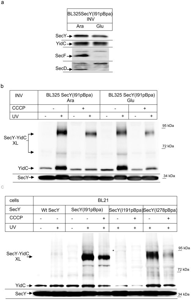
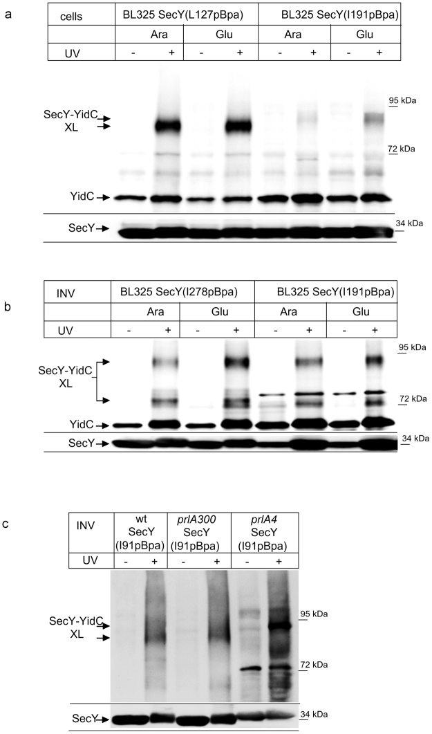
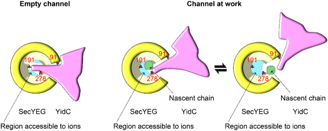
The heterotrimeric SecYEG complex cooperates with YidC to facilitate membrane protein insertion by an unknown mechanism. Here we show that YidC contacts the interior of the SecY channel resulting in a ligand-activated and voltage-dependent complex with distinct ion channel characteristics. The SecYEG pore diameter decreases from 8 Å to only 5 Å for the YidC-SecYEG pore, indicating a reduction in channel cross-section by YidC intercalation. In the presence of a substrate, YidC relocates to the rim of the pore as indicated by increased pore diameter and loss of YidC crosslinks to the channel interior. Changing the surface charge of the pore by incorporating YidC into the channel wall increases the anion selectivity, and the accompanying change in wall hydrophobicity is liable to alter the partition of helices from the pore into the membrane. This could explain how the exit of transmembrane domains from the SecY channel is facilitated by YidC.


Conflict of interest statement

The authors declare that they have no competing interests.

Figures

Figure 1
Figure 1
YidC contacts the channel interior of SecY. (a) Structure of the SecYE complex (left panel; PDB 3J01) and the SecY channel interior and pore ring (right panel). ResiduesI91, L127, I191, I278 and Y332 where pBpa was inserted are indicated in red. The lateral gate is indicated in blue and the residues used for crosslinking are indicated. Residue F383 is at the back of the SecYEG complex and not visible in this front view. (b) In vivo crosslinking of Bl21 (wt) cells expressing SecY, either without pBpa insertion (SecY) or with pBpa inserted at the indicated positions. Crosslinking was induced by UV exposure of whole cells when indicated. SecY and SecY crosslinking products were purified after cell breakage and separated on SDS-PAGE. SecY-YidC crosslinking products were detected after western blotting using α-YidC antibodies. Indicated is the SecY-YidC cross-linking product (upper panel) and the SecY content in these cells, as revealed by α-SecY antibodies (lower panel). (*) indicates a weak UV-dependent SecY-YidC crosslinking product of SecY(I191pBpa). (c) In vivo crosslinking was performed and analyzed as in (b) with pBpa inserted at the indicated residues. (c) In vitro crosslinking with purified inner membrane vesicles (INV) (2.5 nM SecY) derived from BL21 cells expressing the indicated pBpa-containing SecY derivatives. Samples were processed as above. Crosslinking experiments were repeated at least three times and representative blots are shown.
Figure 2
Figure 2
YidC loses contact to the SecY channel interior in the presence of a nascent membrane protein. (a) In vitro crosslinking using SecY(I191pBpa) and SecY(278pBpa) INV (2.5 nM SecY) was performed in the absence or presence of FtsQ-RNCs (2.5 nM) as indicated. Samples were processed as described in Fig. 1 and decorated with antibodies against YidC. The 90 kDa SecY-YidC crosslink product is indicated. (b) The samples described in (a) were decorated with antibodies against SecY after western blotting and the 90 kDa SecY-YidC crosslink product is indicated. (c) Western blot of the material shown in (a) for SecY(I191pBpa) INV using antibodies against the HA-tag present at the N-terminus of the FtsQ-RNCs. The upper band corresponds to FtsQ that is still tRNA-bound (FtsQ-tRNA) and the lower band to FtsQ that was released during sample preparation for SDS-PAGE (FtsQ). Crosslinking experiments were repeated at least three times and representative blots are shown.
Figure 3
Figure 3
Single channel conductance of the ribosome-SecYEG-YidC complex. (a) Conductance measurements in the presence of (i) both bare liposomes and ribosomes (150 nM, upper black trace) or (ii) SecYEG-YidC proteoliposomes (2.5 nM SecYEG) in the absence of ribosomes (lower black trace) showed electrically silent planar bilayers. SecYEG and YidC were present at 1:1 molar ratio and the final protein concentration was 5 μM. Ribosome binding to the YidC-SecYEG complex induced single channel activity. Representative current traces show the subsequent closure of three single channels (blue trace) at −100 mV. Co-reconstitution of YidC with a 3-fold excess of SecYEG yielded a mixture of larger SecYEG and smaller SecYEG-YidC channels (lower black trace). Measurements were obtained from free-standing bilayers formed from E. coli polar lipid extract at symmetrical salt concentrations of 150 mM KCl. (b) Dependence of the single channel current on the transmembrane voltage of the ribosome-SecYEG (black) and the ribosome-YidC-SecYEG complex (blue). The slope of the linear regression gives single channel conductances for SecYEG and YidC-SecYEG of 439 pS and 195 pS, respectively. Ribosomes were present at a concentration of 150 nM.
Figure 4
Figure 4
Single channel conductivity of the RNCFtsQ-SecYEG-YidC complex. Planar bilayers were formed from E. coli lipid. Fusion of proteoliposomes (5 μM final protein concentration, molar ratio SecYEG-YidC 1:1) to the membrane was facilitated by a transmembrane salt gradient of 435:150 mM KCl. (a) Channel activity was measured in the presence of liposomes and FtsQ-RNCs (5 nM, upper black trace), in the presence of SecYEG proteoliposomes (2.5 nM SecYEG; 2nd black trace). Binding of RNCFtsQ (5 nM) to either SecYEG proteoliposomes (3rd black trace) or SecYEG-YidC proteoliposomes (green trace) opened the reconstituted translocons for ions. Channel opening by non-translating ribosomes (150 nM) was also analysed for SecYEG-YidC complexes (red trace). Representative current traces were recorded at the indicated transmembrane potential (b) The corresponding current-voltage characteristics are shown for the RNCFtsQ-SecYEG (black), the RNCFtsQ-YidC-SecYEG (green), and the ribosome-YidC-SecYEG complexes (red). Deriving the corresponding single channel conductances from the slopes of the linear regressions yielded 587, 525, and 324 pS, respectively. The respective ψr values amounted to 6.7, −15, and −9.2 mV. (c) The single channel equivalent conductances of the three complexes are shown in the same color-code.
Figure 5
Figure 5
YidC’s interaction with SecY’s lateral gate is independent of SecDFYajC, but the interaction with both the lateral gate and the channel interior requires the pmf. (a) SecY(I91pBpa) was expressed in the conditional SecDFYajC-depletion strain BL325 and cells were grown either in the presence of arabinose for SecDFYajC induction or in the presence of glucose for SecDFYajC depletion. INV were isolated and the steady-state amounts of SecY, YidC, SecD and SecF were monitored by immune detection using the appropriate antibodies. (b) The INV as in (a) were used for in vitro crosslinking. When indicated, the protonophore CCCP was added at a final concentration of 100 μM. (c) In vivo crosslinking of Bl21 cells expressing either SecY without pBpa (wt) or SecY derivatives where pBpa was inserted at the indicated positions. When indicated, CCCP (100 μM final concentration) was added before UV-induced crosslinking. Experiments were repeated at least three times and representative blots are shown.
Figure 6
Figure 6
The depletion of SecDF enhances the interaction of YidC with the SecY channel interior. (a) In vivo crosslinking of SecDF-containing and –depleted BL325 cells expressing SecY derivatives with pBpa insertion at the indicated conditions. (b) In vitro crosslinking with INV derived from the indicated cells. (c) Crosslinking was performed with INV of E. coli BL21 cells expressing a plasmid-encoded wild type SecY (wt) or the plasmid encoded prlA300 or the prlA4 SecY derivatives, carrying each pBpa at position I91 in TM2b of SecY. Indicated is the SecY-YidC cross-linking product (upper panel) and the SecY content in these INV, as revealed by α-SecY antibodies (lower panel). Experiments were repeated at least three times and representative blots are shown.
Figure 7
Figure 7
Model of the YidC-SecYEG interaction as visualized by crosslinks and electrophysiological experiments. Left Panel: The empty SecYEG-YidC complex allows YidC to crosslink to the three indicated positions (red) at SecYEG’s pore ring (yellow) or the lateral gate. Ribosome binding is required to elicit the formation of a transmembrane channel (cross-section in blue) that allows ion permeation at low membrane potentials. Right Panel: Upon insertion of a nascent chain (green), YidC is expelled to the outer rim of the SecYEG pore, thereby increasing the cross-section (ion conductivity) of the SecYEG channel. Crosslinks to the channel interior (I191) is sterically constrained in the presence of a nascent chain while crosslinks to the peripheral residues 91 and 278 are still observable. In order to release the nascent chain, the lateral gate must open, i.e. conceivably the contact between SecYEG and YidC is further weakened. This is in line with crosslink data showing conformational changes at the SecY(I91)-YidC interface. In summary, YidC facilitates the partitioning of a nascent membrane protein into the lipid environment by reducing the hydrophobicity of the lateral gate.

References

    1. Denks K, et al. The Sec translocon mediated protein transport in prokaryotes and eukaryotes. Molecular membrane biology. 2014;31:58–84. doi: 10.3109/09687688.2014.907455. - DOI - PubMed
    1. Zimmermann R, Eyrisch S, Ahmad M, Helms V. Protein translocation across the ER membrane. Biochimica et biophysica acta. 2011;1808:912–924. doi: 10.1016/j.bbamem.2010.06.015. - DOI - PubMed
    1. Gorlich D, Rapoport TA. Protein translocation into proteoliposomes reconstituted from purified components of the endoplasmic reticulum membrane. Cell. 1993;75:615–630. doi: 10.1016/0092-8674(93)90483-7. - DOI - PubMed
    1. Braig D, et al. Signal sequence-independent SRP-SR complex formation at the membrane suggests an alternative targeting pathway within the SRP cycle. Molecular biology of the cell. 2011;22:2309–2323. doi: 10.1091/mbc.E11-02-0152. - DOI - PMC - PubMed
    1. Van den Berg B, et al. X-ray structure of a protein-conducting channel. Nature. 2004;427:36–44. doi: 10.1038/nature02218. - DOI - PubMed

Publication types

MeSH terms

Substances