. 2017 Mar 8;7(1):115.
doi: 10.1038/s41598-017-00091-1.

ALS-linked FUS exerts a gain of toxic function involving aberrant p38 MAPK activation

Affiliations

ALS-linked FUS exerts a gain of toxic function involving aberrant p38 MAPK activation

Reddy Ranjith K Sama et al. Sci Rep. .

Abstract

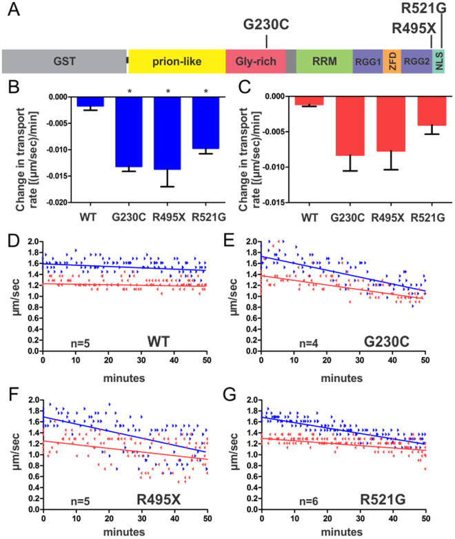
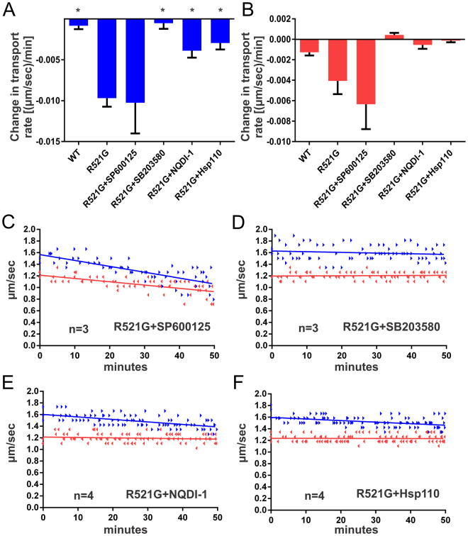
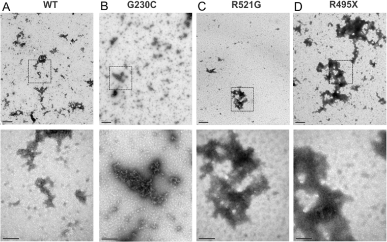
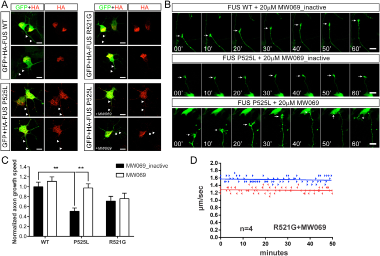
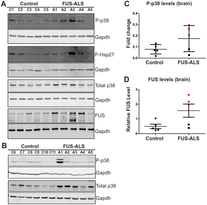
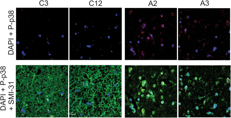
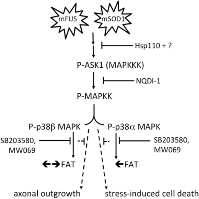
Mutations in Fused in Sarcoma/Translocated in Liposarcoma (FUS) cause familial forms of amyotrophic lateral sclerosis (ALS), a neurodegenerative disease characterized by progressive axonal degeneration mainly affecting motor neurons. Evidence from transgenic mouse models suggests mutant forms of FUS exert an unknown gain-of-toxic function in motor neurons, but mechanisms underlying this effect remain unknown. Towards this end, we studied the effect of wild type FUS (FUS WT) and three ALS-linked variants (G230C, R521G and R495X) on fast axonal transport (FAT), a cellular process critical for appropriate maintenance of axonal connectivity. All ALS-FUS variants impaired anterograde and retrograde FAT in squid axoplasm, whereas FUS WT had no effect. Misfolding of mutant FUS is implicated in this process, as the molecular chaperone Hsp110 mitigated these toxic effects. Interestingly, mutant FUS-induced impairment of FAT in squid axoplasm and of axonal outgrowth in mammalian primary motor neurons involved aberrant activation of the p38 MAPK pathway, as also reported for ALS-linked forms of Cu, Zn superoxide dismutase (SOD1). Accordingly, increased levels of active p38 MAPK were detected in post-mortem human ALS-FUS brain tissues. These data provide evidence for a novel gain-of-toxic function for ALS-linked FUS involving p38 MAPK activation.


Conflict of interest statement

The authors declare that they have no competing interests.

Figures

Figure 1
Figure 1
ALS-linked mutant FUS proteins impair anterograde and retrograde FAT. (A) ALS-linked mutations (G230C, R495X and R521G) investigated in this study are mapped onto the domain structure of GST-FUS. RRM = RNA recognition motif, RGG = arginine-glycine-glycine-rich, ZFD = zinc-finger domain and NLS = nuclear localization signal. (B–G) FUS proteins (2.5 μM) were perfused into isolated squid axoplasm and fast axonal transport (FAT) rates (μm/s) of membrane bounded-organelles measured as a function of time (minutes). For (B,C), slopes of the linear best fits for velocities obtained for each axoplasm (D–G) were averaged and plotted as bar graphs in Graphpad Prism with the standard error of the mean (SEM) for anterograde (blue bars; A) and retrograde (red bars; B) velocities. Those conditions with statistical significance (p < 0.05) relative to FUS WT are denoted by *, determined by one-way Anova and Tukey post-hoc test for multiple comparisons. (D–G) Motility plots comprised of the raw data for every axoplasm (‘n’ denotes the number of axoplasms analyzed for each condition) are shown, where linear best fits of the compiled data are shown for anterograde (blue arrowheads; linear fit shown as a solid blue line) and retrograde (red arrowheads; linear fit shown as a solid red line) directions. Protein obtained from at least two independent protein expression and purifications were included for each FUS variant.
Figure 2
Figure 2
The inhibitory effect of mutant-FUS on FAT is mediated by activated p38 MAPK. FUS R521G was co-perfused with the indicated pharmacological inhibitor or Hsp110 into squid axoplasm and fast axonal transport evaluated as described in Fig. 1. (A–C) Co-perfusion of the pharmacological JNK kinase inhibitor SP600125 (0.5 μM) with R521G failed to rescue FAT inhibition. In contrast, inhibition of the p38 MAPK pathway by co-perfusion of either SB203850 (5 μM) or NQDI-1 (20 μM) prevented the toxic effects of FUS R521G on FAT (A,B,D,E). (F) Similarly, co-perfusion of Hsp110 (0.6 μM) and R521G ameliorated the inhibition of FAT by mutant FUS. Those conditions with statistical significance (p < 0.05) relative to FUS R521G are denoted by *, determined by one-way Anova and Tukey post-hoc test for multiple comparisons.
Figure 3
Figure 3
The morphology of FUS species assessed by electron microscopy. Representative electron microscopy (EM) images of negatively stained FUS WT (A), G230C (B), R521G (C) and R495X (D) proteins. Low magnification images are shown in the top row (scale bar represents 0.5 μm). High magnification images are shown for insets (dashed squares) in the bottom row (scale bar represents 200 nm). FUS exhibits heterogeneous morphologies across all samples.
Figure 4
Figure 4
Mutant FUS impairs axon outgrowth in motor neurons through a mechanism involving p38 MAPK activity. Murine motor neurons were transiently co-transfected with FLAGHA-FUS WT, R521G or P525L and green fluorescent protein (GFP) at 2 days in vitro (DIV). Motor neurons were cultured in 20 μM MW069, a potent and selective p38 MAPK inhibitor, or an inactive analogue, MW069_inactive. (A) FUS WT and R521G are predominately expressed within the nucleus of motor neurons, where as FUS P525L is expressed in the nucleus and within axons (white arrowheads). FUS P525L localization is similar whether motor neurons are treated with MW069_inactive or MW069 (+MW069). (B) Montages corresponding to live cell imaging of axon outgrowth (white arrow) over a 60 min time course for the indicated condition. Note that the growth cone of a FUS P525L expressing motor neuron is stalled in the presence of MW069_inactive (middle panel) relative to the active form of MW069 (bottom panel). See Supplemental Video S1 for live cell imaging of all conditions. (C) Quantification of axon outgrowth speed compiled from n = 3 biological experiments normalized to the WT + MW069_inactive condition. Statistical significance (**p < 0.01) of pertinent comparisons are indicated. Additional significant comparisons include FUS WT + MW069 versus FUS R521G + MW069_inactive (p < 0.05) and FUS WT + MW069 versus FUS P525L + MW069_inactive (p < 0.0001). (D) The inhibition of FAT by FUS R521G is blocked by MW069 in squid axoplasm. The motility plot for FUS R521G in the absence of MW069 is shown in Fig. 1G.
Figure 5
Figure 5
Increased levels of phosphorylated p38 MAPK in human post-mortem CNS tissues derived from individuals with ALS-FUS. Immunoblot analysis of frozen post-mortem brain (A) and spinal cord (B) tissues derived from non-disease control (C1-C11) and ALS individuals harboring FUS mutations (A1-R521G; A2-H517Q; A3- ex14del; A4-P525L and A5- bp1408del) with the indicated antibodies (see Supplemental Table S2 for detailed patient information). (C) Quantification of phosphorylated (active) p38 (p-p38) by immunoblot analysis revealed higher levels of p-p38 MAPK in ALS cases (cases with levels above the control mean are indicated by red squares), compared to control cases. P-p38 values were normalized to total p38 levels. (D) Quantification of FUS levels in brain normalized to Gapdh. ALS cases with FUS levels above the control mean are indicated by red squares. (C,D) Although, there appears to be a trend in the data, differences between ALS cases and controls did not reach statistical significance due to the small sample size and inherent variability of human postmortem samples.
Figure 6
Figure 6
Active p38 MAPK in neurons within post-mortem motor cortex tissues from individuals diagnosed with ALS-FUS. Brain sections from paraffin-embedded tissue samples obtained from two control (C3 and C12) and two ALS-FUS (A2 and A3) cases were probed with antibodies against phosphorylated, catalytically active p38 (P-p38; red), phosphorylated neurofilament H (SMI31; green) as a marker of neuroaxonal integrity, and the nuclear stain DAPI (blue). Scale bar represents 10 μm. Low magnification images are shown in Supplemental Figure S3 . The P-p38 signal is higher, and SMI31 signal lower, in both ALS cases compared to controls; see Supplemental Figure S4 for quantification.
Figure 7
Figure 7
Model for aberrant activation of p38 MAP kinase(s) by mutant and misfolded ALS-associated proteins. Available data (from this study and others, , ) support a model whereby mutant and misfolded forms of FUS (mFUS; left) and SOD1 (mSOD1; right) induce the aberrant activation of the p38 MAPK pathway. Hsp110 likely synergizes with other chaperones to ameliorate the effects of mSOD1 and mFUS, possibly upstream of ASK1 and additional unidentified factors. While mSOD1 and mFUS converge on p38 MAPK activation, perfusion of these proteins into squid axoplasm have differential effects on FAT; mFUS inhibits both anterograde (←) and retrograde (→) FAT (Figs 1 and 2) whereas mSOD1 only inhibits anterograde FAT, , , consistent with inhibition of the beta and alpha isoforms of P38 MAPK, respectively. In addition to FAT inhibition in squid (solid lines), mFUS- and mSOD1-induced activation p38 MAPK can manifest different phenotypes in mammalian systems (dashed lines), including but not limited to inhibition of axon outgrowth (Fig. 4) and enhanced susceptibility to cell stress. This figure is adapted from Song et al. .

References

    1. Kwiatkowski TJ, Jr., et al. Mutations in the FUS/TLS gene on chromosome 16 cause familial amyotrophic lateral sclerosis. Science. 2009;323:1205–8. doi: 10.1126/science.1166066. - DOI - PubMed
    1. Vance C, et al. Mutations in FUS, an RNA processing protein, cause familial amyotrophic lateral sclerosis type 6. Science. 2009;323:1208–11. doi: 10.1126/science.1165942. - DOI - PMC - PubMed
    1. Sama RR, Ward CL, Bosco DA. Functions of FUS/TLS From DNA Repair to Stress Response: Implications for ALS. ASN Neuro. 2014;6:1–18. doi: 10.1177/1759091414544472. - DOI - PMC - PubMed
    1. Scekic-Zahirovic J, et al. Toxic gain of function from mutant FUS protein is crucial to trigger cell autonomous motor neuron loss. EMBO J. 2016;35:1077–97. doi: 10.15252/embj.201592559. - DOI - PMC - PubMed
    1. Sharma A, et al. ALS-associated mutant FUS induces selective motor neuron degeneration through toxic gain of function. Nat Commun. 2016;7:10465. doi: 10.1038/ncomms10465. - DOI - PMC - PubMed

Publication types