

Protective effects of Cassia tora leaves in experimental cataract by modulating intracellular communication, membrane co-transporters, energy metabolism and the ubiquitin-proteasome pathway
- PMID: 28274170
- PMCID: PMC6130452
- DOI: 10.1080/13880209.2017.1299769
Protective effects of Cassia tora leaves in experimental cataract by modulating intracellular communication, membrane co-transporters, energy metabolism and the ubiquitin-proteasome pathway
Abstract
Context: Cataract is the clouding of eye lens which causes impairment in vision and accounts for the leading factor of global blindness. Functional food-based prevention of cataract finds application in vision research because of its availability and easy access to all classes of the society. Cassia tora Linn. (Caesalpinaceae) is an edible plant mentioned in the traditional systems of medicine for whole body health, especially to the eyes.
Objective: The present study evaluates the potential of ethyl acetate fraction of Cassia tora leaves (ECT) on experimental cataract.
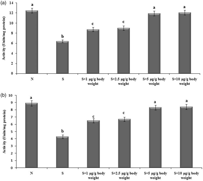
Materials and methods: Cataract was induced by a single subcutaneous injection of sodium selenite (4 μg/g body weight) on 10th day. ECT was supplemented orally from 8th day up to 12th day at a concentration of 5 μg/g body weight and marker parameters were evaluated after 30 days.
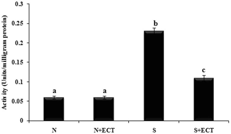
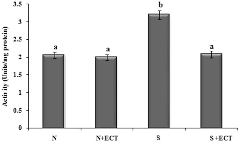
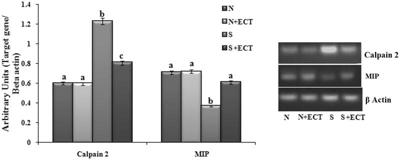
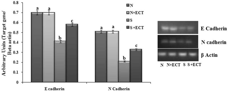
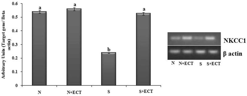
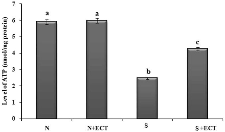
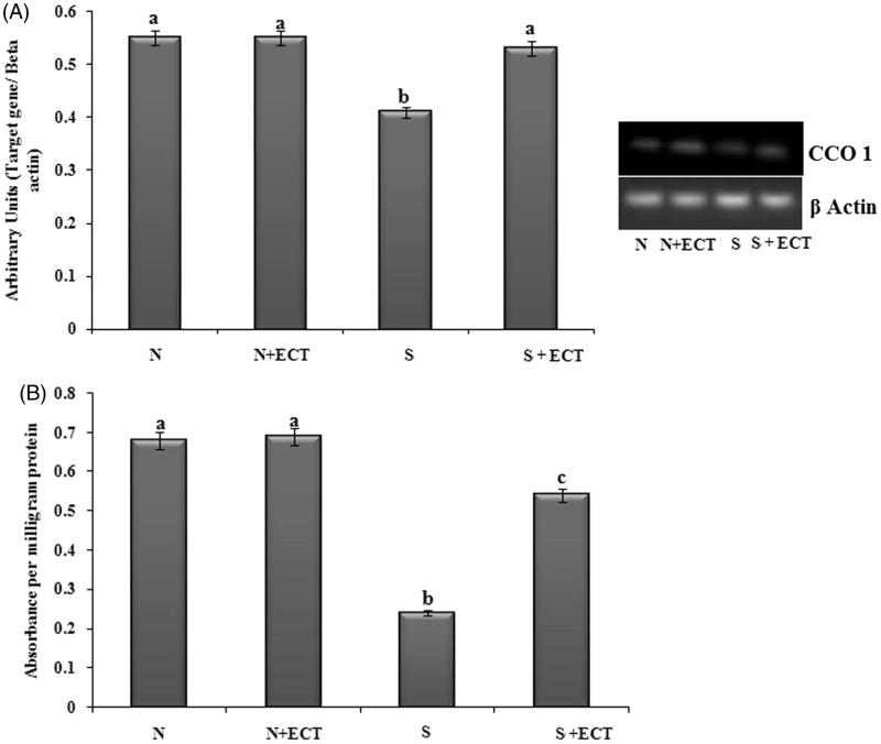
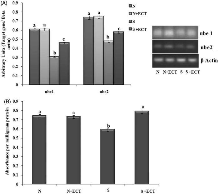
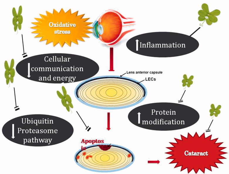
Results: The production of MPO and the activation of calpain were reduced 52.17% and 36.67% by ECT in lens tissue, respectively. It modulated the energy status by significantly increasing the activity of CCO 1 (55.56%) and ATP production (41.88%). ECT maintained the ionic balance in the lens by reducing the level of sodium (50%) and increasing the level of potassium (42.5%). It also reduced cell junction modifications and preserved a functional ubiquitin-proteasome pathway.
Discussion and conclusion: The results reinforce the growing attention on wild plant food resources for preventive protection against cataract. The data suggest the value of Cassia tora leaves as a functional food for ameliorating cataract pathology.
Keywords: Blindness; functional food; oxidative stress; selenite cataract.
Figures
Similar articles
-
Cassia tora leaves modulates selenite cataract by enhancing antioxidant status and preventing cytoskeletal protein loss in lenses of Sprague Dawley rat pups.J Ethnopharmacol. 2016 Feb 3;178:137-43. doi: 10.1016/j.jep.2015.12.012. Epub 2015 Dec 9. J Ethnopharmacol. 2016. PMID: 26692278
-
Anthraquinones and flavonoids of Cassia tora leaves ameliorate sodium selenite induced cataractogenesis in neonatal rats.Food Funct. 2016 Feb;7(2):1087-95. doi: 10.1039/c5fo00905g. Food Funct. 2016. PMID: 26786764
-
Polyphenols of Cassia tora leaves prevents lenticular apoptosis and modulates cataract pathology in Sprague-Dawley rat pups.Biomed Pharmacother. 2016 Jul;81:371-378. doi: 10.1016/j.biopha.2016.04.018. Epub 2016 Apr 26. Biomed Pharmacother. 2016. PMID: 27261615
-
Vitex negundo attenuates calpain activation and cataractogenesis in selenite models.Exp Eye Res. 2009 Mar;88(3):575-82. doi: 10.1016/j.exer.2008.11.020. Epub 2008 Dec 6. Exp Eye Res. 2009. PMID: 19094987
-
Preclinical activities of Cassia tora Linn against aging-related diseases.Expert Rev Mol Med. 2022 Oct 25;24:e43. doi: 10.1017/erm.2022.33. Expert Rev Mol Med. 2022. PMID: 36281483 Review.
Cited by
-
Traditional medicinal plants used for treating emerging and re-emerging viral diseases in northern Nigeria.Eur J Integr Med. 2022 Jan;49:102094. doi: 10.1016/j.eujim.2021.102094. Epub 2021 Nov 27. Eur J Integr Med. 2022. PMID: 36573184 Free PMC article.
-
Network Pharmacology-Based Strategy to Reveal the Mechanism of Cassiae Semen against Cataracts.Comput Math Methods Med. 2022 Jul 11;2022:5654120. doi: 10.1155/2022/5654120. eCollection 2022. Comput Math Methods Med. 2022. PMID: 35860180 Free PMC article.
-
Review on medicinal plants and natural compounds as anti-Onchocerca agents.Parasitol Res. 2018 Sep;117(9):2697-2713. doi: 10.1007/s00436-018-6003-7. Epub 2018 Jul 15. Parasitol Res. 2018. PMID: 30008135 Review.
-
Breaking Barriers: Nanomedicine-Based Drug Delivery for Cataract Treatment.Int J Nanomedicine. 2024 May 6;19:4021-4040. doi: 10.2147/IJN.S463679. eCollection 2024. Int J Nanomedicine. 2024. PMID: 38736657 Free PMC article. Review.
-
Proteostasis in aging-associated ocular disease.Mol Aspects Med. 2022 Dec;88:101157. doi: 10.1016/j.mam.2022.101157. Epub 2022 Nov 29. Mol Aspects Med. 2022. PMID: 36459837 Free PMC article. Review.
References
-
- Aebi H.1984. Catalase in vitro In: Colowick SP, Kaplan NO, editors. Methods in enzymology. New York: Academic Press; p. 121–126. - PubMed
-
- Arulpandi I, Kanimozhi S.. 2011. Antimicrobial activity and phytochemical analysis of Cassia tora Linn. leaves. J Pharm Res. 2:2954–2956.
-
- Balog Z, Sikic J, Vojnikovic B, Balog S. 2001. Senile cataract and the absorption activity of cytochrome C oxidase. Coll Antropol. 25:33–36. - PubMed
-
- Barbusinski K.2009. Fenton reaction-controversy concerning the chemistry. Ecol Chem Eng. 16:347–358.
MeSH terms
Substances
LinkOut - more resources
Full Text Sources
Other Literature Sources
Medical
Research Materials
Miscellaneous