Review
. 2017 May 5;24(5):e00545-16.
doi: 10.1128/CVI.00545-16. Print 2017 May.

Mass Spectrometry Approaches for Identification and Quantitation of Therapeutic Monoclonal Antibodies in the Clinical Laboratory

Affiliations
Review

Mass Spectrometry Approaches for Identification and Quantitation of Therapeutic Monoclonal Antibodies in the Clinical Laboratory

Paula M Ladwig et al. Clin Vaccine Immunol. .

Abstract

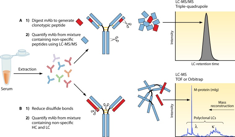
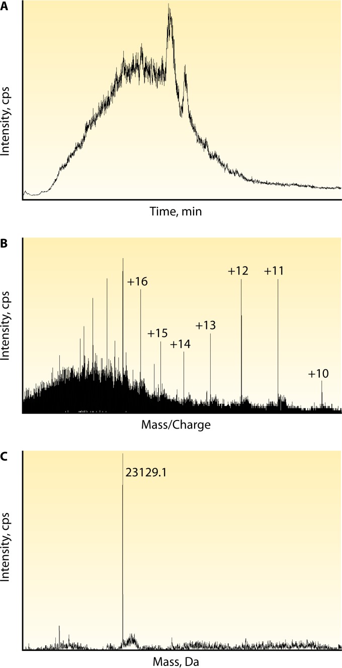
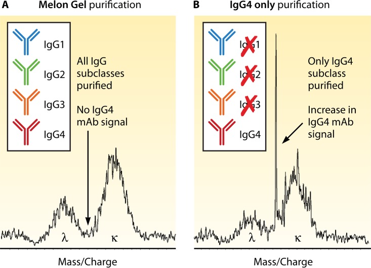
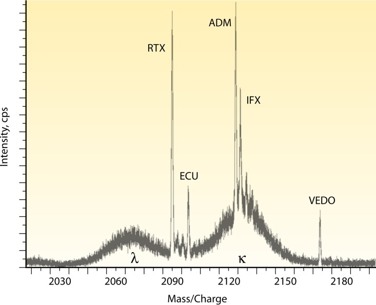
Therapeutic monoclonal antibodies (MAbs) are an important class of drugs used to treat diseases ranging from autoimmune disorders to B cell lymphomas to other rare conditions thought to be untreatable in the past. Many advances have been made in the characterization of immunoglobulins as a result of pharmaceutical companies investing in technologies that allow them to better understand MAbs during the development phase. Mass spectrometry is one of the new advancements utilized extensively by pharma to analyze MAbs and is now beginning to be applied in the clinical laboratory setting. The rise in the use of therapeutic MAbs has opened up new challenges for the development of assays for monitoring this class of drugs. MAbs are larger and more complex than typical small-molecule therapeutic drugs routinely analyzed by mass spectrometry. In addition, they must be quantified in samples that contain endogenous immunoglobulins with nearly identical structures. In contrast to an enzyme-linked immunosorbent assay (ELISA) for quantifying MAbs, mass spectrometry-based assays do not rely on MAb-specific reagents such as recombinant antigens and/or anti-idiotypic antibodies, and time for development is usually shorter. Furthermore, using molecular mass as a measurement tool provides increased specificity since it is a first-order principle unique to each MAb. This enables rapid quantification of MAbs and multiplexing. This review describes how mass spectrometry can become an important tool for clinical chemists and especially immunologists, who are starting to develop assays for MAbs in the clinical laboratory and are considering mass spectrometry as a versatile platform for the task.

Keywords: clinical laboratory; immunogenicity; immunotherapy; intact light chain mass; mass spectrometry; proteomics; therapeutic drug monitoring; therapeutic monoclonal antibodies; tryptic peptides.


Figures

FIG 1
FIG 1
LC-MS versus LC-MS/MS for quantitation of therapeutic MAbs. MAbs can be quantitated by MS. Ig extraction or enrichment techniques (protein crash, Melon Gel IgG enrichment, or affinity matrix for specific IgG subclasses, for example) will help reduce the protein load in the sample. (A) LC-MS/MS method. The LC-MS/MS method includes separation of the light chains from the heavy chains after reduction of the disulfide bonds, followed by trypsin cleavage to generate multiple peptides from the intact Ig. Peptides specific to the variable region of the MAb on either the light or the heavy chains, which do not cross-react with human sequences, are used to quantitate the MAb. Serum samples are extracted/enriched to reduce the protein load. Samples are denatured (protein is unfolded), reduced (cysteine reduction breaks the disulfide bonds connecting MAb light and heavy chains), alkylated (alkylation of cysteines prevents the disulfide bonds from reforming), and digested by trypsin into smaller peptides. The peptide mixture is separated by LC before analysis by MS/MS. The mass of the peptide of interest (parent ion) is recorded, the peptide is further cleaved inside the mass spectrometer (fragment ion), and the ion pair transition is utilized for quantitation, preferably on triple-quadrupole instruments. (B) LC-MS method. The LC-MS method separates the light chains (LC) from the heavy chains (HC) through reduction, and the intact light chains are not further processed or cleaved. Instead, their accurate mass is measured. In healthy individuals, the polyclonal repertoire of lambda and kappa light chains follows a Gaussian distribution at a 1:2 lambda-to-kappa ratio, and MAbs present in significant amounts stand out of the polyclonal endogenous background as peaks or spikes.
FIG 2
FIG 2
Data overview of LC-MS for miRAMM. The analysis of intact light chains by MS may be visualized in different ways. In this example, ECU at 100 μg/ml was used to spike normal human serum enriched for Ig with Melon Gel. (A) The total ion chromatogram from which a known MAb can be extracted. Light chains elute in a 5- to 8-min retention time window with a gradient mobile phase chromatographic protocol (9). (B) Once the retention time window is selected, the extracted ion spectrum shows all of the proteins eluted in that time frame, peak intensities, and the multiple charge states acquired. Visualization and quantitation are best at the +12, +11, and +10 charge states for Ig light chains by the published method. Depending on the software used, one or more charge states can be used for quantitation. (C) Deconvolution software will reconstruct the intact accurate mass from the acquired multiple charge states and may be used for MAb confirmation or quantitation.
FIG 3
FIG 3
Ig enrichment strategies impact the LOQ. Observation of a unique MAb accurate mass in a serum matrix of patients undergoing therapy is accomplished among 1 g/dl of human endogenous circulating Igs. Depending on the MAb molecular mass, the LOQ can be an issue, as shown in this miRAMM example. When studying ECU, an IgG2/IgG4 hybrid MAb, different enrichment strategies were employed. (A) When Melon Gel enrichment was used, an ECU concentration of 5 μg/ml was not detectable. (B) Selective enrichment for IgG4 with an affinity matrix allowed for significant reduction of the endogenous Ig repertoire by removing all non-IgG4 Ig from the background and increased the analytical sensitivity of the assay by approximately 10-fold, with an increased signal-to-noise ratio.
FIG 4
FIG 4
Therapeutic MAbs can be measured simultaneously by miRAMM. In this example, several therapeutic MAbs were used to spike commercially available normal human serum. A given therapeutic MAb's specific light chain will have a unique mass and be distinguishable from the endogenous Ig background when present in large concentrations. The extracted ion spectrum shows the +11 charge state of five MAbs' specific light chains detected above the human serum polyclonal Ig background. Although it is highly unlikely that a patient would not be undergoing therapy with five or six different MAbs at the same time, the multiplex capability allows application of the technique for screening methods, such as the differentiation of a therapeutic MAb from a disease-causing endogenous monoclonal protein.
FIG 5
FIG 5
Multiple charge states acquired by high-resolution MS for ECU. Normal human serum was spiked with ECU at 100 μg/ml and enriched for Ig with Melon Gel. After ESI, the light chains acquired multiple charge states and were detected with an Orbitrap (Thermo Fisher) instrument. (A) Mass spectrum of the +12, +11, and +10 charge states. Utilizing low-resolution scanning, the areas under the curve of one or multiple charge states are added together for quantitation of the MAb. By using a high-resolution view, the area under the curve of multiple isotopes of a given charge state can be analyzed. (B) Isotopic distribution of a +11 charge state.
None
None
None

References

    1. Berry JD. 2014. Introduction to therapeutic Fc-fusion proteins, p 217–232. In Chamow SM, Ryll T, Lowman HB, Farson D (ed), Therapeutic Fc-Fusion Proteins. Wiley-VCH Verlag GmbH & Co. KGaA, Weinheim, Germany.
    1. Rajpal A, Strop P, Yeung YA, Chaparro-Riggers J, Pons J. 2014. Introduction: antibody structure and function, p 1–44. In Chamow SM, Ryll T, Lowman HB, Farson D (ed), Therapeutic Fc-fusion proteins. Wiley-VCH Verlag GmbH & Co. KGaA, Weinheim, Germany.
    1. Jones TD, Carter PJ, Pluckthun A, Vasquez M, Holgate RG, Hotzel I, Popplewell AG, Parren PW, Enzelberger M, Rademaker HJ, Clark MR, Lowe DC, Dahiyat BI, Smith V, Lambert JM, Wu H, Reilly M, Haurum JS, Dubel S, Huston JS, Schirrmann T, Janssen RA, Steegmaier M, Gross JA, Bradbury AR, Burton DR, Dimitrov DS, Chester KA, Glennie MJ, Davies J, Walker A, Martin S, McCafferty J, Baker MP. 2016. The INNs and outs of antibody nonproprietary names. MAbs 8:1–9. doi:10.1080/19420862.2015.1114320. - DOI - PMC - PubMed
    1. Abbas AK, Lichtman AH, Pillai S. 2007. Cellular and molecular immunology, 6th ed, p 75–96. Saunders Elsevier, Philadelphia, PA.
    1. Kunert R, Reinhart D. 2016. Advances in recombinant antibody manufacturing. Appl Microbiol Biotechnol 100:3451–3461. doi:10.1007/s00253-016-7388-9. - DOI - PMC - PubMed

MeSH terms

Substances