. 2017 Apr 28;91(10):e00137-17.
doi: 10.1128/JVI.00137-17. Print 2017 May 15.

Identification of SERINC5-001 as the Predominant Spliced Isoform for HIV-1 Restriction

Affiliations

Identification of SERINC5-001 as the Predominant Spliced Isoform for HIV-1 Restriction

Xianfeng Zhang et al. J Virol. .

Abstract

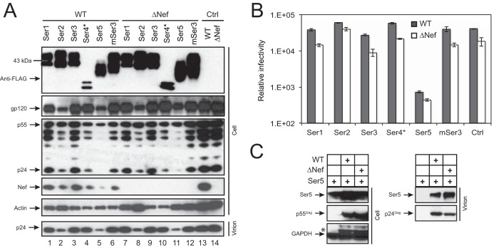
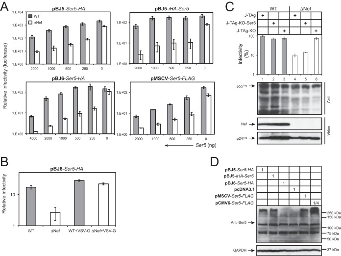
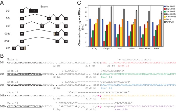
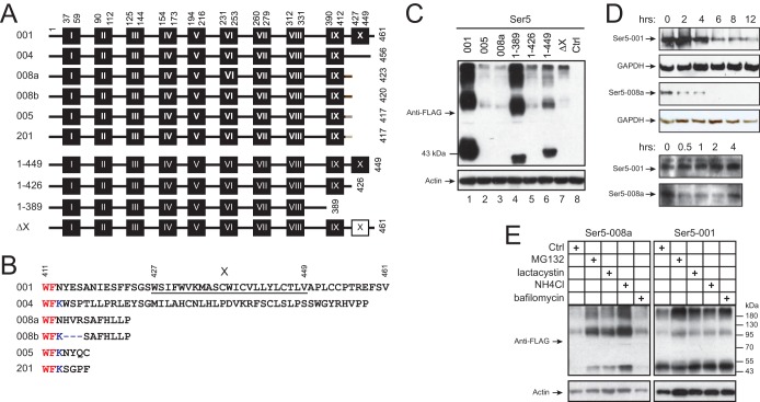
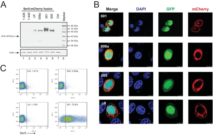
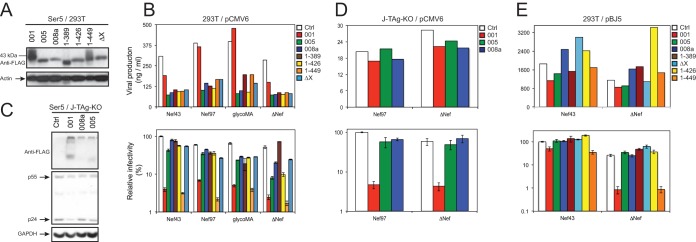
Among the five serine incorporator (SERINC) family members, SERINC5 (Ser5) was reported to strongly inhibit HIV-1 replication, which is counteracted by Nef. Ser5 produces 5 alternatively spliced isoforms: Ser5-001 has 10 putative transmembrane domains, whereas Ser5-004, -005, -008a, and -008b do not have the last one. Here, we confirmed the strong Ser5 anti-HIV-1 activity and investigated its isoforms' expression and antiviral activities. It was found that Ser5-001 transcripts were detected at least 10-fold more than the other isoforms by real-time quantitative PCR. When Ser5-001 and its two isoforms Ser5-005 and Ser5-008a were expressed from the same mammalian expression vector, only Ser5-001 was stably expressed, whereas the others were poorly expressed due to rapid degradation. In addition, unlike the other isoforms, which are located mainly in the cytoplasm, Ser5-001 is localized primarily to the plasma membrane. To map the critical determinant, Ser5 mutants bearing C-terminal deletions were created. It was found that the 10th transmembrane domain is required for Ser5 stable expression and plasma membrane localization. As expected, only Ser5-001 strongly inhibits HIV-1 infectivity, whereas the other Ser5 isoforms and mutants that do not have the 10th transmembrane domain show very poor activity. It was also observed that the Nef counteractive activity could be easily saturated by Ser5 overexpression. Thus, we conclude that Ser5-001 is the predominant antiviral isoform that restricts HIV-1, and the 10th transmembrane domain plays a critical role in this process by regulating its protein stability and plasma membrane targeting.IMPORTANCE Human immunodeficiency virus (HIV) and simian immunodeficiency virus (SIV) express a small protein, Nef, to enhance viral pathogenesis in vivo Nef has an important in vitro function, which is to make virus particles more infectious, but the mechanism has been unclear. Recently, Nef was reported to counteract a novel anti-HIV host protein, SERINC5 (Ser5). Ser5 has five alternatively spliced isoforms, Ser5-001, -004, -005, -008a, and -008b, and only Ser5-001 has an extra C-terminal transmembrane domain. We now show that the Ser5-001 transcripts are produced at least 10-fold more than the others, and only Ser5-001 produces stable proteins that are targeted to the plasma membrane. Importantly, only Ser5-001 shows strong anti-HIV-1 activity. We further demonstrate that the extra transmembrane domain is required for Ser5 stable expression and plasma membrane localization. These results suggest that plasma membrane localization is required for Ser5 antiviral activity, and Ser5-001 is the predominant isoform that contributes to the activity.

Keywords: HIV-1; Nef; SERINC3; SERINC5; infectivity; restriction factor.


Figures

FIG 1
FIG 1
Analysis of the anti-HIV-1 activity of Ser family members. (A) 293T cells were transfected with an HIV-1 WT or ΔNef proviral vector in the presence of the indicated pCMV6-Ser expression vectors at a 1:1 ratio. After 48 h, virions were collected from the culture supernatants and purified by ultracentrifugation. The purified virions and cells were lysed and analyzed by Western blotting using the indicted antibodies. (B) After virions produced from 293T cells were quantified by p24Gag ELISA, equal amounts of viruses were used to infect the HIV-1 luciferase reporter cell line TZM-b1 to determine viral infectivity. The error bars represent standard deviations (SD) from three experiments. (C) 293T cells were transfected with pCMV6-Ser5 in the presence or absence of WT or ΔNef HIV-1 vector. Viral particles were purified from culture supernatants by ultracentrifugation. Cell and virion lysates were analyzed by Western blotting. The asterisk indicates remnant protein bands from a previous Western blot. The GenBank accession numbers for the Ser proteins are as follows: NM_020755 (human Ser1), NM_178865 (human Ser2), NM_006811 (human Ser3), NM_001258032 (human Ser4*), NM_001174072 (human Ser5), and NM_012032 (murine Ser3).
FIG 2
FIG 2
Saturation of HIV-1 Nef counteractive activity by Ser5 overexpression. (A) 293T cells were transfected with a fixed amount of HIV-1 WT or ΔNef proviral vector and the indicated amounts of 4 different Ser5 expression vectors, pBJ6-Ser5-HA, pBJ5-Ser5-HA, pBJ5-iHA-Ser5, and pMSCV-Ser5-FLAG. After 48 h, viruses were collected and normalized by p24Gag ELISA; viral infectivity was measured by infecting TZM-b1 cells. (B) The anti-HIV-1 activity of Ser5 expressed from the pBJ6 vector was determined in the presence or absence of VSV G pseudotyping. (C) J-TAg, J-TAg-KO, and J-TAg-KO-Ser5 cells were infected with WT or ΔNef HIV-1 by spinoculation. The viruses produced were normalized by p24Gag ELISA, and viral infectivity was determined after infection of TZM-b1 cells. Viruses were also purified by ultracentrifugation, and viral protein expression in the Jurkat cells and purified virions was analyzed by Western blotting. (D) The Ser5 expression levels in 293T cells from the different expression vectors were compared by Western blotting using an anti-Ser5 antibody. Numbers in the boxes indicate relative amounts of each vector used for transfection of 293T cells. The error bars represent SD from three experiments.
FIG 3
FIG 3
Detection of human Ser5 gene expression by RT-qPCR. (A) Human Ser5 gene alternatively spliced isoforms. Isoform numbers are shown on the left, and the differentially spliced exons are shown on the right. Exons are shown as black boxes with numerals, but the first 10 exons, which are constitutively spliced, are not shown (except for the putative isoform 201). Exons and introns are not drawn to scale, although exon 12 for isoform 001 is shown as a longer exon. The positions for the stop codons for each isoform are indicated by asterisks. The gray regions for single exons for isoforms 008a and 201 indicate the alternative splice points for the two isoforms. The question mark after exon 11 indicates that the end of the 3′ untranslated region for the isoform has not been identified. (B) Locations of forward and reverse RT-qPCR primers for each Ser5 isoform. The locations of the forward primers (boldface underlined letters) are the same for each primer set. Uppercase letters represent exonic regions, and lowercase letters represent intronic regions. The ellipses indicate sequences that are not shown (the numbers of bases that are not displayed are given below). The dashed lines indicate the intronic regions crossed by the reverse primers. Exons 11, 12, 13, 14, 15a, and 15b are shown in different colors. Instead of the canonical AG normally found for a U2 splice acceptor sequence, 008a and 008b have the rare TG acceptors, which are italicized and shown in magenta. (C) Ser3 and Ser5 copy numbers in J-TAg, J-TAg-KO, and 293T cells; MDMs; PBMCs; and PBMCs activated with PHA. GAPDH was used as a control. The GenBank accession numbers for Ser5-001, -004, -005, -008a, and -008b are NM_001174072.2, BC101280, BC101281, AF498273, and BC101280, respectively. No GenBank accession number was found to be associated with Ser5-201.
FIG 4
FIG 4
Analysis of Ser5 isoform protein expression. (A) Schematic diagram of the putative transmembrane domains of Ser5 isoforms and mutants. The domains were predicted by TransMembrane prediction using hidden Markov models (TMHMM) at the public server (http://www.cbs.dtu.dk/services/TMHMM/) and are shown as black boxes labeled with Roman numerals (not drawn to scale). The Arabic numerals indicate the amino acid positions for the transmembrane domains, as well as the first and the last amino acids of the isoforms. The white box indicates the deleted transmembrane domain. (B) C-terminal amino acid sequences of Ser5 isoforms from residue 411 to the last residue. The 10th transmembrane domain of Ser5-001 is underlined and marked with the Roman numeral X. The red letters indicate the last two residues in the 9th transmembrane domain. (C) The Ser5 proteins were expressed in 293T cells from the pCMV6 vector, and their expression levels were compared by Western blotting. (D) Ser5-001 and Ser5-008a were expressed in 293T cells. The cells were treated with 50 μM cycloheximide, and protein expression was analyzed by Western blotting at the indicated times after treatment. (E) Ser5-001 and Ser5-008a were expressed in 293T cells. The cells were either untreated (control [Ctrl]) or treated with MG132 (2.5 μM), lactacystin (20 μM), NH4Cl (15 mM), or bafilomycin (100 nm). Ser5 expression was analyzed by Western blotting.
FIG 5
FIG 5
Analysis of Ser5 subcellular localization. (A) The indicated Ser5 isoforms and mutants were fused with a C-terminal mCherry tag, and their expression levels were determined by Western blotting. (B) Then, Ser5-001-, 008a-, 005-, and ΔX-mCherry fusion proteins were expressed in HeLa cells in the presence of GFP, and their subcellular localization was determined. (C) Ser5-001, Ser5-005, and Ser5-ΔX with an HA tag inserted between residues 290 and 291 were expressed in 293T cells, and their expression was determined by flow cytometry using an Alexa Fluor 647-labeled anti-HA antibody (BioLegend).
FIG 6
FIG 6
Analysis of Ser5 splicing isoform anti-HIV-1 activities. (A) The expression levels of Ser5 isoforms and mutants from the pCMV6 vector were normalized to similar levels by adjusting the amounts of the expression vectors during transfection of 293T cells and detected by Western blotting. (B) Similar levels of the indicated Ser5 isoforms and mutants from the pCMV6 vector were expressed in 293T cells during production of ΔNef HIV-1 or HIV-1 expressing NL4-3 Nef (Nef43), 97ZA012 Nef (Nef97), or glycoMA. (Top) Virus production was measured by p24Gag ELISA. (Bottom) Then, equal amounts of viruses were used to infect TZM-b1 cells to determine viral infectivity. (C) Similar levels of the indicated Ser5 variants from the pCMV6 vector were expressed in J-TAg-KO cells in the presence of HIV-1, which was verified by Western blotting. (D) After viral production from the Jurkat cells was measured by p24Gag ELISA (top), equal amounts of viruses were used to measure viral infectivity (bottom). (E) The indicated Ser5 isoforms and mutants were cloned into the pBJ5 vector and expressed in 293T cells. Then, viral production and infectivity were determined similarly. The error bars represent SD from three experiments.

References

    1. Basmaciogullari S, Pizzato M. 2014. The activity of Nef on HIV-1 infectivity. Front Microbiol 5:232. doi:10.3389/fmicb.2014.00232. - DOI - PMC - PubMed
    1. Geyer M, Fackler OT, Peterlin BM. 2001. Structure–function relationships in HIV-1 Nef. EMBO Rep 2:580–585. doi:10.1093/embo-reports/kve141. - DOI - PMC - PubMed
    1. Wang JK, Kiyokawa E, Verdin E, Trono D. 2000. The Nef protein of HIV-1 associates with rafts and primes T cells for activation. Proc Natl Acad Sci U S A 97:394–399. doi:10.1073/pnas.97.1.394. - DOI - PMC - PubMed
    1. Zheng YH, Plemenitas A, Linnemann T, Fackler OT, Peterlin BM. 2001. Nef increases infectivity of HIV via lipid rafts. Curr Biol 11:875–879. doi:10.1016/S0960-9822(01)00237-8. - DOI - PubMed
    1. Malim MH, Emerman M. 2008. HIV-1 accessory proteins—ensuring viral survival in a hostile environment. Cell Host Microbe 3:388–398. doi:10.1016/j.chom.2008.04.008. - DOI - PubMed

LinkOut - more resources