. 2016 Nov;8(Suppl 1):75-87.
doi: 10.1007/s12551-016-0215-9. Epub 2016 Jul 13.

DNA supercoiling during transcription

Affiliations

DNA supercoiling during transcription

Jie Ma et al. Biophys Rev. 2016 Nov.

Abstract

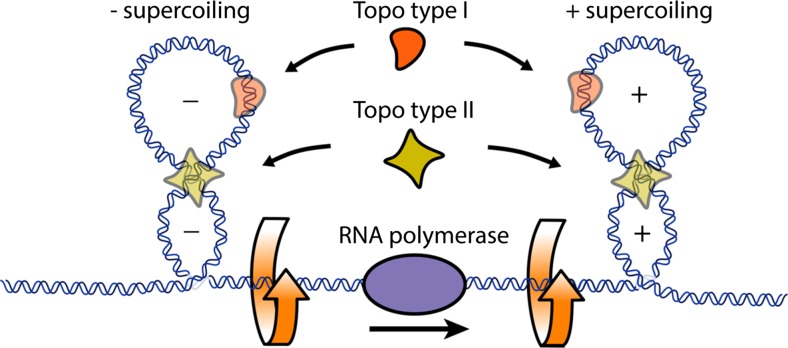
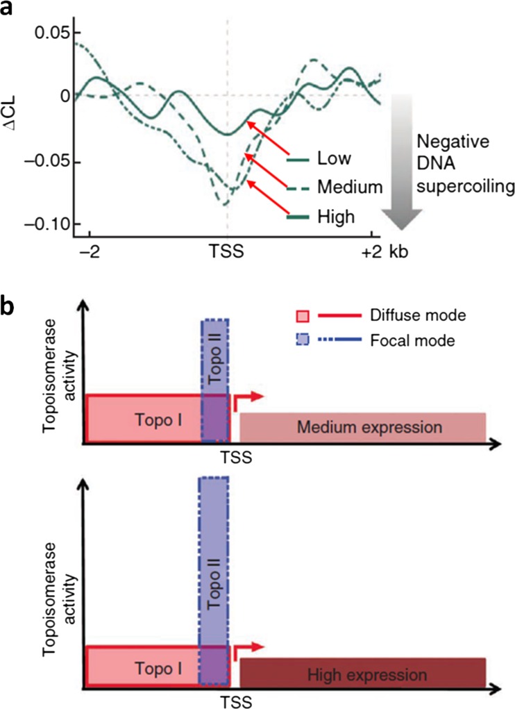
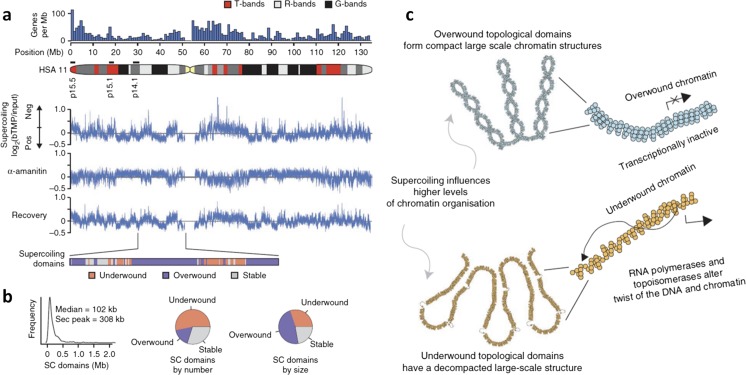
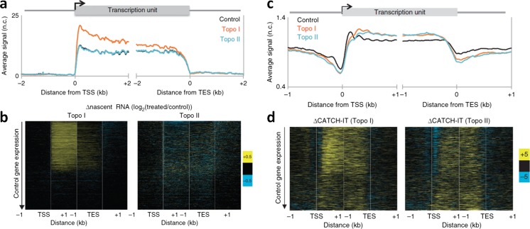
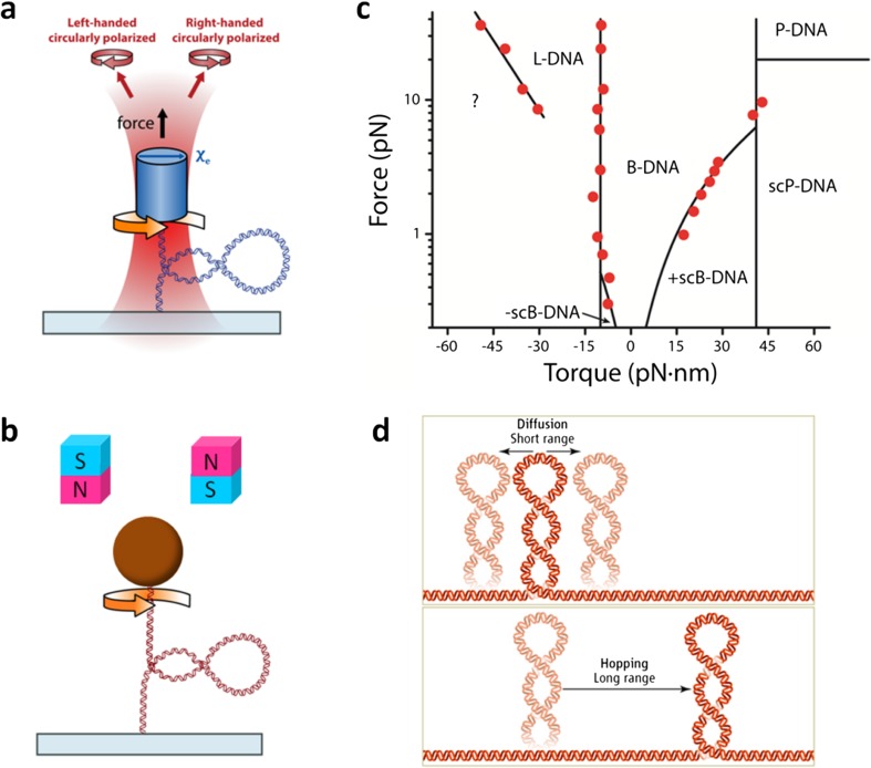
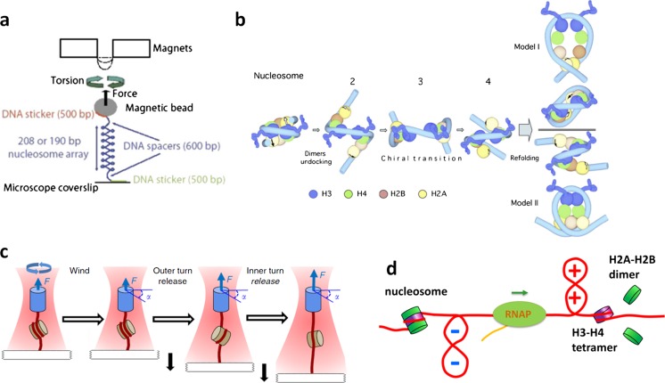
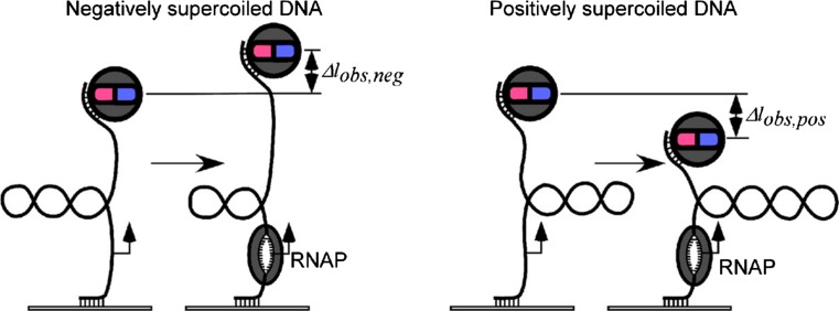
The twin-supercoiled-domain model describes how transcription can drive DNA supercoiling, and how DNA supercoiling, in turn plays an important role in regulating gene transcription. In vivo and in vitro experiments have disclosed many details of the complex interactions in this relationship, and recently new insights have been gained with the help of genome-wide DNA supercoiling mapping techniques and single molecule methods. This review summarizes the general mechanisms of the interplay between DNA supercoiling and transcription, considers the biological implications, and focuses on recent important discoveries and technical advances in this field. We highlight the significant impact of DNA supercoiling in transcription, but also more broadly in all processes operating on DNA.

Keywords: chromatin; gene regulation; mechanics; supercoiling; torque; transcription.


Conflict of interest statement

Conflict of interest

None.

Ethical approval

This article does not contain any studies with human or animal subjects performed by the authors.

Figures

Fig. 1
Fig. 1
A schematic of the twin-supercoiled-domain model. During transcription, RNA polymerase must rotate relative to its helical DNA track. Owing to the size and typical confinement of the polymerase and associated machinery, the DNA is (+) supercoiled in front and (−) supercoiled behind. Adapted from Forth et al. (2013) with permission
Fig. 2
Fig. 2
Genome-wide coupling between transcription and DNA topology. a Dynamic (−) DNA supercoiling around transcription start sites (TSSs) for genes with low, medium, or high expression levels. The vertical axis shows the ∆CL, which is the difference between CL values derived from transcription inhibited and uninhibited cells, and reflects DNA supercoiling induced by transcription. b A model for DNA supercoiling regulation by Topo I and Topo II. In this model, Topo I is diffusely recruited to the DNA supercoiling regions (diffuse mode), while Topo II is recruited in a focused manner to the most dynamic (−) supercoiling region (focal mode). For a moderately expressed gene, dynamic supercoiling is mainly managed by Topo I, which is recruited to a broad range upstream of the TSSs. For a highly transcribed gene, dynamic supercoiling is resolved efficiently by Topo II, which is recruited focally to the TSSs. Adapted from Kouzine et al. (2013) with permission
Fig. 3
Fig. 3
High-resolution mapping of DNA supercoiling reveals hundreds of supercoiling domains in the human chromosome (HSA) 11. a Microarray data of biotin-tagged psoralen bTMP binding across HSA 11 to assay the level of DNA supercoiling. α-Amanitin is used to inhibit transcription. Supercoiling domains are categorized as underwound, overwound, or stable regions. b The distributions of supercoiling domains. c Model of the large-scale chromatin structures. An overwound domain corresponds to transcriptionally inactive chromatin and is compact over a large scale, whereas an underwound domain corresponds to a transcriptionally active region and a decompacted chromatin structure. Adapted from Naughton et al. (2013) with permission
Fig. 4
Fig. 4
Transcription-generated torsional stress can destabilize nucleosomes. a, b Altered Pol II elongation kinetics upon Topo I or Topo II inhibition. a The average transcription profiles surrounding the transcription start sites (TSSs) and transcription end site (TESs) of all genes. Topo I inhibition increases the overall nascent-RNA production near TSSs, but not near TESs, suggesting Pol II stalling before the completion of the transcription, whereas Topo II inhibition showed no overall change relative to the control. b Heat maps of the log ratio of nascent RNA for Topo I inhibited samples over the control (left) and Topo II inhibited samples over the control (right), with genes ordered by decreasing expression in control samples. Topo I inhibition primarily affects transcribed genes, whereas Topo II inhibition results in heterogeneous changes in nascent-RNA levels, suggesting that Topo II plays only a secondary role in transcription. c, d Altered nucleosome turnover under torsion. c Nucleosome turnover is measured by CATCH-IT, followed by mapping of the positions of captured nucleosomes. d Heat maps showing changes in CATCH-IT signals, after Topo I (left) and Topo II (right) inhibition, from the controls. Adapted from Teves and Henikoff (2014) with permission
Fig. 5
Fig. 5
Primary single-molecule torsional manipulation tools for studies of DNA supercoiling. a A schematic of an angular optical trap (AOT) setup (Forth et al. , , ; Inman et al. ; La Porta and Wang ; Ma et al. ; Sheinin et al. , ; Sheinin and Wang 2009). A DNA molecule is torsionally anchored at one end to the surface of a coverglass and at the other end to a nanofabricated quartz cylinder held in an optical trap of linear polarization. The optical trap exerts both a force and torque on the cylinder. Rotation of the cylinder via rotation of the laser polarization introduces supercoiling into DNA. During a measurement, torque, rotation, force, and DNA extension are simultaneously measured. b A schematic of a magnetic tweezers (MTs) setup. DNA is anchored in a similar fashion as in an AOT. To introduce DNA supercoiling, the magnetic bead is rotated via rotation of the magnetic field. In most magnetic tweezers, only force and DNA extension may be measured. Recent enhancement of magnetic tweezers also permits torque detection (Celedon et al. ; Janssen et al. ; Lipfert et al. , ; Mosconi et al. 2011). c DNA force–torque phase diagram. Phase transitions between specific states of DNA are represented by solid black lines (Bryant et al. ; Marko ; Sarkar et al. 2001). The red points indicate torque values measured during phase transitions using an angular optical trap (Deufel et al. ; Forth et al. ; Sheinin et al. ; Sheinin and Wang 2009). Adapted, with permission, from Forth et al. (2013). d A drawing depicting plectoneme migration via diffusion or hopping along DNA. Adapted from Sheinin and Wang (2012) with permission
Fig. 6
Fig. 6
Nucleosomes under DNA supercoiling. a A schematic of the magnetic tweezers experiments to study nucleosome chiral transition. A nucleosome array was twisted and held under constant force while the DNA was supercoiled. b A model of the nucleosome chiral transition from left- to right-handed configuration. Two alternative routes for the refolding of the dimers are shown. c A drawing depicting the nucleosome-stretching assay with an AOT. A DNA containing a single nucleosome was stretched under a defined torsion. d A model for nucleosome turnover during transcription. (+) DNA supercoiling in front of a transcribing RNAP may induce H2A-H2B dimer loss, while (−) DNA supercoiling behind the RNAP facilitates nucleosome assembly and stabilizes assembled nucleosomes. a and b are adapted from Bancaud et al. (2007) and c from Sheinin et al. (2013) with permission
Fig. 7
Fig. 7
A schematic depicting the method of detection for promoter opening during transcription initiation using magnetic tweezers. Prior to a measurement, the DNA molecule was either (−) or (+) supercoiled under a small and constant force. Promoter opening by an RNAP increased or decreased the DNA extension for the (−) or (+) supercoiled DNA respectively. Adapted from (Revyakin et al. 2004) with permission
Fig. 8
Fig. 8
Determination of the torque generated by RNAP during transcription. a A drawing depicting the “twin-supercoiled-domain” model and experimental configuration to measure transcription against (−) supercoiling upstream (behind the RNAP) or (+) supercoiling downstream (in front of the RNAP). Escherichia coli RNAP was torsionally anchored to the surface of a coverglass while either the downstream end or upstream end of the DNA template was torsionally constrained to a quartz cylinder held in an AOT. The AOT monitored the translocation of the RNAP along DNA and the torque generated by RNAP in real-time. RNAP elongation accumulated (+) or (−) DNA supercoiling, respectively. As torque increased, RNAP was eventually stalled and the AOT reported the value of the stall torque. b Distributions of the measured downstream (left) and upstream (right) stall torques of RNAP. Adapted from Ma et al. (2013) with permission
Fig. 9
Fig. 9
Single-molecule observations of transcription bursting. a A drawing depicting how multiple rounds of transcription on a circular template led to an accumulation of significant (+) torsional stress and inhibition of transcription in the presence of Topo I and absence of gyrase. The subsequent addition of gyrase recovered transcription. b Time dependence of T7 transcription initiation rate (blue) under the conditions shown in a. Transcription initiation was inhibited by (+) supercoiling, but then recovered after the addition of gyrase. Adapted from Chong et al. (2014) with permission

References

    1. Allemand JF, Bensimon D, Lavery R, Croquette V. Stretched and overwound DNA forms a pauling-like structure with exposed bases. Proc Natl Acad Sci U S A. 1998;95:14152–14157. doi: 10.1073/pnas.95.24.14152. - DOI - PMC - PubMed
    1. Ashkin A, Dziedzic JM, Bjorkholm JE, Chu S. Observation of a single-beam gradient force optical trap for dielectric particles. Opt Lett. 1986;11:288–290. doi: 10.1364/OL.11.000288. - DOI - PubMed
    1. Ashkin A, Dziedzic JM, Yamane T. Optical trapping and manipulation of single cells using infrared laser beams. Nature. 1987;330:769–771. doi: 10.1038/330769a0. - DOI - PubMed
    1. Bai L, Fulbright RM, Wang MD. Mechanochemical kinetics of transcription elongation. Phys Rev Lett. 2007;98:068103. doi: 10.1103/PhysRevLett.98.068103. - DOI - PubMed
    1. Bancaud A, Conde e Silva N, Barbi M, Wagner G, Allemand JF, Mozziconacci J, Lavelle C, Croquette V, Victor JM, Prunell A, Viovy JL. Structural plasticity of single chromatin fibers revealed by torsional manipulation. Nat Struct Mol Biol. 2006;13:444–450. doi: 10.1038/nsmb1087. - DOI - PubMed

LinkOut - more resources