Case Reports
. 2017 Feb;9(2):E121-E125.
doi: 10.21037/jtd.2017.02.31.

Chronic pulmonary mucormycosis: an emerging fungal infection in diabetes mellitus

Affiliations
Case Reports

Chronic pulmonary mucormycosis: an emerging fungal infection in diabetes mellitus

Nousheen Iqbal et al. J Thorac Dis. 2017 Feb.

Abstract

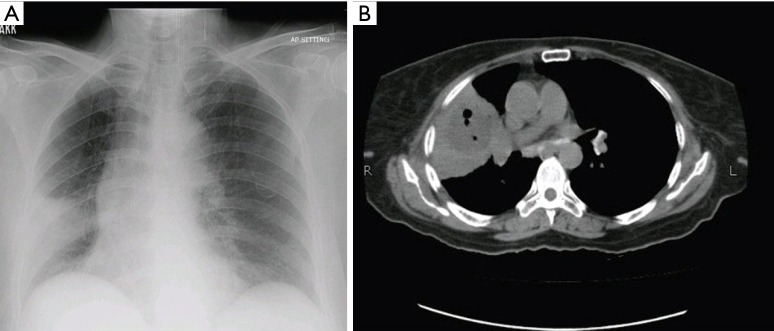
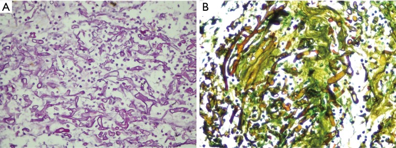
Mucormycosis commonly affects immunocompromised individuals with defects in neutrophil function or count. Diabetes mellitus is an important risk factor due to impair innate and acquired immunity for mucormycosis, with rhino-orbital-cerebral involvement as a common presentation. Pulmonary mucormycosis (PM) although a rare presentation in diabetic patients but is associated with high mortality and morbidity. An early diagnosis of PM is difficult, due to rarity of the disease and clinical and radiological features resembling tuberculosis (TB) which is common in Pakistan. Here we present three cases of chronic PM in patients with diabetes and with no other apparent risk factors.

Keywords: Diabetes; immunocompromised; pulmonary mucormycosis (PM).


Conflict of interest statement

Conflicts of Interest: The authors have no conflicts of interest to declare.

Figures

Figure 1
Figure 1
Right non resolving consolidation. (A) Chest X-rays showing consolidation in right lower lung zone; (B) CT scan chest showing a consolidation with a cavitation in right middle lobe. CT, computed tomographic.
Figure 2
Figure 2
Histopathology of Mucor. (A) Histopathology showing large collections of non-septate branching hyphae (H&E, 400× magnification); (B) non-septate fungal hyphae (Mucor) on Gomori methenamine silver stains (400× magnification).
Figure 3
Figure 3
Left hilar soft tissue mass. (A) Chest X-ray showing left hilar soft tissue mass; (B) HRCT showing consolidation in the lingular segment and left upper lobe of lung. HRCT, high-resolution computed tomographic.
Figure 4
Figure 4
CT chest showing fibrocavitary changes on right side of lung. CT, computed tomographic.

References

    1. Lee FY, Mossad SB, Adal KA. Pulmonary mucormycosis: the last 30 years. Arch Intern Med 1999;159:1301-9. 10.1001/archinte.159.12.1301 - DOI - PubMed
    1. Agarwal R, Kumar V, Gupta D. Pulmonary mucormycosis: two of a kind. Eur J Intern Med 2006;17:63-5. 10.1016/j.ejim.2005.08.009 - DOI - PubMed
    1. Garg R, Marak RS, Verma SK, et al. Pulmonary mucormycosis mimicking as pulmonary tuberculosis: a case report. Lung India 2008;25:129-31. 10.4103/0970-2113.59595 - DOI - PMC - PubMed
    1. Panigrahi MK, Manju R, Kumar SV, et al. Pulmonary mucormycosis presenting as nonresolving pneumonia in a patient with diabetes mellitus. Respir Care 2014;59:e201-5. 10.4187/respcare.03205 - DOI - PubMed
    1. Aggarwal D, Chander J, Janmeja AK, et al. Pulmonary tuberculosis and mucormycosis co-infection in a diabetic patient. Lung India 2015;32:53-5. 10.4103/0970-2113.148452 - DOI - PMC - PubMed

Publication types

LinkOut - more resources