

Review
doi: 10.21037/atm.2016.11.47.
Cannulation techniques for extracorporeal life support
Affiliations
- PMID: 28275615
- PMCID: PMC5337209
- DOI: 10.21037/atm.2016.11.47
Item in Clipboard
Review
Cannulation techniques for extracorporeal life support
Ann Transl Med.
2017 Feb.
Abstract
The article reviews cannulation strategy for different modes of extracorporeal life support. Technical aspects, pitfalls and complications are discussed for central and peripheral extracorporeal membrane oxygenation (VA, VV, VAV, VVA), biventricular assist device support and extracorporeal CO2 removal.
Keywords: BiVAD; Cannulation; ECLS; ECMO; biventricular assist device; extracorporeal CO2 removal; extracorporeal life support; extracorporeal membrane oxygenation.
Conflict of interest statement
Conflicts of Interest: The authors have no conflicts of interest to declare.
Figures
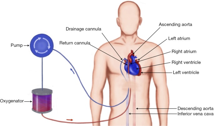
Diagram of central ECMO: the blood is drained via a large venous cannula from the right atrium under negative pressure and transferred through oxygenator, following which returned to the body via a cannula into the ascending aorta. The cannula can be tunnelled under the skin and chest closed if longer than few days’ support is anticipated.
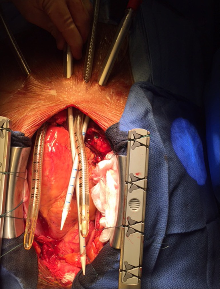
Cannulae for BiVAD tunnelled and ready for cannulation.
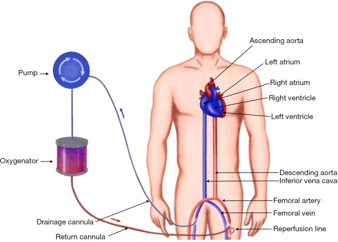
Peripheral VA ECMO cannulation: the blood is drained from the IVC via femoral cannulation. It passes through a centrifugal pump and oxygenator and is returned to the patient via femoral artery. On this diagram, a reperfusion line is used to ensure distal limb perfusion.
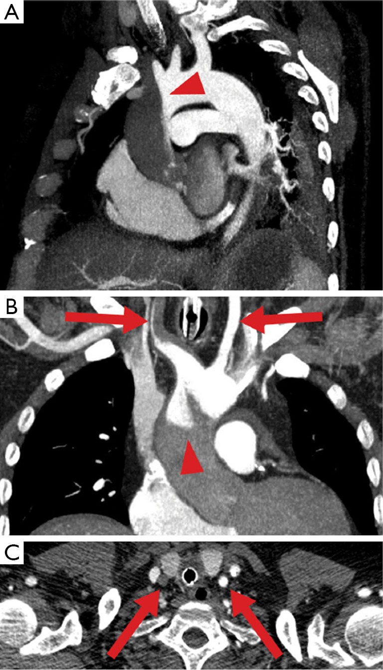
Watershed phenomenon during veno-arterial ECMO visualized by computed tomography. Antegrade blood flow (low contrast) from the heart competes with retrograde blood flow (high contrast) from the ECMO in the aorta, resulting in a watershed phenomenon (arrowhead). Here computed tomography of a patient with pulmonary embolism and reduced cardiac output demonstrates a rather proximal watershed, leading to perfusion of the right carotid artery with “heart blood” (dark) and the left carotid artery with “ECMO blood” (bright, arrows). Upper panel sagittal oblique maximum intensity projection (MIP), middle panel coronal oblique MIP, lower panel transverse plane (14).
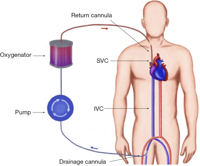
Veno-venous ECMO: blood is drained from the IVC and passes through an oxygenator, then returned to the venous system via the right atrium.
References
-
- Extracorporeal life support organization (ELSO guidelines). 2013. Available online: http://www.elso.org/resources/Guidelines.aspx
Publication types
LinkOut - more resources
Full Text Sources
Other Literature Sources
Miscellaneous