. 2017 May;6(5):1356-1365.
doi: 10.1002/sctm.16-0441. Epub 2017 Mar 9.

Ascorbic Acid Promotes the Stemness of Corneal Epithelial Stem/Progenitor Cells and Accelerates Epithelial Wound Healing in the Cornea

Affiliations

Ascorbic Acid Promotes the Stemness of Corneal Epithelial Stem/Progenitor Cells and Accelerates Epithelial Wound Healing in the Cornea

Jialin Chen et al. Stem Cells Transl Med. 2017 May.

Abstract

High concentration of ascorbic acid (vitamin C) has been found in corneal epithelium of various species. However, the specific functions and mechanisms of ascorbic acid in the repair of corneal epithelium are not clear. In this study, it was found that ascorbic acid accelerates corneal epithelial wound healing in vivo in mouse. In addition, ascorbic acid enhanced the stemness of cultured mouse corneal epithelial stem/progenitor cells (TKE2) in vitro, as shown by elevated clone formation ability and increased expression of stemness markers (especially p63 and SOX2). The contribution of ascorbic acid on the stemness enhancement was not dependent on the promotion of Akt phosphorylation, as concluded by using Akt inhibitor, nor was the stemness found to be dependent on the regulation of oxidative stress, as seen by the use of two other antioxidants (GMEE and NAC). However, ascorbic acid was found to promote extracellular matrix (ECM) production, and by using two collagen synthesis inhibitors (AzC and CIS), the increased expression of p63 and SOX2 by ascorbic acid was decreased by around 50%, showing that the increased stemness by ascorbic acid can be attributed to its regulation of ECM components. Moreover, the expression of p63 and SOX2 was elevated when TKE2 cells were cultured on collagen I coated plates, a situation that mimics the in vivo situation as collagen I is the main component in the corneal stroma. This study shows direct therapeutic benefits of ascorbic acid on corneal epithelial wound healing and provides new insights into the mechanisms involved. Stem Cells Translational Medicine 2017;6:1356-1365.

Keywords: Colony formation; Microenvironment; Proliferation; Stem cell-microenvironment interactions; Stem/progenitor cell; Tissue regeneration.


Figures

Figure 1
Figure 1
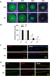
Ascorbic acid (i.e., VC) accelerates corneal epithelial wound healing and increases ΔNP63 expression in vivo. 10% A2‐P eye drops were applied after the scrape of corneal epithelium. (A): The defect area of corneal epithelium was evaluated after 24, 48, and 72 hours by 0.25% fluorescein sodium under a BQ900 slit lamp. (B): Residual epithelial defect is presented as the percentage of the original wound size. (C): Immunofluorescence staining of p‐Akt between A2‐P treated group and Ctrl at 48 hours after epithelial debridement. The right column is the merged picture of left column and DAPI staining. (D): Immunofluorescence staining was carried out to show the expression of Ki67 and ΔNP63 in both groups at 48 hours after epithelial debridement. The right column is the merged picture of left column and middle column. Ascorbic acid treatment was performed using the vitamin C derivate A2‐P (VC). **Significant difference between two groups at p < .001. Abbreviations: Ctrl, control group; VC, vitamin C; p‐Akt, Akt phosphorylation.
Figure 2
Figure 2
Ascorbic acid (VC) promotes the stemness of corneal epithelial stem/progenitor cells. (A): Mouse TKE2 cells were incubated with or without 50 μg/ml A2‐P for 10–12 days. The colonies formed were stained with 1% crystal violet. (B): The number of all colonies with diameters >0.5 mm was counted and compared between the two groups. (C, D): TKE2 cells were treated with 50 μg/ml A2‐P for 12 hours. The expression of stemness markers for corneal epithelial stem cells (p63, ABCG2, and TCF4) and pluripotent stem cells (SOX2 and OCT4) was analyzed by Western blot (C) and quantified relative to β‐Actin (D). *Significant difference between two groups at p < .05. N.S,.No significant difference between two groups at p ≥ .05. Abbreviations: Ctrl, control group; VC, vitamin C.
Figure 3
Figure 3
Ascorbic acid (VC) activates p‐Akt and enhances cell proliferation. (A): The proliferation rate was compared after TKE2 cells were cultured for 1, 3, 5, and 7 days with or without 50 μg/ml of A2‐P. (B): Immunofluorescence staining was performed to analyze the expression of Ki67. The right column is the merged picture of left column and middle column. (C): The expression of PCNA was analyzed by Western blot after A2‐P treatment for 0, 1, 6, 12, and 24 hours, and quantified relative to β‐Actin. (D): The p‐Akt level was analyzed by Western blot after A2‐P treatment for 0, 1, 6, 12, and 24 hours, and quantified relative to Akt. In (C, D), 0 hours was set as 1 in the quantified data, and all time points were compared to 0 hours in the statistical analysis; only statistically significant differences are marked. (E): The p‐Akt level was analyzed by Western blot after Akti treatment, and quantified relative to Akt. The A2‐P treated group was set as 1 in the quantified data. (F): The proliferation rate was compared between A2‐P+Akti treated group and A2‐P group after 0 and 2 days. (G): The expression of p63 and SOX2 was evaluated by Western blot after Akti treatment, and quantified relative to β‐Actin. A2‐P treated group was set as 1 in the quantified data. *Significant difference between two groups at p < .05. **Significant difference between two groups at p < .001. N.S,.no significant difference between two groups at p ≥ .05. Abbreviations: Akti, Akt inhibitor; Ctrl, control group; VC, vitamin C; p‐Akt, Akt phosphorylation.
Figure 4
Figure 4
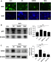
Ascorbic acid (VC) acts as an antioxidant in corneal epithelial stem/progenitor cells, but other antioxidants do not promote stemness in these cells. (A): Mouse TKE2 cells were treated with 50 μg/ml of A2‐P, or two other antioxidants (GMEE and NAC), for 24 hours. The staining density of intracellular ROS (green) and GSH (blue) was compared to the Ctrl. (B): The expression of p63 was analyzed by Western blot after treatment with A2‐P, GMEE or NAC and quantified relative to β‐Actin. (C): The expression of SOX2 was analyzed by Western blot after treatment with A2‐P, GMEE, or NAC and quantified relative to β‐Actin. *Significant difference between two groups at p < .05. N.S,.no significant difference between two groups at p ≥ .05. All experimental groups were compared to 0 hours. Abbreviations: Ctrl, control group; ROS, reactive oxygen species; GSH, glutathione; VC, vitamin C.
Figure 5
Figure 5
Ascorbic acid (VC) regulates extracellular matrix (ECM) components which accounts for the stemness enhancement. (A): Immunofluorescence staining was performed to analyze the expression of ECM components, with or without 50 μg/ml of A2‐P treatment. “COL I” indicates collagen type I; “COL IV” indicates collagen type IV; “FN” indicates fibronectin. (B): TKE2 cells were not treated (Ctrl) or treated with 50 μg/ml of A2‐P, alone or together with one of two collagen synthesis inhibitors (AzC and CIS) for 3 days. Collagen content was assessed for all groups and compared to the VC group. (C): The expression of p63 and SOX2 was analyzed by Western blot after treatment with A2‐P alone or together with AzC or CIS, and quantified relative to β‐Actin. VC group was set as 1 in the quantified data, and all other groups were compared to the VC group in the statistical analysis. (D): TKE2 cells were cultured on collagen I coated plates or normal culture plates for 3 days. The expression of p63 and SOX2 was analyzed by Western blot and quantified relative to β‐Actin. Group with cells cultured on normal culture plates was set as 1 in the quantified data. *Significant difference between two groups at p < .05. N.S,.no significant difference between two groups at p ≥ .05. Abbreviations: COL I, collagen type I; COL IV, collagen type IV; FN, fibronectin.

References

    1. Blanchard J, Tozer TN, Rowland M. Pharmacokinetic perspectives on megadoses of ascorbic acid. Am J Clin Nutr 1997;66:1165–1171. - PubMed
    1. Ichibe Y, Ishikawa S. [Optic neuritis and vitamin C]. Nippon Ganka Gakkai zasshi 1996;100:381–387. - PubMed
    1. Vitetta L, Sali A, Paspaliaris B et al. Megadose vitamin C in treatment of the common cold: A randomised controlled trial. Med J Aust 2002;176:298–299. - PubMed
    1. Iqbal Z, Midgley JM, Watson DG et al. Effect of oral administration of vitamin C on human aqueous humor ascorbate concentration. Zhongguo Yao Li Xue Bao 1999;20:879–883. - PubMed
    1. Savini I, Rossi A, Pierro C et al. SVCT1 and SVCT2: Key proteins for vitamin C uptake. Amino acids 2008;34:347–355. - PubMed

Publication types