

The acute airway inflammation induced by PM2.5 exposure and the treatment of essential oils in Balb/c mice
- PMID: 28276511
- PMCID: PMC5343586
- DOI: 10.1038/srep44256
The acute airway inflammation induced by PM2.5 exposure and the treatment of essential oils in Balb/c mice
Abstract
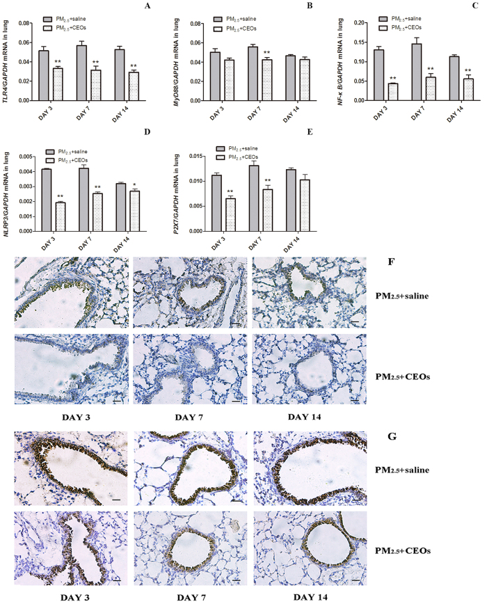
PM2.5 is the main particulate air pollutant whose aerodynamic diameter is less than 2.5 micron. The inflammation of various respiratory diseases are associated with PM2.5 inhalation. Pro-inflammatory cytokine IL-1β generated from effected cells usually plays a crucial role in many kinds of lung inflammatory reactions. The exacerbation of Th immune responses are identified in some PM2.5 related diseases. To elucidate the underlying mechanism of PM2.5-induced acute lung inflammation, we exposed Balb/c mice to PM2.5 intratracheally and established a mice model. Acute lung inflammation and increased IL-1β expression was observed after PM2.5 instillation. Regulatory factors of IL-1β (TLR4/MyD88 signaling pathway and NLRP3 inflammasome) participated in this lung inflammatory response as well. Treatment with compound essential oils (CEOs) substantially attenuated PM2.5-induced acute lung inflammation. The decreased IL-1β and Th immune responses after CEOs treatment were significant. PM2.5 may increase the secretion of IL-1β through TLR4/MyD88 and NLRP3 pathway resulting in murine airway inflammation. CEOs could attenuate the lung inflammation by reducing IL-1β and Th immune responses in this model. This study describes a potentially important mechanism of PM2.5-induced acute lung inflammation and that may bring about novel therapies for the inflammatory diseases associated with PM2.5 inhalation.
Conflict of interest statement
The authors declare no competing financial interests.
Figures


Similar articles
-
Involvement of EGF receptor signaling and NLRP12 inflammasome in fine particulate matter-induced lung inflammation in mice.Environ Toxicol. 2017 Apr;32(4):1121-1134. doi: 10.1002/tox.22308. Epub 2016 Jul 5. Environ Toxicol. 2017. PMID: 27377055
-
Effects on IL-1β signaling activation induced by water and organic extracts of fine particulate matter (PM2.5) in vitro.Environ Pollut. 2018 Jun;237:592-600. doi: 10.1016/j.envpol.2018.02.086. Epub 2018 Mar 15. Environ Pollut. 2018. PMID: 29525626
-
PM2.5-induced pulmonary inflammation via activating of the NLRP3/caspase-1 signaling pathway.Environ Toxicol. 2021 Mar;36(3):298-307. doi: 10.1002/tox.23035. Epub 2020 Sep 30. Environ Toxicol. 2021. PMID: 32996690 Free PMC article.
-
NLRP3 inflammasome activation and lung fibrosis caused by airborne fine particulate matter.Ecotoxicol Environ Saf. 2018 Nov 15;163:612-619. doi: 10.1016/j.ecoenv.2018.07.076. Epub 2018 Aug 6. Ecotoxicol Environ Saf. 2018. PMID: 30092543
-
Liver injury induced in Balb/c mice by PM2.5 exposure and its alleviation by compound essential oils.Biomed Pharmacother. 2018 Sep;105:590-598. doi: 10.1016/j.biopha.2018.06.010. Epub 2018 Jun 8. Biomed Pharmacother. 2018. PMID: 29890467
Cited by
-
Fine particulate matter (PM2.5): The culprit for chronic lung diseases in China.Chronic Dis Transl Med. 2018 Aug 28;4(3):176-186. doi: 10.1016/j.cdtm.2018.07.002. eCollection 2018 Sep. Chronic Dis Transl Med. 2018. PMID: 30276364 Free PMC article.
-
Antibacterial, Immunomodulatory, and Lung Protective Effects of Boswelliadalzielii Oleoresin Ethanol Extract in Pulmonary Diseases: In Vitro and In Vivo Studies.Antibiotics (Basel). 2021 Nov 25;10(12):1444. doi: 10.3390/antibiotics10121444. Antibiotics (Basel). 2021. PMID: 34943656 Free PMC article.
-
PM2.5 Aggravated OVA-Induced Epithelial Tight Junction Disruption Through Fas Associated via Death Domain-Dependent Apoptosis in Asthmatic Mice.J Asthma Allergy. 2021 Nov 20;14:1411-1423. doi: 10.2147/JAA.S335590. eCollection 2021. J Asthma Allergy. 2021. PMID: 34848976 Free PMC article.
-
Ni2+-Assisted Hydrolysis May Affect the Human Proteome; Filaggrin Degradation Ex Vivo as an Example of Possible Consequences.Front Mol Biosci. 2022 Mar 10;9:828674. doi: 10.3389/fmolb.2022.828674. eCollection 2022. Front Mol Biosci. 2022. PMID: 35359602 Free PMC article.
-
lncRNA Gm16410 Mediates PM2.5-Induced Macrophage Activation via PI3K/AKT Pathway.Front Cell Dev Biol. 2021 Mar 16;9:618045. doi: 10.3389/fcell.2021.618045. eCollection 2021. Front Cell Dev Biol. 2021. PMID: 33796524 Free PMC article.
References
-
- Li M. & Zhang L. Haze in China: current and future challenges. Environ Pollut. 189, 85–6 (2014). - PubMed
-
- Brüggemann E., Gerwig H., Gnauk Th., Müller K. & Herrmann H. Influence of seasons, air mass origin and day of the week on size-segregated chemical composition of aerosol particles at a kerb side. Atmos Environ. 43, 2456–2463 (2009).
Publication types
MeSH terms
Substances
LinkOut - more resources
Full Text Sources
Other Literature Sources
Medical
