. 2017 Mar 9:7:44332.
doi: 10.1038/srep44332.

Antimicrobial peptide cWFW kills by combining lipid phase separation with autolysis

Affiliations

Antimicrobial peptide cWFW kills by combining lipid phase separation with autolysis

Kathi Scheinpflug et al. Sci Rep. .

Abstract

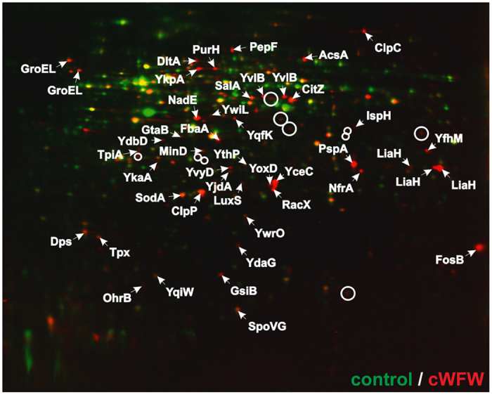
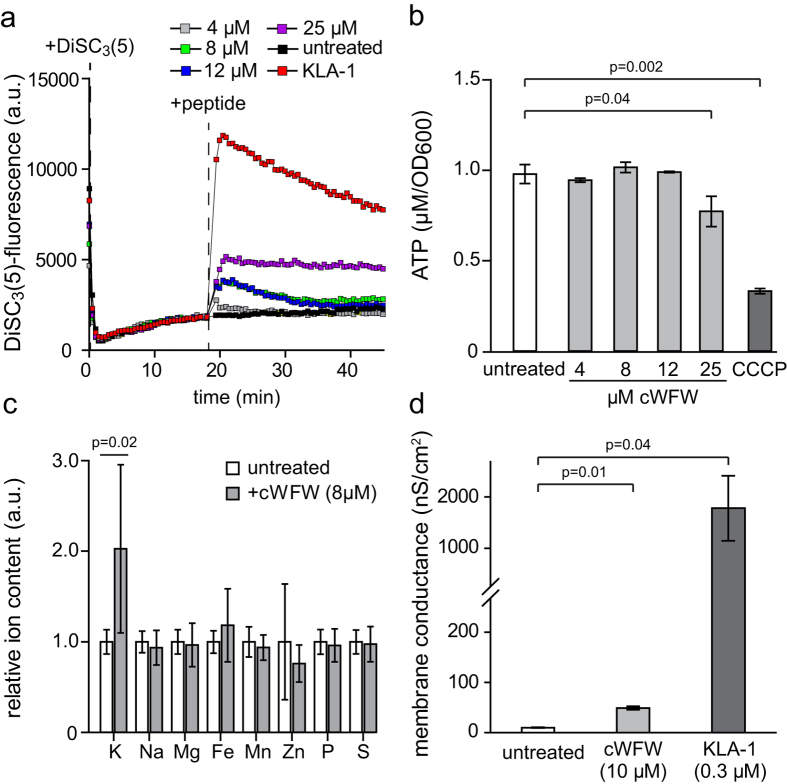
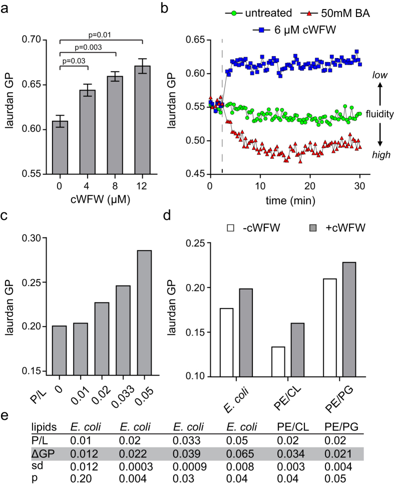
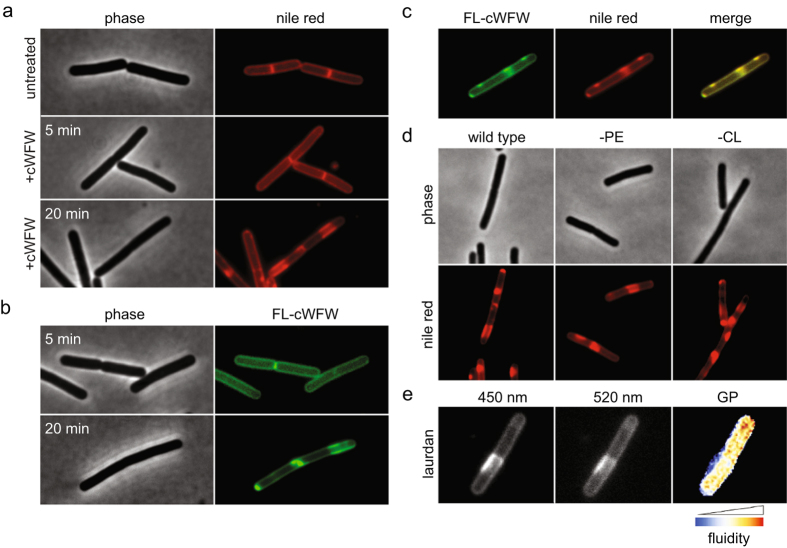
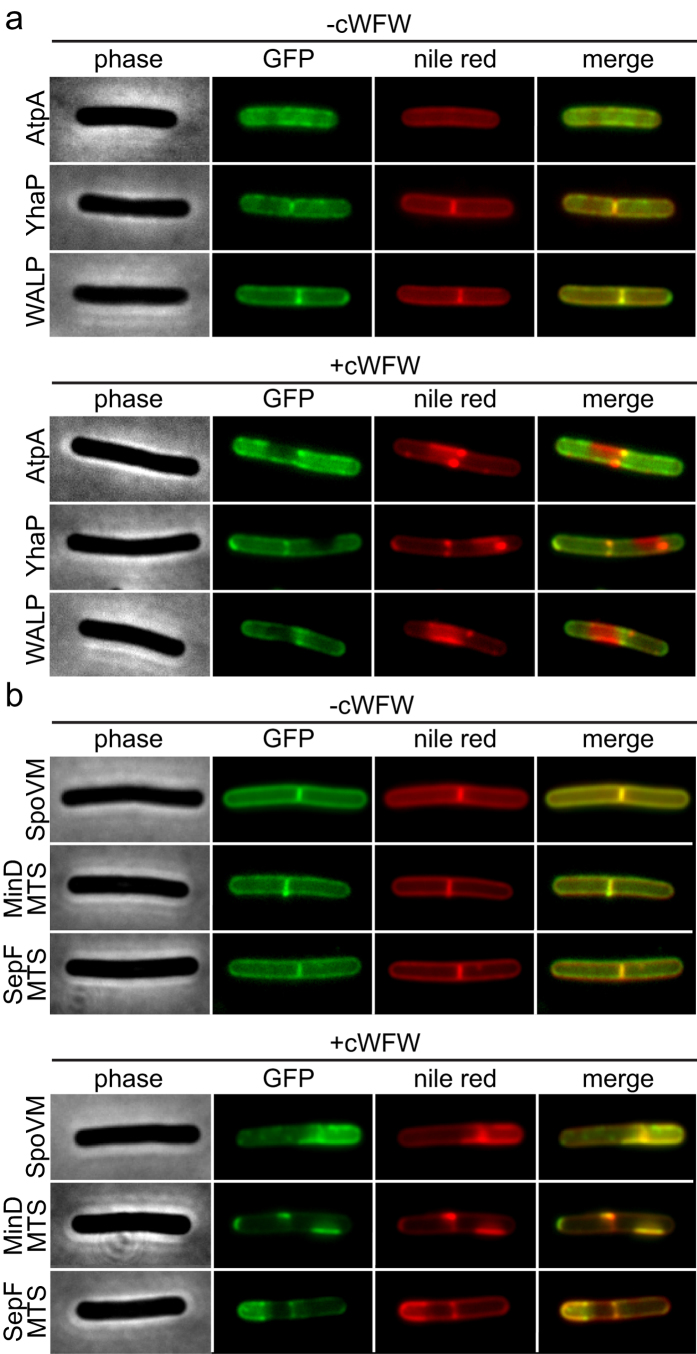
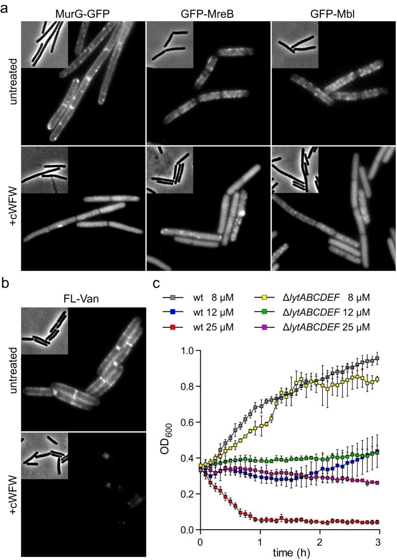
The synthetic cyclic hexapeptide cWFW (cyclo(RRRWFW)) has a rapid bactericidal activity against both Gram-positive and Gram-negative bacteria. Its detailed mode of action has, however, remained elusive. In contrast to most antimicrobial peptides, cWFW neither permeabilizes the membrane nor translocates to the cytoplasm. Using a combination of proteome analysis, fluorescence microscopy, and membrane analysis we show that cWFW instead triggers a rapid reduction of membrane fluidity both in live Bacillus subtilis cells and in model membranes. This immediate activity is accompanied by formation of distinct membrane domains which differ in local membrane fluidity, and which severely disrupts membrane protein organisation by segregating peripheral and integral proteins into domains of different rigidity. These major membrane disturbances cause specific inhibition of cell wall synthesis, and trigger autolysis. This novel antibacterial mode of action holds a low risk to induce bacterial resistance, and provides valuable information for the design of new synthetic antimicrobial peptides.


Conflict of interest statement

The authors declare no competing financial interests.

Figures

Figure 1
Figure 1. Cytosolic proteome response profile of B. subtilis to cWFW.
Autoradiographs of antibiotic-treated cells (red) were overlaid with those of untreated controls (green). Upregulated proteins in response to cWFW treatment (8 μM) appear red, down-regulated proteins green. Proteins synthesised at equal rates appear yellow. Proteins upregulated more than 2-fold in three biological replicates were defined as marker proteins and identified by mass spectrometry. Unidentified marker proteins are indicated by circles. See supplementary Figure 1a for the growth inhibition of B. subtilis observed in BMM with different cWFW concentrations. Strain used: B. subtilis 168/DSM 402.
Figure 2
Figure 2. Impact of cWFW on cell energy state and membrane permeability.
(a) Membrane potential levels of B. subtilis upon addition of different cWFW concentrations were measured using fluorescent voltage-sensitive dye DiSC3(5). For positive control, cells were depolarised by addition of the helical pore-forming antimicrobial peptide KLA-1 (40 μM). The time points of DiSC3(5) and peptide additions are highlighted with dashed lines. See supplementary Figure 1b for growth inhibition at identical cWFW-concentrations and cell densities. The graph depicts a representative measurement of three independent replicates. (b) ATP levels in B. subtilis after 20 min incubation with different cWFW concentrations were measured using a Luciferase-based luminescence assay. For positive control, cells were incubated with the proton ionophore CCCP (100 μM). Cell densities are comparable to the data shown in panel A and supplementary Figure 1b. The diagram depicts the average and standard deviation values of three independent replicates. No significant changes (p ≥ 0.05) were observed for samples treated with 4, 8, and 12 μM cWFW. (c) Changes in relative ion content of B. subtilis upon 15 min incubation with 8 μM cWFW were determined using inductively-coupled plasma optical emission spectroscopy (ICP-OES). Phosphorus, mainly prevalent in DNA-bound form, served as internal control for cell mass. The diagram depicts the average and standard deviation values of three independent measurements. No significant changes (p ≥ 0.05) were observed ions other than K+. See supplementary Figure S1a for the growth inhibition of B. subtilis observed in BMM with different cWFW concentrations. (d) Conductivity measurements on planar lipid membranes formed of E. coli lipid extract upon addition of 10 μM cWFW. The pore-forming helical peptide KLA-1 (0.3 μM) served as positive control. The diagram depicts the average and standard error of two independent measurements. The statistical significances were calculated using unpaired (panels b/d) and paired (panel c) two-tailed Student t test. Strains used: (a/b) B. subtilis 168, (c) B. subtilis 168/DSM 402.
Figure 3
Figure 3. cWFW reduces membrane fluidity in vivo and in vitro.
(a) The fluidity of the cytoplasmic membrane was measured for B. subtilis cells upon 10 min incubation with increasing concentrations of cWFW using the fluidity-sensitive fluorescent dye laurdan. Please note that high laurdan generalised polarisation (GP) correlates with low membrane fluidity. The diagram depicts the average and standard deviation of three replicate measurements. (b) Time-resolved laurdan generalised polarisation (GP) was measured upon addition of 6 μM cWFW. As a positive control, 50 mM of the membrane fluidiser benzyl alcohol (BA) was added to a replicate sample. The time point of addition is indicated with a dashed line. The graph depicts a representative measurement of three independent replicates. (c) Laurdan GP was measured for large unilamellar vesicles (LUVs) formed of E. coli polar lipid extract in the presence of increasing concentrations of cWFW. The peptide-to-lipid molar ratios (P/L) are indicated below the graph. See supplementary Figure S3b for examples of the recorded spectra. (d) Laurdan GP in the presence and absence of cWFW (P/L = 0.02) was measured for LUVs with varying lipid compositions (lipid molar ratios: PE/CL 87.5/12.5, PE/PG 75/25). See supplementary Figure 3c–f for recorded spectra. PE: palmitoyl-oleoyl-phosphatidylethanolamine, PG: palmitoyl-oleoyl-phosphatidylglycerol, CL: cardiolipin and E. coli: E. coli polar lipid extract. (e) Average and standard deviation of cWFW-induced changes in laurdan GP (ΔGP) are shown for the different lipid compositions and peptide-to-lipid molar ratios (P/L) from two independent experiments. The statistical significances were calculated using unpaired (panels a) and paired (panel e) two-tailed Student t test.
Figure 4
Figure 4. cWFW triggers large-scale lipid domain formation in vivo.
(a) Phase contrast and fluorescence images of B. subtilis cells stained with fluorescent membrane dye nile red before addition (upper panels) and after 5 min (middle panels) and 20 min (lower panels) incubation with cWFW (12 μM). (b) Phase contrast and fluorescence images of B. subtilis cells stained with a 1:5 mix of NBD-labelled (FL-cWFW) and unlabelled cWFW (combined concentration of 12 μM) after 5 min (upper panels) and 20 min (lower panels) incubation. (c) Fluorescence images of B. subtilis cells stained with NBD-labelled peptide (FL-cWFW; left panel) and nile red (middle panel) upon 20 min incubation. The right panel depicts a colour overlay of the images shown in left and middle panels. (d) Phase contrast and fluorescence images of B. subtilis cells stained with fluorescent membrane dye nile red after 20 min incubation with cWFW (12 μM). Depicted are wild type cells (left panels), cells deficient for phosphatidylethanolamine (-PE, middle panels), and cells deficient for cardiolipin (-CL, right panels). (e) Fluorescence images of B. subtilis cell stained with laurdan upon 20 min incubation with cWFW (12 μM). Depicted is the laurdan emission at 450 nm (left panel), 520 nm (middle panel) and a colour-coded laurdan GP map calculated from the images shown in left and middle panels, respectively. Strains used: (ae) B. subtilis 168 (wild type), (d) B. subtilis HB5343 (Δpsd, PE-deficient) and B. subtilis SDB206 (ΔclsA, ΔclsB, ΔywiE, CL-deficient).
Figure 5
Figure 5. cWFW-triggered lipid domain formation segregates membrane proteins.
(a) Phase contrast, GFP-fluorescence, nile red-fluorescence and fluorescent colour overlays are depicted for cells expressing different integral membrane proteins in the absence (upper panels) and presence (lower panels) of cWFW (20 min incubation with 12 μM). (b) Comparable images are depicted for cells expressing different peripheral membrane proteins. Strains used: (a) B. subtilis BS23 (AtpA-GFP), B. subtilis HS41 (YhaP-GFP), B. subtilis HS64 (WALP23-GFP), (b) B. subtilis KR318 (SpoVM-GFP), B. subtilis HS65 (GFP-MinDMTS) and B. subtilis HS208 (SepFMTS-GFP).
Figure 6
Figure 6. cWFW inhibits cell wall synthesis and triggers autolysis.
(a) Phase contrast and fluorescence images of B. subtilis cells expressing MurG-GFP, GFP MreB and GFP-Mbl are depicted in the absence (upper panels) and presence (lower panels) of cWFW (20 min incubation with 12 μM). (b) Phase contrast and fluorescence images of B. subtilis wild type cells stained with fluorescent vancomycin (FL-Van) in the absence (upper panel) and presence (lower panel) of cWFW (20 min incubation with 12 μM). (c) Changes in optical density of B. subtilis wild type cells (wt), and cells deficient for the autolytic enzymes LytCDEF upon incubation with different concentrations of cWFW. The diagram depicts the average and standard deviation of three replicate cultures. Please note that the lytABC operon encodes the amidase LytC and accessory proteins LytAB which are involved in regulation and secretion of LytC, respectively. Strains used: (a) B. subtilis 168, B. subtilis KS69 (GFP-MreB), B. subtilis KS70 (GFP-Mbl), and B. subtilis TNVS175 (MurG-GFP), (b) B. subtilis 168, and (c) B. subtilis 168 (wt) and B. subtilis KS19 (ΔlytABCDEF).

References

    1. Baltzer S. A. & Brown M. H. Antimicrobial peptides: Promising alternatives to conventional antibiotics. J. Mol. Microbiol. Biotechnol. 20, 228–235 (2011). - PubMed
    1. Appelt C., Wessolowski A., Dathe M. & Schmieder P. Structures of cyclic, antimicrobial peptides in a membrane-mimicking environment define requirements for activity. J. Pept. Sci. 14, 524–527 (2008). - PubMed
    1. Blondelle S. E., Perez-Paya E. & Houghten R. A. Synthetic combinatorial libraries: Novel discovery strategy for identification of antimicrobial agents. Antimicrob. Agents Chemother. 40, 1067–1071 (1996). - PMC - PubMed
    1. Scheinpflug K., Nikolenko H., Komarov I., Rautenbach M. & Dathe M. What goes around comes around-a comparative study of the influence of chemical modifications on the antimicrobial properties of small cyclic peptides. Pharmaceuticals 6, 1130–1144 (2013). - PMC - PubMed
    1. Junkes C. et al.. Cyclic antimicrobial R-, W-rich peptides: The role of peptide structure and E. coli outer and inner membranes in activity and the mode of action. Eur. Biophys. J. 40, 515–528 (2011). - PubMed

Publication types

MeSH terms

LinkOut - more resources