. 2017 May;26(5):1060-1069.
doi: 10.1002/pro.3155. Epub 2017 Mar 24.

Structural and functional characterization of a ubiquitin variant engineered for tight and specific binding to an alpha-helical ubiquitin interacting motif

Affiliations

Structural and functional characterization of a ubiquitin variant engineered for tight and specific binding to an alpha-helical ubiquitin interacting motif

Noah Manczyk et al. Protein Sci. 2017 May.

Abstract

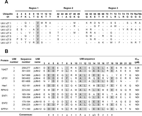
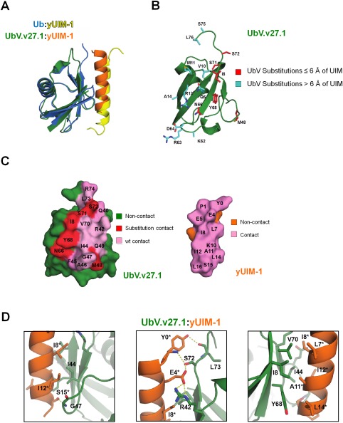
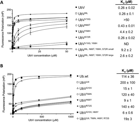
Ubiquitin interacting motifs (UIMs) are short α-helices found in a number of eukaryotic proteins. UIMs interact weakly but specifically with ubiquitin conjugated to other proteins, and in so doing, mediate specific cellular signals. Here we used phage display to generate ubiquitin variants (UbVs) targeting the N-terminal UIM of the yeast Vps27 protein. Selections yielded UbV.v27.1, which recognized the cognate UIM with high specificity relative to other yeast UIMs and bound with an affinity more than two orders of magnitude higher than that of ubiquitin. Structural and mutational studies of the UbV.v27.1-UIM complex revealed the molecular details for the enhanced affinity and specificity of UbV.v27.1, and underscored the importance of changes at the binding interface as well as at positions that do not contact the UIM. Our study highlights the power of the phage display approach for selecting UbVs with unprecedented affinity and high selectivity for particular α-helical UIM domains within proteomes, and it establishes a general approach for the development of inhibitors targeting interactions of this type.

Keywords: phage display; protein engineering; ubiquitin; ubiquitin interacting motif.


Figures

Figure 1
Figure 1
Selective binding of UbV.v27.1 to yUIM‐1. (A) Sequence alignment of UbV.v27.1 and other UbVs selected for binding to yUIM‐1. The alignment shows only those positions that were diversified in the UbV library, and positions that were conserved as the wt sequence are shown as dashes. Sequences showing conservation across selected UbVs are highlighted in grey. (B) Sequence alignment of yeast UIMs and IC50 values for inhibition of UbV.v27.1 binding to immobilized yUIM‐1. Residues (arbitrarily numbered from 3 to 22) that conform to the UIM consensus are highlighted in grey, and the consensus is shown below (e is an acidic residue, φ is a hydrophobic residue, z is a bulky hydrophobic or polar residue with high aliphatic content, A is alanine, S is serine, and x is a helix‐favoring residue). “Sequence number” denotes the position of each UIM domain in the full‐length protein sequence according to the UniProt database.14 IC50 values were defined as the concentration of solution‐phase GST‐UIM fusion protein that inhibited 50% of the UbV.v27.1 binding to immobilized yUIM‐1. “NDI” denotes “no detectable inhibition” with 60 µM GST‐UIM.
Figure 2
Figure 2
The crystal structure of the UbV.v27.1‐yUIM‐1 complex. (A) Superposition of the crystal structure of UbV.v27.1 bound to yUIM‐1 with the NMR structure of Ub.wt bound to yUIM‐1 (PDB entry: 1Q0W). (B) Substitutions in UbV.v27.1 relative to Ub.wt. The UbV.v27.1 backbone is shown as a green ribbon with side‐chains of substitutions colored red or cyan for residues ≤6 Å or >6 Å from yUIM‐1, respectively. Of note, the entirety of the side chains for Lys62 and Ser75 could not be modeled. (C) Contact surfaces at the interface between UbV.v27.1 and yUIM‐1. The complex is shown in an open book view with wt and substituted contact residues colored pink or red, respectively, and non‐contact residues on UbV.v27.1 and yUIM‐1 colored green or orange, respectively. (D) Details of the molecular interactions between UbV.v27.1 and yUIM‐1. Polar contacts are shown between Ser15* and Gly47 (left panel), between Tyr0* and Ser72 and Leu73, and between Glu4* and Arg42 and Ser72 (middle panel). Hydrophobic interactions are shown between Ile44 and Ile8* and Ile12* (left panel) and between Leu7*, Ile8*, Ala11*, Ile12* and Leu14*, and Ile8, Ile44, Tyr68 and Val70 (right panel). Asterisks indicate yUIM‐1 residues. An interactive view is available in the electronic version of the article.
Figure 3
Figure 3
Effects of substitutions on UbV.v27.1 and Ub.wt binding to yUIM‐1. Fluorescence polarization binding experiments are shown for the binding of yUIM‐1 to UbVs harboring the indicated substitutions in the background of (A) UbV.v27.1 or (B) Ub.wt. Fluorescence polarization (y‐axis) was measured for varying concentrations of UbVs (x‐axis) and 25 nM yUIM‐1 peptide (mean of triplicate ±1 SD). For the quadruple substitution in the UbV.v27.1 background, “large” and “small” indicate measurements for the first or second peak that eluted from a SEC column, respectively. ND indicates no detectable binding.

Similar articles

Cited by

References

    1. Pickart CM (2001) Mechanisms underlying ubiquitination. Annu Rev Biochem 70:503–533. - PubMed
    1. Spence J, Sadis S, Haas AL, Finley D (1995) A ubiquitin mutant with specific defects in DNA repair and multiubiquitination. Mol Cell Biol 15:1265–1273. - PMC - PubMed
    1. Huang TT, D'Andrea AD (2006) Regulation of DNA repair by ubiquitylation. Nat Rev Mol Cell Biol 7:323–334. - PubMed
    1. Glotzer M, Murray AW, Kirschner MW (1991) Cyclin is degraded by the ubiquitin pathway. Nature 349:132–138. - PubMed
    1. Nakayama KI, Nakayama K (2006) Ubiquitin ligases: cell‐cycle control and cancer. Nat Rev Cancer 6:369–381. - PubMed

MeSH terms

Substances

LinkOut - more resources