

Adverse effects of antiretroviral therapy on liver hepatocytes and endothelium in HIV patients: An ultrastructural perspective
- PMID: 28277148
- PMCID: PMC6077994
- DOI: 10.1080/01913123.2017.1282066
Adverse effects of antiretroviral therapy on liver hepatocytes and endothelium in HIV patients: An ultrastructural perspective
Abstract
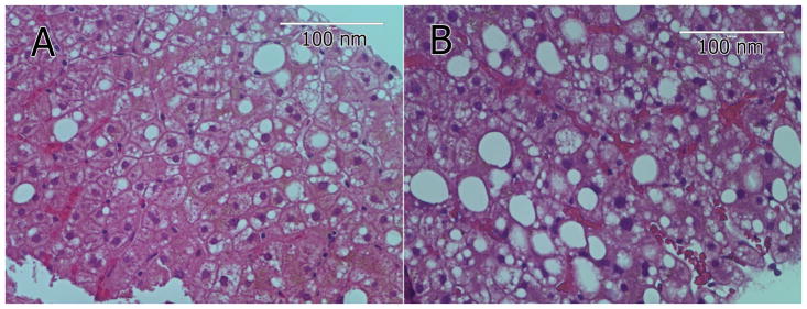
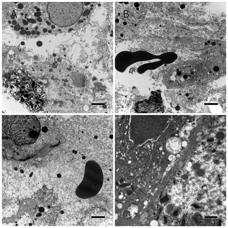
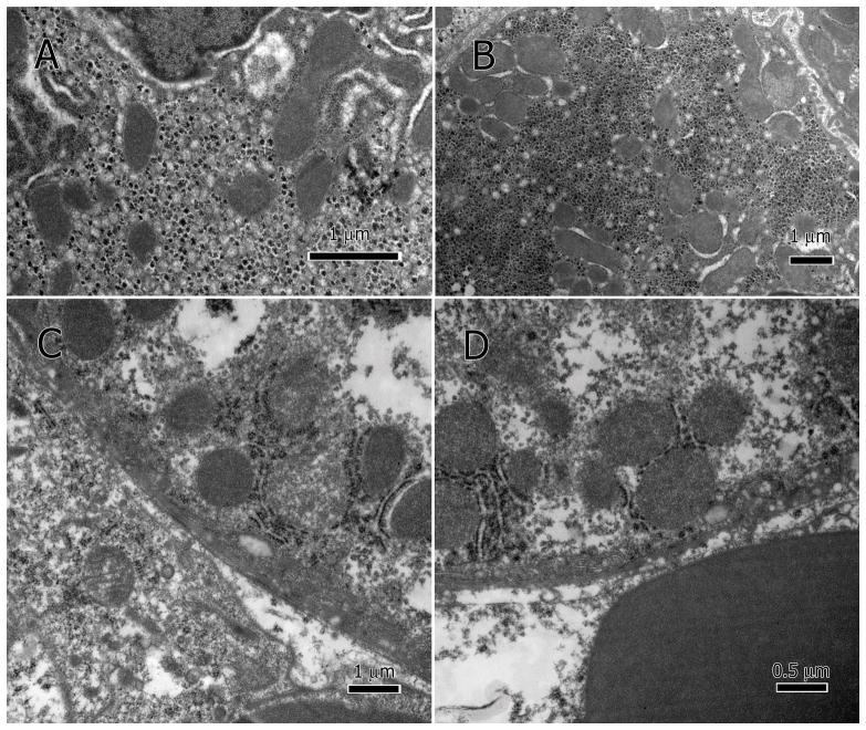
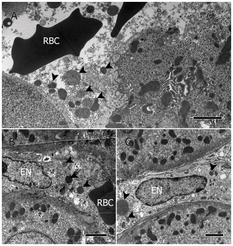
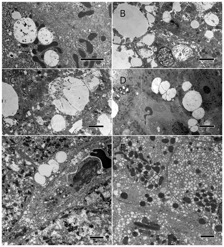
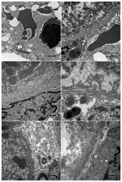
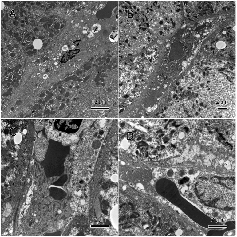
Human immunodeficiency virus and antiretroviral therapy (ART) together can be far more detrimental to liver cells than either of the two unaided. However, ultrastructural aspects of the synergistic effects of HIV and ART have been understudied. In a patient cohort receiving ART, this study characterizes ultrastructurally sinusoidal degeneration, hepatocytic aberrations, mitochondrial dysfunction, accumulation of bulky lipid droplets (steatosis), and occlusion of sinusoidal lumina. Mitochondrial dysfunction causes the accumulation of acetyl-CoA which leads to insulin upregulation and resistance, lipid synthesis, and steatosis. Lipid droplets deposited in the sinusoids could be the source of the blood's lipid profile alterations in HIV patients on ART.
Keywords: Antiretroviral therapy; HIV; endothelium; hepatotoxicity; liver; mitochondria; sinusoid; steatosis.
Figures
Similar articles
-
Nevirapine-induced liver lipid-SER inclusions and other ultrastructural aberrations.Ultrastruct Pathol. 2018 Mar-Apr;42(2):108-115. doi: 10.1080/01913123.2017.1422831. Epub 2018 Feb 9. Ultrastruct Pathol. 2018. PMID: 29424579 Free PMC article.
-
Ultrastructural hepatocytic alterations induced by silver nanoparticle toxicity.Ultrastruct Pathol. 2016;40(2):92-100. doi: 10.3109/01913123.2016.1150377. Epub 2016 Mar 2. Ultrastruct Pathol. 2016. PMID: 26934218
-
Liver as a target of human immunodeficiency virus infection.World J Gastroenterol. 2018 Nov 14;24(42):4728-4737. doi: 10.3748/wjg.v24.i42.4728. World J Gastroenterol. 2018. PMID: 30479460 Free PMC article. Review.
-
Modelling hepatotoxicity and antiretroviral therapeutic effect in HIV/HBV coinfection.Math Biosci. 2018 Aug;302:67-79. doi: 10.1016/j.mbs.2018.05.012. Epub 2018 May 22. Math Biosci. 2018. PMID: 29800563
-
Deciphering the Molecular Mechanisms of HAART-Induced Hepatotoxicity.J Biochem Mol Toxicol. 2025 Feb;39(2):e70174. doi: 10.1002/jbt.70174. J Biochem Mol Toxicol. 2025. PMID: 39959953 Review.
Cited by
-
HIV-1 Latency and Viral Reservoirs: Existing Reversal Approaches and Potential Technologies, Targets, and Pathways Involved in HIV Latency Studies.Cells. 2021 Feb 23;10(2):475. doi: 10.3390/cells10020475. Cells. 2021. PMID: 33672138 Free PMC article. Review.
-
Protective and Ameliorative Effects of Hydroethanolic Extract of Piper nigrum (L.) Stem against Antiretroviral Therapy-Induced Hepatotoxicity and Dyslipidemia in Wistar Rats.J Toxicol. 2024 Feb 7;2024:5811080. doi: 10.1155/2024/5811080. eCollection 2024. J Toxicol. 2024. PMID: 38357682 Free PMC article.
-
Evaluating Hepatotoxicity: A Comparative Analysis of New Generation versus Historical Antiretroviral Agents.Infect Dis Rep. 2024 Apr 24;16(3):423-434. doi: 10.3390/idr16030031. Infect Dis Rep. 2024. PMID: 38804441 Free PMC article.
-
Mitochondrial stress response in drug-induced liver injury.Mol Biol Rep. 2021 Oct;48(10):6949-6958. doi: 10.1007/s11033-021-06674-6. Epub 2021 Aug 25. Mol Biol Rep. 2021. PMID: 34432218 Review.
-
Nevirapine-induced liver lipid-SER inclusions and other ultrastructural aberrations.Ultrastruct Pathol. 2018 Mar-Apr;42(2):108-115. doi: 10.1080/01913123.2017.1422831. Epub 2018 Feb 9. Ultrastruct Pathol. 2018. PMID: 29424579 Free PMC article.
References
-
- Prevention CfDCa. Diagnoses of HIV Infection in the United States and Dependent Areas, 2014. Centers for Disease Control and Prevention; 2015. p. 26.
-
- Joshi D, O’Grady J, Dieterich D, Gazzard B, Agarwal K. Increasing burden of liver disease in patients with HIV infection. Lancet. 2011;377(9772):1198–1209. - PubMed
MeSH terms
Substances
Grants and funding
LinkOut - more resources
Full Text Sources
Other Literature Sources
Medical