. 2017 Mar 9;8(3):e2663.
doi: 10.1038/cddis.2017.75.

miR-377 induces senescence in human skin fibroblasts by targeting DNA methyltransferase 1

Affiliations

miR-377 induces senescence in human skin fibroblasts by targeting DNA methyltransferase 1

Hong-Fu Xie et al. Cell Death Dis. .

Erratum in

Abstract

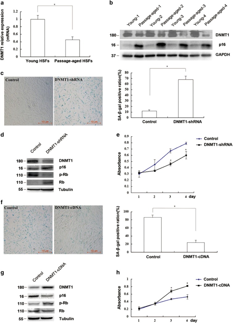
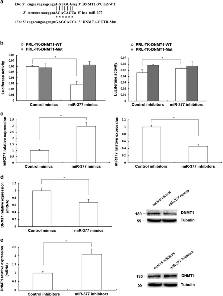
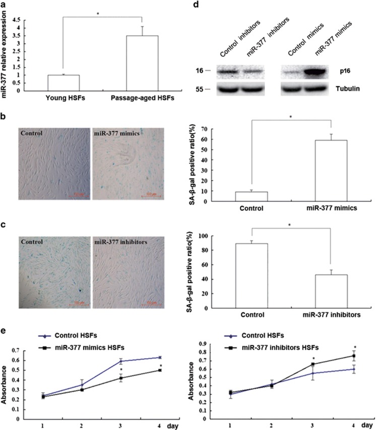
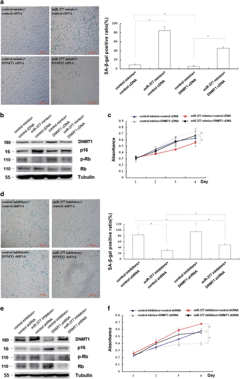
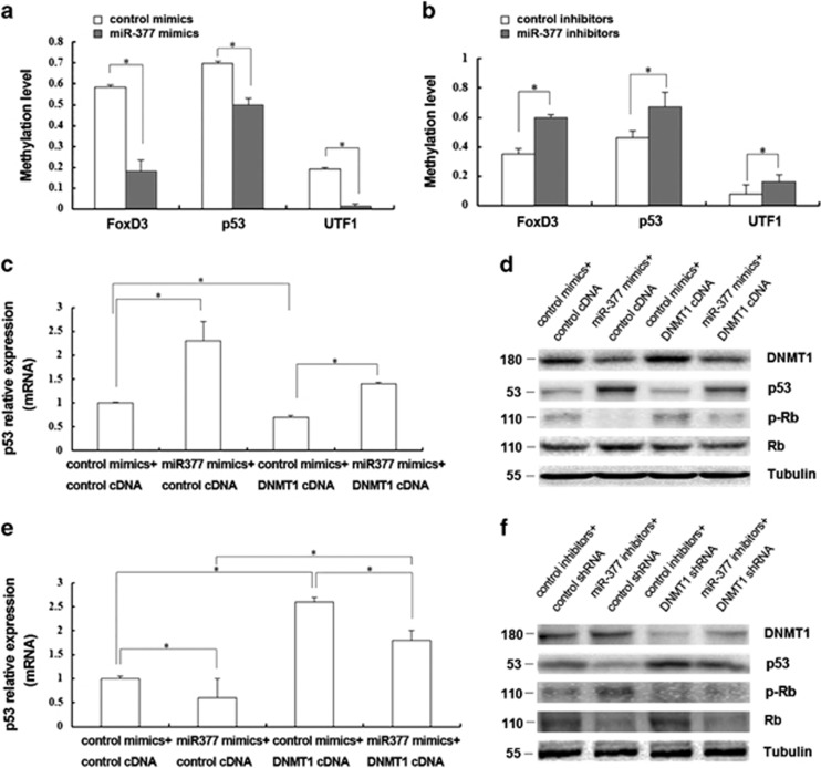
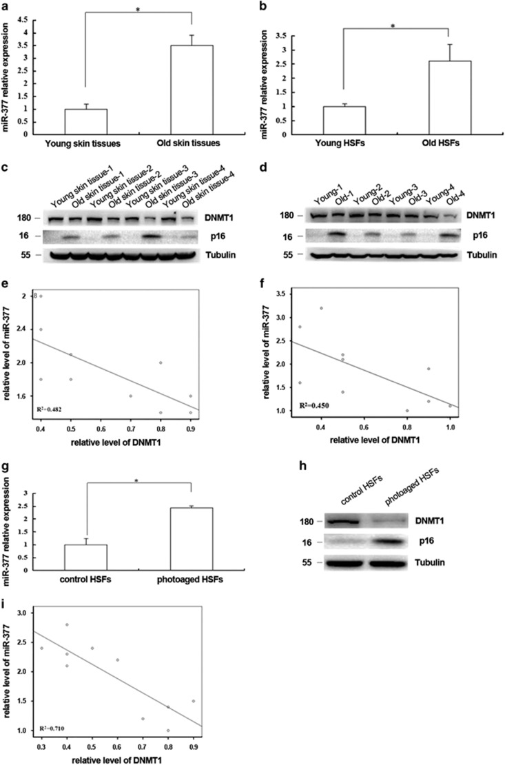
Skin aging is a complicated physiological process and epigenetic feature, including microRNA-mediated regulation and DNA methylation, have been shown to contribute to this process. DNA methylation is regulated by DNA methyltransferase, of which DNA methyltransferase 1 (DNMT1) is the most abundantly known. But evidence supporting its role in skin aging remains scarce, and no report regards its specifical upstream-regulating molecules in the process of skin aging so far. Here, we found that DNMT1 expression was markedly higher in young human skin fibroblasts (HSFs) than that in passage-aged HSFs, and DNMT1 knockdown significantly induced the senescence phenotype in young HSFs. We predicted the upstream miRNAs which could regulate DNMT1 with miRNA databases and found miR-377 had high homology with a sequence in the 3'-UTR of human DNMT1 mRNA. We confirmed that miR-377 was a potential regulator of DNMT1 by luciferase reporter assays. miR-377 expression in passage-aged HSFs was markedly higher than that in the young HSFs. miR-377 overexpression promoted senescence in young HSFs, and inhibition of miR-377 reduced senescence in passage-aged HSFs. Moreover, these functions were mediated by targeting DNMT1. Microfluidic PCR and next-generation bisulfite sequencing of 24 senescent-associated genes' promoters revealed alterations of the promoter methylation levels of FoxD3, p53, and UTF1 in HSFs treated with miR-377 mimics or inhibitors. We also verified that the miR-377-mediated changes in p53 expression could be reversed by regulation of DNMT1 in HSFs. Similarly, there was a negative correlation between miR-377 and DNMT1 expression in young and photoaged HSFs, HSFs, or skin tissues from UV-unexposed areas of different aged donors. Our results highlight a novel role for miR-377-DNMT1-p53 axis in HSF senescence. These findings shed new light on the mechanisms of skin aging and identify future opportunities for its therapeutic prevention.


Conflict of interest statement

The authors declare no conflict of interest.

Figures

Figure 1
Figure 1
The expression and role of DNMT1 in HSFs senescence. (a) DNMT1 mRNA in passage-aged (PD>50) and young (PD<10) HSFs was detected by RT-qPCR (Data represented as the mean±S.E.M. n=10, *P<0.05). Representative data was shown. (b) DNMT1 and p16 protein levels in passage-aged (PD>50) and young (PD<10) HSFs were detected by western blot (n=10, *P<0.05). Representative data was shown. (c) SA-β-gal-positive cells in young HSFs (PD<10) transfected with control shRNA or DNMT1-shRNA were detected by using kit (left). The SA-β-gal-positive ratio was shown (right; Data represented as the mean±S.E.M. n=3, *P<0.05). (d) DNMT1, p16, and Rb protein expressions and phosphorylation of Rb in young HSFs (PD<10) transfected with control shRNA or DNMT1-shRNA were detected by western blot (*P<0.05). Representative data was shown. (e) Absorbance at 490 nm in young HSFs (PD<10) transfected with control shRNA or DNMT1-shRNA was detected by MTS assays (Data represented as the mean±S.E.M. n=3 at each time point, *P<0.05). (f) SA-β-gal-positive cells in the passage-aged HSFs (PD>50) transfected with control cDNA or DNMT1 cDNA were detected by using kit (left). The SA-β-gal-positive ratio was shown (right; data represented as the mean±S.E.M. n=3, *P<0.05). (g) DNMT1, p16, and Rb protein expressions and phosphorylation of Rb were detected by western blot in the passage-aged HSFs (PD>50) after being transfected with control cDNA or DNMT1 cDNA (*P<0.05). Representative data was shown. (h) Absorbance at 490 nm was detected in the passage-aged HSFs (PD>50) after being transfected with control cDNA or DNMT1 cDNA by MTS assays. (Data represented as the mean±S.E.M. n=3 at each time point, *P<0.05)
Figure 2
Figure 2
miR-377 could regulate DNMT1 expression by directly targeting DNMT1 in HSFs. (a) Though bioinformatics prediction, the sequence of the miR-377 binding site in the 3′-UTR of DNMT1 was shown at the upper site. Mutated residues were shown at the lower site. (b) Luciferase activity change of the wild-type 3′-UTR reporters and the mutant 3′-UTR reporters in 293T cells treated with control mimics or miR-377 mimics (left) and 293T cells treated with control inhibitors or miR-377 inhibitors (right) was shown, respectively (Data represented as the mean±S.E.M. n=3, *P<0.05, respectively). (c) miR-377 level in young HSFs (PD<10) treated with control mimics or miR-377 mimics (left) and in passage-aged HSFs (PD>50) treated with control inhibitors or miR-377 inhibitors (right) was respectively detected by RT-qPCR (Data represented as the mean±S.E.M. n=3, *P<0.05, respectively). (d) DNMT1 mRNA and protein expression in the young HSFs (PD<10) treated with control mimics or miR-377 mimics was detected by RT-qPCR and western blot, respectively (Data represent the mean±S.E.M. n=3, *P<0.05). (e) DNMT1 mRNA and protein expression in the passage-aged HSFs (PD>50) treated with control inhibitors or miR-377 inhibitors was detected by RT-qPCR and western blot, respectively (Data represent the mean±S.E.M. n=3, *P<0.05)
Figure 3
Figure 3
miR-377 mediated senescence in HSFs. (a) miR-377 level in the young (PD<10) and passage-aged (PD>50) HSFs was detected by RT-qPCR (Data represented as the mean±S.E.M. n=10, *P<0.05). (b) SA-β-gal-positive cells in young HSFs (PD<10) treated with control mimics or miR-377 mimics were detected by using kit (left). The SA-β-gal-positive ratio was shown (right; data represented as the mean±S.E.M. n=3, *P<0.05). (c) SA-β-gal-positive cells in passage-aged HSFs (PD>50) treated with control inhibitors or miR-377 inhibitors were detected by using kit (left). The SA-β-gal-positive ratio was shown (right; data represented as the mean±S.E.M. n=3, *P<0.05). (d) p16 protein expression was respectively detected by western blot in passage-aged HSFs (PD>50) treated with control inhibitors or miR-377 inhibitors (left) and in young HSFs treated with control mimics or miR-377 mimics (right; n=3, *P<0.05). Representative data was shown. (e) Absorbance at 490 nm was respectively detected by MTS assays in young HSFs (PD<10) treated with control mimics or miR-377 mimics (left) and in passage-aged HSFs treated with control inhibitors or miR-377 inhibitors (right; data represented as the mean±S.E.M. n=3 at each time point, *P<0.05)
Figure 4
Figure 4
miR-377 promoted senescence by suppressing DNMT1 expression in HSFs. (a) Cellular senescence was detected by evaluating SA-β-gal-positive cells in young HSFs (PD<10) treated with miR-377 mimics together with control cDNA or DNMT1 cDNA as indicated (left). The positive cell quantification was shown (right; data represent the mean±S.E.M. n=3, *P<0.05). (b) DNMT1, p16, and Rb expressions and phosphorylation level of Rb were detected by western blot in young HSFs (PD<10) treated with miR-377 mimics together with control cDNA or DNMT1 cDNA as indicated (*P<0.05). Representative data was shown. (c) Absorbance at 490 nm was detected by MTS assays in young HSFs (PD<10) treated with miR-377 mimics together with control cDNA or DNMT1 cDNA as indicated (data represented as the mean±S.E.M. n=3 at each time point, *P<0.05). (d) Cellular senescence was detected by evaluating SA-β-gal-positive cells in passage-aged HSFs (PD>50) treated with miR-377 inhibitors together with control shRNA or DNMT1 shRNA as indicated (left). The SA-β-gal-positive ratio was shown (right; data represented as the mean±S.E.M. n=3, *P<0.05). (e) DNMT1, p16, and Rb expressions and phosphorylation level of Rb were detected by western blot in passage-aged HSFs (PD>50) treated with miR-377 inhibitors together with control cDNA or DNMT1 cDNA as indicated (*P<0.05). Representative data was shown. (f) Absorbance at 490 nm was detected by MTS assays in passage-aged HSFs (PD>50) treated with miR-377 inhibitors together with control cDNA or DNMT1 cDNA as indicated (data represented as the mean±S.E.M. n=3 at each time point, *P<0.05)
Figure 5
Figure 5
Role of miR-377 in modulating promoter methylation levels of senescent-associated genes in HSFs. (a) The promoter methylation levels of FoxD3, p53 and UTF1 were analyzed in young HSFs (PD<10) transfected with control mimics or miR-377 mimics through microfluidic PCR and next-generation bisulfite sequencing (data represented as the mean±S.E.M. *P<0.05). (b) The promoter methylation levels of FoxD3, p53 and UTF1 were analyzed in passage-aged HSFs (PD>50) transfected with control inhibitors or miR-377 inhibitors through microfluidic PCR and next-generation bisulfite sequencing (data represented as the mean±S.E.M. *P<0.05). (c) p53 mRNA was detected by RT-qPCR in young HSFs (PD<10) treated with miR-377 mimics together with control cDNA or DNMT1 cDNA as indicated (data represented as the mean±S.E.M. n=3, *P<0.05). (d) DNMT1, p53, and Rb expressions and phosphorylation of Rb were detected by western blot in young HSFs (PD<10) treated with miR-377 mimics together with control cDNA or DNMT1 cDNA as indicated (*P<0.05). Representative data was shown. (e) p53 mRNA was detected by RT-qPCR in passage-aged HSFs (PD>50) treated with miR-377 inhibitors together with control cDNA or DNMT1 cDNA as indicated (data represented as the mean±S.E.M. n=3, *P<0.05). (f) DNMT1, p53, and Rb expressions and phosphorylation of Rb were detected by western blot in passage-aged HSFs (PD>50) treated with miR-377 inhibitors together with control shRNA or DNMT1 shRNA as indicated (*P<0.05). Representative data was shown
Figure 6
Figure 6
miR-377 and DNMT1 expression in vivo and photoaged HSFs. (a) miR-377 level was detected by RT-qPCR in the young and the old skin tissues. (Data represented as the mean±S.E.M. n=10, *P<0.05). (b) miR-377 level was detected by RT-qPCR in HSFs from the young and the old skin tissues (Data represented as the mean±S.E.M. n=10, *P<0.05). (c) DNMT1 and p16 expressions were detected by western blot in the young and the old skin tissues (n=10, *P<0.05). Representative data was shown. (d) DNMT1 and p16 expressions were detected by western blot in HSFs from the young and the old skin tissues (n=10, *P<0.05). Representative data was shown. (e and f) The correlations between miR-377 and DNMT1 levels in different couples of young and old skin tissues and in HSFs from young and old skin tissues were shown respectively (n=10, R2=0.482,0.450, respectively, *P<0.05). (g) miR-377 level was detected in control and photoaged HSFs by RT-qPCR (Data represented as the mean±S.E.M. n=3, *P<0.05). (h) DNMT1 and p16 expression levels were detected by western blot in control and photoaged HSFs (*P<0.05). Representative data was shown. (i) The correlation between miR-377 and DNMT1 levels was analyzed in several couples of UVA-untreated and UVA-treated HSFs (n=10, R2=0.471, *P<0.05)

Similar articles

Cited by

References

    1. Farage MA, Miller KW, Elsner P, Maibach HI. Characteristics of the aging Skin. Adv Wound Care 2013; 2: 5–10. - PMC - PubMed
    1. Kammeyer A Luiten. Oxidation events and skin aging. Ageing Res Rev 2015; 21: 16–29. - PubMed
    1. Thangapazham RL, Darling TN, Meyerle J. Alteration of skin properties with autologous dermal fibroblasts. Int J Mol Sci 2014; 15: 8407–8427. - PMC - PubMed
    1. Rinnerthaler M, Bischof J, Streubel MK, Trost A, Richter K. Oxidative stress in aging human skin. Biomolecules 2015; 5: 545–589. - PMC - PubMed
    1. Menon GK, Dal Farra C, Botto JM, Domloge N. Mitochondria: a new focus as an anti-aging target in skin care. J Cosmet Dermatol 2010; 9: 122–131. - PubMed

Publication types

MeSH terms