. 2017 Mar 8;9(3):778-789.
doi: 10.18632/aging.101191.

Microvesicles from the plasma of elderly subjects and from senescent endothelial cells promote vascular calcification

Affiliations

Microvesicles from the plasma of elderly subjects and from senescent endothelial cells promote vascular calcification

Matilde Alique et al. Aging (Albany NY). .

Abstract

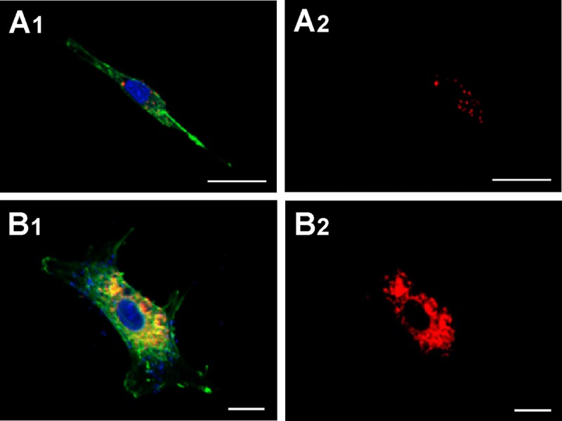
Vascular calcification is commonly seen in elderly people, though it can also appear in middle-aged subjects affected by premature vascular aging. The aim of this work is to test the involvement of microvesicles (MVs) produced by senescent endothelial cells (EC) and from plasma of elderly people in vascular calcification. The present work shows that MVs produced by senescent cultured ECs, plus those found in the plasma of elderly subjects, promote calcification in vascular smooth muscle cells. Only MVs from senescent ECs, and from elderly subjects' plasma, induced calcification. This ability correlated with these types of MVs' carriage of: a) increased quantities of annexins (which might act as nucleation sites for calcification), b) increased quantities of bone-morphogenic protein, and c) larger Ca contents. The MVs of senescent, cultured ECs, and those present in the plasma of elderly subjects, promote vascular calcification. The present results provide mechanistic insights into the observed increase in vascular calcification-related diseases in the elderly, and in younger patients with premature vascular aging, paving the way towards novel therapeutic strategies.

Keywords: aging; endothelial cells; microvesicles; senescence; vascular calcification; vascular smooth muscle cells.


Conflict of interest statement

CONFLICTS OF INTEREST

The authors declare no conflict of interests.

Figures

Figure 1
Figure 1. Plasma MVs assessed by flow cytometry
(A) Representative dot plot showing log forward scatter (FSC) vs. log side scatter (SSC) localization of MVs in a young subject. The upper right gate shows the bead flow count, used as an index to count MVs in absolute terms. The lower left gate shows MVs smaller than 1 μm. (B) Representative dot plot showing localization of MVs in an elderly subject.
Figure 2
Figure 2. HUVEC-derived MVs assessed by flow cytometry
Representative dot plots showing log forward scatter (FSC) vs. log side scatter (SSC) from young (A1) and senescent (B1) HUVEC. Representative dot plots showing annexin A5 and CD31 phenotype from young (A2) and senescent (B2) HUVEC. (C) Absolute number of MVs per μL of sample from young and senescent HUVEC; *p<0.05. (D) Absolute number of CD31+/annexin A5+ MVs per μL of sample; *p<0.05. Results are presented as the mean ± SD of 6 independent experiments.
Figure 3
Figure 3. MVs from senescent HUVEC cells induced calcification in HASMC
Cells were cultured with 50,000 MVs/mL from young (Y MV) or senescent (S MV) HUVEC for different times. (A) Qualitative calcification was stained with alizarin red. A representative experiment from six different experiments per duplicate is show. (B) Calcium content was determined by spectrophotometer by phenolsulphonephthalein dye at 6 and 9 days of treatment. The graphs present calcium content of the cells expressed as μg/mg protein. The data represent means ± SD of 3 independent experiments. *** p<0.001 vs control and Y MV at the same time.
Figure 4
Figure 4. Capture of MVs by HASMC
Cells were incubated for 48 h with MVs from young (panels A1 and A2) or senescent HUVEC (panels B1 and B2) stained with CellTracker CM-Dil PI (red) and then were stained with phalloidin (PKH67, green). Nuclei were stained with DAPI (blue). Fluorescence was evaluated by confocal microscopy. (A) Scale bar 25μm, and (B) scale bar 10 μm. A representative experiment from three different experiments is shown.
Figure 5
Figure 5. Scanning electron microscopy
(A) Gold-palladium coated senescent MVs. Scale bar 1μm. Note the poly-L-lysine layer. (B and C) Young MVs prepared without metal coating. Note the heterogeneous size and brightness. Scale bar 5μm. The background noise of some pictures a consequence of the fast scanning speed.
Figure 6
Figure 6
Spot mode microanalysis of an MV (A1) with its corresponding plot (A2); arrows indicate Ca spikes. Scale bar 5μm. Larger MVs are usually brighter and have a higher Ca content. Ca mapping of MVs from (B1) young and (B2) senescent HUVEC. Ca is seen as grey on the black background.
Figure 7
Figure 7
TEM analysis of MVs from (A) young and (B) senescent HUVEC.
Figure 8
Figure 8
(A) Western blot showing that BMP2, annexin A6 and annexin A2 carriage to be higher in MVs from senescent than from young HUVEC. Equal protein loading was confirmed using a HSP90 antibody. Total protein expression was obtained via densitometric analysis and expressed as the ratio protein/HSP90 in arbitrary units. n=3 pools; *p<0.05; **p<0.01. (B) Representative BMP2, annexin A6 and annexin A2 western blot of young and senescent HUVEC pools. Equal protein loading was confirmed probing with GAPDH or β-Actin. The graphs present densitometric band analysis normalized to GAPDH or β-Actin in arbitrary units. n=3 pools.

References

    1. Wayhs R, Zelinger A, Raggi P. High coronary artery calcium scores pose an extremely elevated risk for hard events. J Am Coll Cardiol. 2002;39:225–30. doi: 10.1016/S0735-1097(01)01737-5. - DOI - PubMed
    1. Shanahan CM. Mechanisms of vascular calcification in CKD-evidence for premature ageing? Nat Rev Nephrol. 2013;9:661–70. doi: 10.1038/nrneph.2013.176. - DOI - PubMed
    1. Leopold JA. Vascular calcification: mechanisms of vascular smooth muscle cell calcification. Trends Cardiovasc Med. 2015;25:267–74. doi: 10.1016/j.tcm.2014.10.021. - DOI - PMC - PubMed
    1. Li X, Yang HY, Giachelli CM. BMP-2 promotes phosphate uptake, phenotypic modulation, and calcification of human vascular smooth muscle cells. Atherosclerosis. 2008;199:271–77. doi: 10.1016/j.atherosclerosis.2007.11.031. - DOI - PMC - PubMed
    1. Yáñez-Mó M, Siljander PR, Andreu Z, Zavec AB, Borràs FE, Buzas EI, Buzas K, Casal E, Cappello F, Carvalho J, Colás E, Cordeiro-da Silva A, Fais S, et al. Biological properties of extracellular vesicles and their physiological functions. J Extracell Vesicles. 2015;4:27066. doi: 10.3402/jev.v4.27066. - DOI - PMC - PubMed

Publication types