. 2017 Mar 9;12(3):e0172311.
doi: 10.1371/journal.pone.0172311. eCollection 2017.

LipidFrag: Improving reliability of in silico fragmentation of lipids and application to the Caenorhabditis elegans lipidome

Affiliations

LipidFrag: Improving reliability of in silico fragmentation of lipids and application to the Caenorhabditis elegans lipidome

Michael Witting et al. PLoS One. .

Abstract

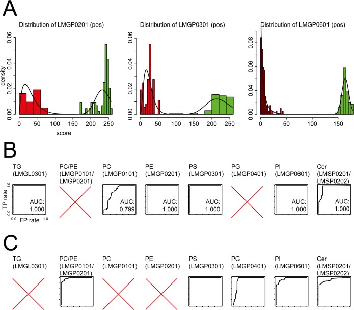
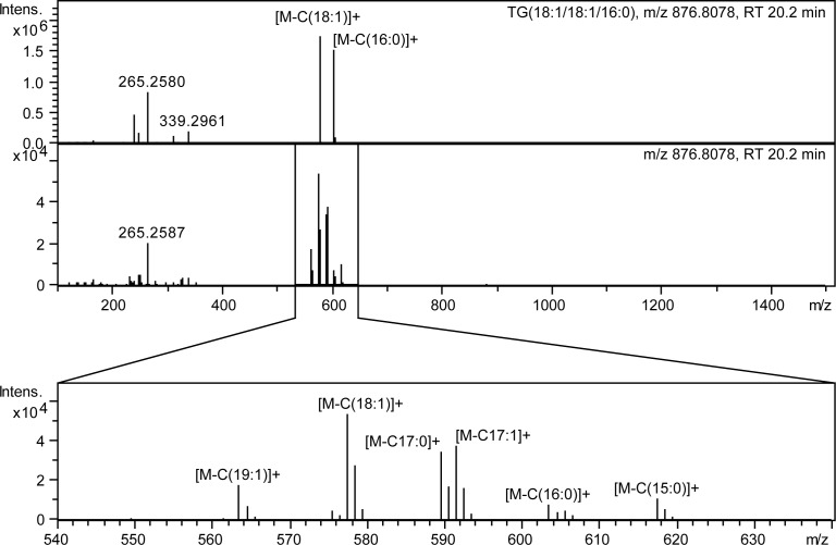
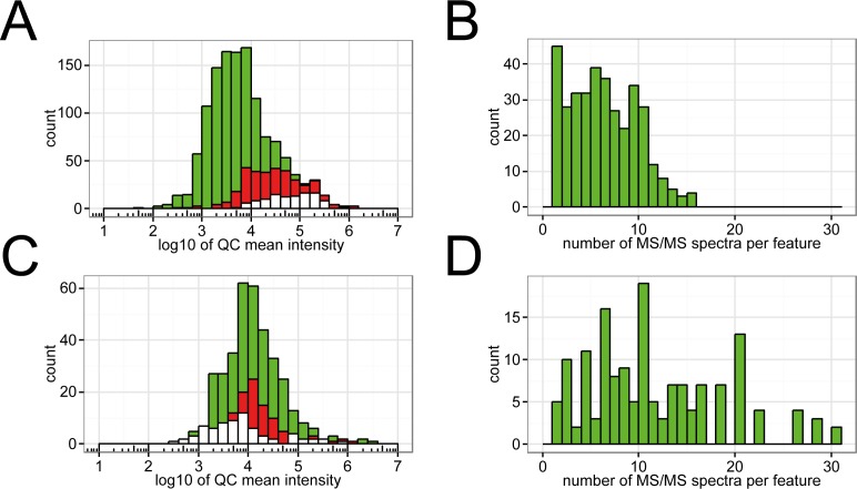
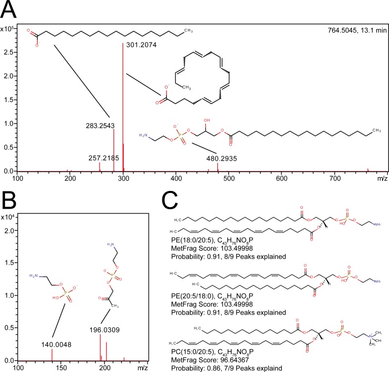
Lipid identification is a major bottleneck in high-throughput lipidomics studies. However, tools for the analysis of lipid tandem MS spectra are rather limited. While the comparison against spectra in reference libraries is one of the preferred methods, these libraries are far from being complete. In order to improve identification rates, the in silico fragmentation tool MetFrag was combined with Lipid Maps and lipid-class specific classifiers which calculate probabilities for lipid class assignments. The resulting LipidFrag workflow was trained and evaluated on different commercially available lipid standard materials, measured with data dependent UPLC-Q-ToF-MS/MS acquisition. The automatic analysis was compared against manual MS/MS spectra interpretation. With the lipid class specific models, identification of the true positives was improved especially for cases where candidate lipids from different lipid classes had similar MetFrag scores by removing up to 56% of false positive results. This LipidFrag approach was then applied to MS/MS spectra of lipid extracts of the nematode Caenorhabditis elegans. Fragments explained by LipidFrag match known fragmentation pathways, e.g., neutral losses of lipid headgroups and fatty acid side chain fragments. Based on prediction models trained on standard lipid materials, high probabilities for correct annotations were achieved, which makes LipidFrag a good choice for automated lipid data analysis and reliability testing of lipid identifications.


Conflict of interest statement

Competing Interests: The authors have declared that no competing interests exist.

Figures

Fig 1
Fig 1. LipidFrag workflow and related lipid sub-classes.
(A) Schematic drawing of LipidFrag workflow. MS/MS spectra from known lipids derived from lipid standard materials and from unknown lipids are subjected to MetFrag in silico fragmentation, whereby all possible precursor structures are taken into consideration. During training phase true positive identity and decoy candidates are used to calculate a 2-class classifier by which reliable results from unknown lipids can be identified. (B) Structures of detected phospholipid classes, phosphatidylethanolamines (PE, LMGP0201), phosphatidylcholines (PC, LMGP0101), phosphatidylglycerols (PG, LMGP0401), phosphatidylserines (PS, LMGP0301) and phosphatidylinositols (PI, LMGP0601) (C) Structure of triacylglycerols (TG, LMGL0301) (D) Structure of ceramides (Cer, LMSP0201) and dihydroceramides (Cer, LMSP0202).
Fig 2
Fig 2. Visualization of input data and results obtained by LipidFrag.
(A) Examples of histograms showing distribution of raw MetFrag score for the back- and foreground training dataset. (B) Receiver-Operator characteristics (ROC) derived from 10-fold cross-validation of MS/MS spectra from lipid standard materials detected negative ion mode. (C) Receiver-Operator characteristics (ROC) derived from 10-fold cross-validation of MS/MS spectra from lipid standard materials detected positive ion mode. In both panels, plots having no AUC value indicate that this lipid class was not detected in this ion mode concluding that there was not training data for classifiers available. All axes have the same scale.
Fig 3
Fig 3. Example of co-elution and overlapping of different TG species in C. elegans.
Analysis of spectra derived from TGs is complicated in real samples due to overlap of several isomeric and isobaric species. The upper panel shows the MS/MS spectrum of TG(18:1/18:1/16:0) standard and the lower of the same chromatographic peak in a C. elegans lipid extract.
Fig 4
Fig 4. Histogram of intensities of features detected in positive ion mode.
(A) The green histogram represents all features, in red are features with one or more associated MS/MS spectra and the white features having a FCP > 0.95 in LipidFrag. (B) Histogram of MS/MS spectra per feature in positive ion mode across all 5 technical replicates. (C) and (D) show the same for negative ion mode.
Fig 5
Fig 5. Example of a LipidFrag identification in C. elegans data.
(A) MS/MS spectrum of m/z 764.5045 at 13.1 minutes detected in C. elegans with fragment structures annotated. (B) Close up of lower mass region (m/z 100–250). (C) Structures of the best three candidates obtained from MetFrag with result filtering using foreground class probabilities. Name, formula, MetFrag score and probability are indicated below each structure.

References

    1. van Meer G. Cellular lipidomics. EMBO J. 2005;24(18):3159–65. 10.1038/sj.emboj.7600798 - DOI - PMC - PubMed
    1. Caffrey M, Hogan J. LIPIDAT: A database of lipid phase transition temperatures and enthalpy changes. DMPC data subset analysis. Chemistry and Physics of Lipids. 1992;61(1):1–109. - PubMed
    1. Watanabe K, Yasugi E, Oshima M. How to Search the Glycolipid data in “LIPIDBANK for Web”, the Newly Developed Lipid Database in Japan. Trends in Glycoscience and Glycotechnology. 2000;12(65):175–84.
    1. Sud M, Fahy E, Cotter D, Brown A, Dennis EA, Glass CK, et al. LMSD: LIPID MAPS structure database. Nucleic Acids Research. 2007;35(suppl 1):D527–D32. - PMC - PubMed
    1. Foster JM, Moreno P, Fabregat A, Hermjakob H, Steinbeck C, Apweiler R, et al. LipidHome: A Database of Theoretical Lipids Optimized for High Throughput Mass Spectrometry Lipidomics. PLoS ONE. 2013;8(5):e61951 10.1371/journal.pone.0061951 - DOI - PMC - PubMed

MeSH terms

LinkOut - more resources