. 2017 Mar 9;8(1):61.
doi: 10.1186/s13287-017-0506-5.

Pulp regeneration by transplantation of dental pulp stem cells in pulpitis: a pilot clinical study

Affiliations

Pulp regeneration by transplantation of dental pulp stem cells in pulpitis: a pilot clinical study

Misako Nakashima et al. Stem Cell Res Ther. .

Abstract

Background: Experiments have previously demonstrated the therapeutic potential of mobilized dental pulp stem cells (MDPSCs) for complete pulp regeneration. The aim of the present pilot clinical study is to assess the safety, potential efficacy, and feasibility of autologous transplantation of MDPSCs in pulpectomized teeth.

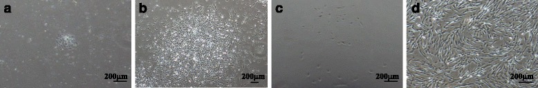
Methods: Five patients with irreversible pulpitis were enrolled and monitored for up to 24 weeks following MDPSC transplantation. The MDPSCs were isolated from discarded teeth and expanded based on good manufacturing practice (GMP). The quality of the MDPSCs at passages 9 or 10 was ascertained by karyotype analyses. The MDPSCs were transplanted with granulocyte colony-stimulating factor (G-CSF) in atelocollagen into pulpectomized teeth.

Results: The clinical and laboratory evaluations demonstrated no adverse events or toxicity. The electric pulp test (EPT) of the pulp at 4 weeks demonstrated a robust positive response. The signal intensity of magnetic resonance imaging (MRI) of the regenerated tissue in the root canal after 24 weeks was similar to that of normal dental pulp in the untreated control. Finally, cone beam computed tomography demonstrated functional dentin formation in three of the five patients.

Conclusions: Human MDPSCs are safe and efficacious for complete pulp regeneration in humans in this pilot clinical study.

Keywords: Autologous cell transplantation; Clinical study; Good manufacturing practice (GMP); Granulocyte colony-stimulating factor (G-CSF); Mobilized dental pulp stem cells (Mobilized DPSCs); Pulp regeneration; Pulpectomy.


Figures

Fig. 1
Fig. 1
A sequence of illustrations describing step-by-step the sequences of the clinical study, including caries treatment with composite resin wall restoration followed by pulpectomy, cell processing, and cell transplantation, followed by final restoration. CBCT cone beam computed tomography, CPC Cell Processing Center, GMP good manufacturing practice, MDPSC mobilized dental pulp stem cell, MRI magnetic resonance imaging
Fig. 2
Fig. 2
Isolation of MDPSCs from an autologous discarded tooth. a Primary DPSCs forming a small colony on day 3. b The DPSCs on day 7. The colony increased in size. c MDPSCs at passage 2 of culture on day 3. d MDPSCs at passage 7 of culture on day 5 before cryopreservation
Fig. 3
Fig. 3
Radiological analyses. a X-ray photographic analysis to show the changes and evolution of periapical tissues and apical and/or lateral dentin formation in the root canal at the first visit (FV), pre-transplantation just before cell transplantation (Pre), and 4, 12, and 24/28 weeks (wks) after autologous transplantation of MDPSCs with G-CSF in pulpectomized teeth in five patients. No significant changes were seen in the periapical areas, except in patient 4 who showed widening of periodontal ligament space at 12 weeks and periapical radiolucency at 24 and 32 weeks. Patient 2 preoperatively had periapical radiolucent lesion decreased in area size with a little radiographic periodontal ligament space widening at 24 weeks. b Cone beam computed tomography evaluation of apical/lateral dentin formation in the root canal in coronal and axial slices in three patients at 16 and 28/32 weeks. Arrows indicate newly formed dentin. The 3D cone beam computed tomography images by the OsiriX program demonstrated a decrease in low-density areas at 28 weeks compared with 16 weeks
Fig. 4
Fig. 4
The changes of fat-suppressed T2-weighted (FST2W) MRI in cell-transplanted root canal in five patients. Squares indicate the teeth with cell transplantation. ac Twelve weeks and df 24 weeks after transplantation of MDPSCs and G-CSF with collagen. a,d Sagittal slice; b,e axial slice at the coronal part of the root canal; c,f axial slice at the apical part of the root canal. g The relative SI of MRI. There are significant differences in the relative SI between pulpectomized root canal and cell transplanted root canal at 12 and 24 weeks both in the coronal part and apical part (**P < 0.01); data are expressed as mean ± SD (n = 3). There is a significant difference in the relative SI of root canal between 12 weeks and 24 weeks in the coronal part (*P <0.05); data are expressed as mean ± SD (n = 4)

References

    1. Nakashima M, Akamine A. The application of tissue engineering to regeneration of pulp and dentin in endodontics. J Endod. 2005;31:711–8. doi: 10.1097/01.don.0000164138.49923.e5. - DOI - PubMed
    1. Nixdorf DR, Moana-Filho EJ, Law AS, McGuire LA, Hodges JS, John MT. Frequency of nonodontogenic pain after endodontic therapy: a systematic review and meta-analysis. J Endod. 2010;36:1494–98. doi: 10.1016/j.joen.2010.06.020. - DOI - PMC - PubMed
    1. Gale MS. Coronal microleakage. Ann R Australas Coll Dent Surg. 2000;15:299–305. - PubMed
    1. Sritharan A. Discuss that the coronal seal is more important than the apical seal for endodontic success. Aust Endod J. 2002;28:112–5. doi: 10.1111/j.1747-4477.2002.tb00404.x. - DOI - PubMed
    1. Moule AJ, Kahler B. Diagnosis and management of teeth with vertical root fractures. Aust Dent J. 1999;44:75–87. doi: 10.1111/j.1834-7819.1999.tb00205.x. - DOI - PubMed

Publication types

MeSH terms

Substances