. 2017 Mar 8;21(3):356-366.
doi: 10.1016/j.chom.2017.02.008.

Betacoronavirus Adaptation to Humans Involved Progressive Loss of Hemagglutinin-Esterase Lectin Activity

Affiliations

Betacoronavirus Adaptation to Humans Involved Progressive Loss of Hemagglutinin-Esterase Lectin Activity

Mark J G Bakkers et al. Cell Host Microbe. .

Abstract

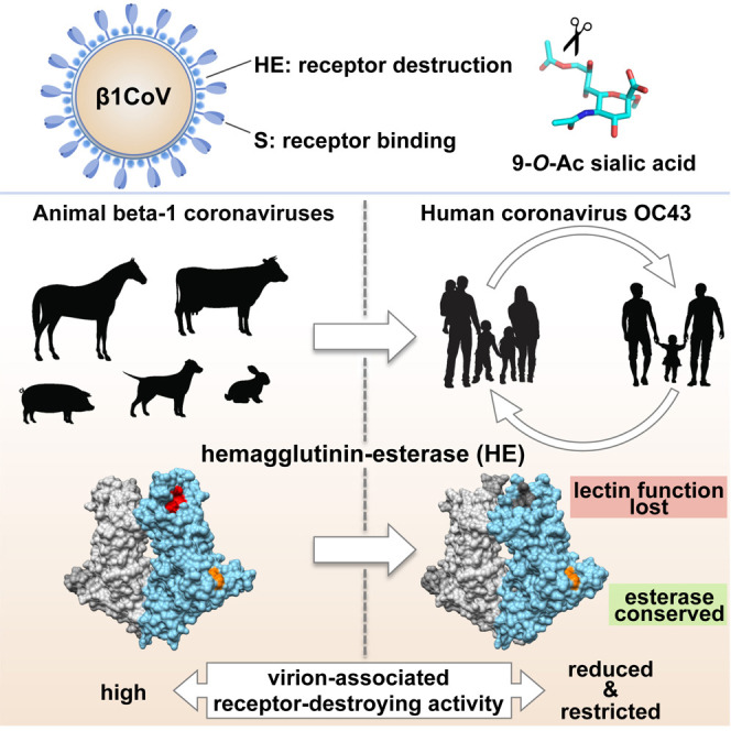
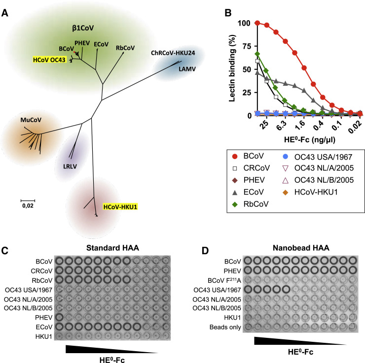
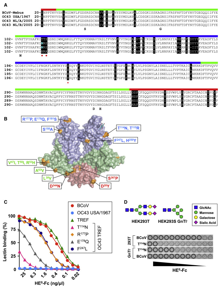
Human beta1-coronavirus (β1CoV) OC43 emerged relatively recently through a single zoonotic introduction. Like related animal β1CoVs, OC43 uses 9-O-acetylated sialic acid as receptor determinant. β1CoV receptor binding is typically controlled by attachment/fusion spike protein S and receptor-binding/receptor-destroying hemagglutinin-esterase protein HE. We show that following OC43's introduction into humans, HE-mediated receptor binding was selected against and ultimately lost through progressive accumulation of mutations in the HE lectin domain. Consequently, virion-associated receptor-destroying activity toward multivalent glycoconjugates was reduced and altered such that some clustered receptor populations are no longer cleaved. Loss of HE lectin function was also observed for another respiratory human coronavirus, HKU1. This thus appears to be an adaptation to the sialoglycome of the human respiratory tract and for replication in human airways. The findings suggest that the dynamics of virion-glycan interactions contribute to host tropism. Our observations are relevant also to other human respiratory viruses of zoonotic origin, particularly influenza A virus.


Figures

None
Graphical abstract
Figure 1
Figure 1
Loss of HE-Mediated Receptor Binding in Human Betacoronaviruses (A) Evolutionary relationships among lineage A betacoronaviruses. Neighbor-joining tree based on βCoV lineage A ORF1b sequences in the NCBI database (n = 206), with 100% bootstrap support for all major branches. Evolutionary distances were computed using the Maximum Composite Likelihood method in MEGA6 (Tamura et al., 2013). The positions of human coronaviruses OC43 and HKU1 are highlighted relative to those of animal lineage A betacoronaviruses, including the various β1CoV subspecies and classical mouse hepatitis virus-type murine coronavirus (MuCoV). ChRCoV-HKU24, Chinese rat coronavirus HKU24 (Lau et al., 2015); LAMV, Longquan Aa mouse coronavirus (Wang et al., 2015); LRLV, Longquan RI rat coronavirus (Wang et al., 2015). The position of CRCoV, a recent split-off of BCoV, is indicated by a red dot. (See also Figure S1.) (B) HE0-Fc lectins (2-fold serial dilutions, starting at 50 ng/μL) were compared by sp-LBA for relative binding to BSM (with 50 ng/μL BCoV-HE0-Fc set at 100%). (C) Conventional HAA with 2-fold serial dilutions of HE0-Fc fusion proteins of β1CoV members and HCoV-HKU1 (starting at 25 ng/well). Wells positive for hemagglutination are encircled. (D) High-sensitivity nanobead HAA. Non-complexed nanobeads (“beads only”) and nanobeads complexed with lectin-inactive mutant BCoV HE0 F211A were included as negative controls.
Figure 2
Figure 2
Loss of OC43 USA/1967 HE0 Lectin Affinity Attributed to a Combination of Four Mutations (A) Alignment of BCoV, OC43 USA/1967, OC43 NL/A/2005, and OC43 NL/B/2005 HE with sequences color coded by domain (membrane-proximal domain, red; esterase domain, green; lectin domain, blue). Residues crucial for esterase activity (SGNDH) are annotated. Amino acid differences are marked in black. N114 substituting for Thr is colored in cyan, and the resulting N-linked glycosylation site (NRS) marked with red dots. D220 is marked with a red asterisk. (B) Overall structure of BCoV HE (PDB: 3CL5) with mutations in OC43 USA/1967 HE indicated. Domain coloring as in (A). (C) Comparison of binding affinities of BCoV HE0, OC43 USA/1967 HE0, and derivatives by sp-LBA. BCoV, BCoV-Mebus HE0; OC43 TREF, OC43 USA/1967 HE0 with N114, P177, Q178 and L247 replaced by BCoV-Mebus HE orthologs; T114N, R177P, E178Q, and F247L, TREF derivatives with indicated residues re-converted to the autologous OC43 orthologs. (D) Conventional HAA with BCoV-Mebus HE0 T114N expressed in HEK293T or HEK293S GnTI cells. (See also Figure S2.)
Figure 3
Figure 3
Complete Loss of HE Lectin Function during OC43 Evolution due to Progressive Accumulation of Mutations (A) Close up of the BCoV-Mebus HE CBS in complex with α-Neu5,9Ac22Me (in sticks). Residues comprising the metal-binding site (MBS) are also shown in sticks, the potassium ion is shown as a magenta sphere, and interactions between the metal ion and coordinating amino acid residues as red dashed lines. (B) Disruption of the MBS in BCoV HE leads to loss of lectin affinity. sp-LBA as in Figure 1B. (C) Disruption of the BCoV HE lectin MBS through D220Y substitution renders receptor binding non-detectable even by high-sensitivity nanobead HAA. (D) The Y220D mutation partially restores lectin affinity of OC43 NL/B/2005 HE0. (E) D220H substitution in BCoV HE results in thermolability of receptor binding. Conventional HAA before (4°C) and after (37°C) a temperature shift up. (F) Receptor binding of OC43 NL/A/2005 HE0 and derivatives as determined by sp-LBA. In OC43 NL/A/2005 HE0, N114, P177, Q178, and L247 were replaced by BCoV-Mebus HE orthologs (TREF), in combination with (1) H220D substitution (TREF, H220D), (2) repair of the β5-β6 loop by substituting D183YYY186 for IIT (TREF, −IIT), or (3) H220D and repair of the β5-β6 loop (TREF, H220D, −IIT).
Figure 4
Figure 4
Crystal Structure of OC43 NL/A/2005 HE (A and B) Side-by-side cartoon representations of the overall crystal structures of BCoV-Mebus HE (A) and OC43 NL/A/2005 HE (B). HE monomers are colored gray or by domain (as in Figure 2A). (C) BCoV and OC43 HE have identical esterase catalytic sites. Overlay of BCoV (blue) and OC43 NL/A/2005 (green) HE esterase domains. Cartoon representations with residues crucial for activity indicated as sticks. (D) Overlay of BCoV and OC43 NL/A/2005 HE lectin domains. F211 indicated with sticks. (E) 9-O-Ac-Sia (with carbon atoms in cyan) binding in the BCoV-Mebus HE lectin CBS as observed in the crystal complex (PDB: 3CL5). Close up with contacting amino acid side chains shown in stick representation. Hydrogen bonds are shown as black dashed lines, and hydrophobic interactions with the Sia-9-O- and −5-N-methyl groups as thin gray lines. P1 and P2 indicate the pocket and hydrophobic depression, which accommodate the methyl groups of the Sia-9-O- and −5-N-acetyls, respectively. (F) Close up of the inactivated CBS of OC43 NL/A/2005 HE. Residues corresponding to those in (E) are in stick representation and colored by atom type. NAG, N-acetylglucosamine attached to N114, is shown in stick representation. (See also Figure S3 and Table S1.)
Figure 5
Figure 5
Loss of HE Lectin-Mediated Receptor Binding Alters Sialate-9-O-Acetylesterase Receptor, Destroying Activity toward Multivalent Substrates (A) Esterase activity of soluble recombinant HE+-Fc fusion proteins (sHE+) toward monovalent substrate pNPA. Enzymatic activity is shown as percentage of BCoV HE wild-type activity. WT, sHE+ with wild-type HE ectodomains of BCoV-Mebus, OC43 USA/1967, or HCoV-HKU1 as indicated; F211A, BCoV-Mebus sHE+ derivative with the lectin CBS inactivated through a F211A substitution (Zeng et al., 2008); TREF, OC43 USA/1967 HE with the lectin CBS repaired (as in Figure 2C). Data are represented as mean ± SEM. (B) Loss of sHE+-mediated receptor binding results in reduced esterase activity toward high-multivalency substrates as determined by on-the-plate O-Ac-Sia depletion assay. BSM, coated on maxisorp plates, was incubated for 1 hr with 2-fold serial dilutions of sHE+. Receptor destruction was assessed by sp-LBA with BCoV HE0-Fc at fixed concentration (2 ng/μL). (C) Whole-virion receptor-destruction assays with 2-fold serial dilutions of purified viruses (starting at 15 mU). BCoV and OC43 sHE+s were included for comparison. Receptor destruction was measured after 1 hr or 4 hr incubation. Black dotted lines indicate 100% receptor destruction as determined with excess amounts of BCoV or OC43 sHE+. (D) Whole-virus-mediated receptor destruction over time with fixed amounts of BCoV-Mebus and OC43 USA/1967 virions (15 mU). To exclude “exhaustion” (inactivation of OC43 esterase and/or changes in the physicochemical properties of the virions over time), we removed BCoV and OC43 at t = 24 and replaced them with equal amounts of freshly thawed aliquots of the virus stocks (black arrow). (E) Scaled side-by-side representations of the structures of coronavirus S (Walls et al., 2016) and HE proteins. Indicated are the estimated heights of S and HE and the approximate distance separating S receptor-binding sites and HE catalytic sites. (See also Figure S4 and S5 and Table S2.)
Figure 6
Figure 6
Hypothetical Model for the Interaction of HCoV and BCoV Virions with Multivalent Glycoconjugates Schematically depicted are portions of HCoV (OC43 or HKU1) and BCoV virions, with large spikes comprised of S (in gray) and smaller protrusions comprised of HE (yellow) extending from the viral membrane (blue). Functional HE lectin CBSs are indicated by black holes (only one shown per HE dimer for reasons of simplicity). Also shown schematically are membrane-anchored (bottom) and non-anchored (top) mucin-type glycoconjugates of bottle-brush filamentous appearance with clustered receptors (9-O-Ac-Sias, red dots) arranged in linear arrays and with absence of red dots indicating receptor-destruction. The model, based on the size difference between S and HE, visualizes how loss of HE lectin function might alter virion-associated receptor-destroying activity, reducing the specific activity of virions as well as the rate and selectivity of receptor destruction. In virions with lectin-deficient HEs, clustered substrates will be largely kept at a distance from the HE esterase catalytic pocket as a result of S-glycoconjugate interaction. In contrast, HEs with intact lectin CBS may draw in portions of the glycoconjugates (or will draw the virion-associated HEs toward them), aided by cooperativity of binding between adjacent HEs within the viral envelope. Thus, clustered glycotopes become fixed within reach of the esterase catalytic sites and receptor destruction is promoted.

Similar articles

Cited by

References

    1. Blumenkrantz D., Roberts K.L., Shelton H., Lycett S., Barclay W.S. The short stalk length of highly pathogenic avian influenza H5N1 virus neuraminidase limits transmission of pandemic H1N1 virus in ferrets. J. Virol. 2013;87:10539–10551. - PMC - PubMed
    1. Boraston A.B., Bolam D.N., Gilbert H.J., Davies G.J. Carbohydrate-binding modules: fine-tuning polysaccharide recognition. Biochem. J. 2004;382:769–781. - PMC - PubMed
    1. Castrucci M.R., Kawaoka Y. Biologic importance of neuraminidase stalk length in influenza A virus. J. Virol. 1993;67:759–764. - PMC - PubMed
    1. de Groot R.J. Structure, function and evolution of the hemagglutinin-esterase proteins of corona- and toroviruses. Glycoconj. J. 2006;23:59–72. - PMC - PubMed
    1. de Groot R.J., Baker S.C., Baric R., Enjuanes L., Gorbalenya A.E., Holmes K.V., Perlman S., Poon L., Rottier P.J.M., Talbot P.J. Family Coronaviridae. In: King A., Lefkowitz E., Adams M.J., Carstens E.B., editors. Virus Taxonomy, Ninth Report of the International Committee on Taxonomy of Viruses. Elsevier; 2011. pp. 806–828.

LinkOut - more resources