. 2017 Mar 8;21(3):403-414.
doi: 10.1016/j.chom.2017.02.009.

Structure-Guided Identification of a Family of Dual Receptor-Binding PfEMP1 that Is Associated with Cerebral Malaria

Affiliations

Structure-Guided Identification of a Family of Dual Receptor-Binding PfEMP1 that Is Associated with Cerebral Malaria

Frank Lennartz et al. Cell Host Microbe. .

Abstract

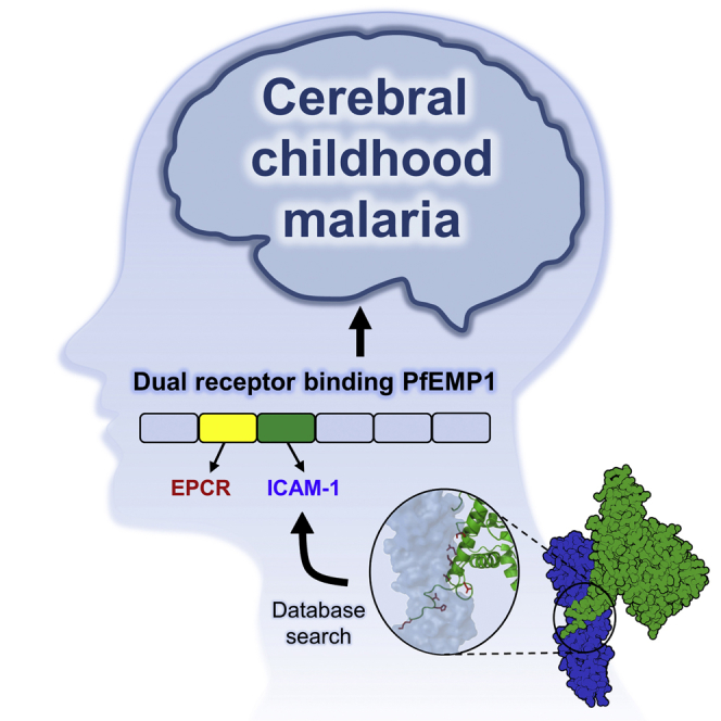
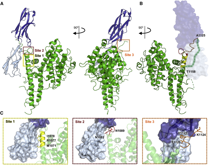
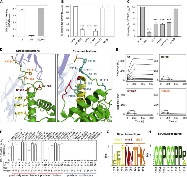
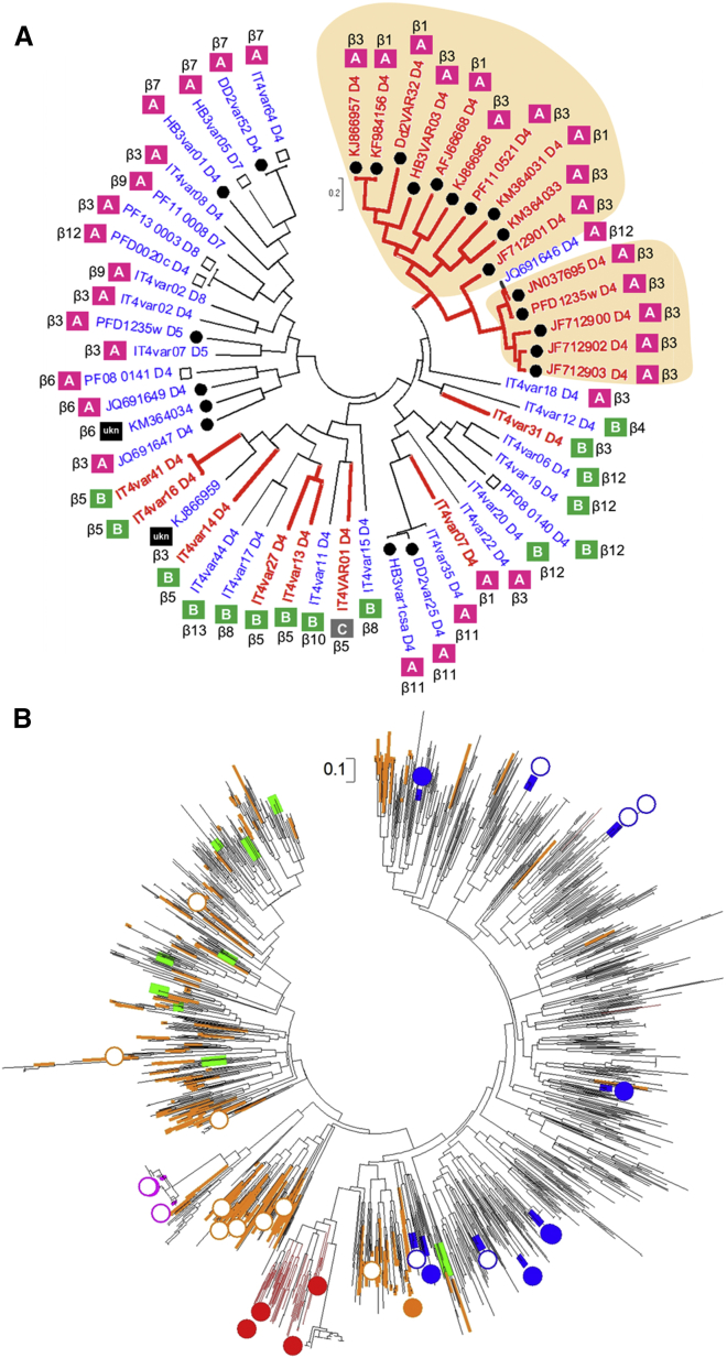
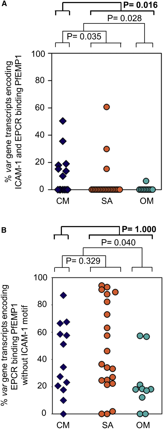
Cerebral malaria is a deadly outcome of infection by Plasmodium falciparum, occurring when parasite-infected erythrocytes accumulate in the brain. These erythrocytes display parasite proteins of the PfEMP1 family that bind various endothelial receptors. Despite the importance of cerebral malaria, a binding phenotype linked to its symptoms has not been identified. Here, we used structural biology to determine how a group of PfEMP1 proteins interacts with intercellular adhesion molecule 1 (ICAM-1), allowing us to predict binders from a specific sequence motif alone. Analysis of multiple Plasmodium falciparum genomes showed that ICAM-1-binding PfEMP1s also interact with endothelial protein C receptor (EPCR), allowing infected erythrocytes to synergistically bind both receptors. Expression of these PfEMP1s, predicted to bind both ICAM-1 and EPCR, is associated with increased risk of developing cerebral malaria. This study therefore reveals an important PfEMP1-binding phenotype that could be targeted as part of a strategy to prevent cerebral malaria.

Keywords: EPCR; ICAM-1; PfEMP1; Plasmodium falciparum; cerebral malaria.


Figures

None
Graphical abstract
Figure 1
Figure 1
The Structural Basis for ICAM-1 Binding by Group A PfEMP1s (A) Front and side views of the PF11_0521 DBLβ_D4 domain (green) bound to ICAM-1D1D2 (D1, light blue; D2, dark blue). Dashed boxes highlight three sites that make direct contact with ICAM-1. (B) Back view of PF11_0521 DBLβ_D4 domain (green) bound to ICAM-1D1D2. Dashed lines represent residues not visible in the electron density map with the disordered regions near site 3 (orange) of the ICAM-1-binding site highlighted. (C) Three distinct sites in the PF11_0521 DBLβ_D4 domain directly interact with ICAM-1 with residues that mediate this interaction indicated. See also Figures S1 and S2 and Tables S1 and S2.
Figure 2
Figure 2
Structure-Guided Identification of a Motif that Predicts ICAM-1 Binding (A) ICAM-1 binding (ELISA OD 490 nm; ± SD) of recombinant 3D7 PFD1235w DBLβ3_D4, PFD1235w DBLβ3_D5, and a chimeric DBLβ3_D5 containing the ICAM-1-binding motif region of DBLβ3_D4 (D5_motif). (B and C) Inhibition of 3D7 PFD1235W infected erythrocyte adhering to ICAM-1 under flow in the presence of anti-ICAM-1 Abs, affinity-purified anti-PFD1235w DBLβ3_D4 (anti-D4) IgG, anti-PFD1235w_motif IgG, anti-ITVAR13, and control IgG. IgG antibodies affinity purified from (B) rat anti-serum and (C) human serum. ± SD of a minimum of three independent experiments done in triplicate. Data were analyzed using one-way ANOVA. Asterisks indicate significance (p = 0.0004). (D) The three sites within the PF11_0521 DBLβ3_D4-binding site, indicating residues that directly contact ICAM-1 (site 1, yellow; site 2, red; site 3, orange). Structural residues important for positioning of interacting residues are highlighted in teal. (E) Surface plasmon resonance curves for injection of 2-fold dilution series of DBLβ wild-type and binding site mutants over ICAM-1D1D5. (F) ICAM-1 binding (ELISA OD 490 nm; ± SD three replicates) of 26 recombinant group A DBLβ domains (4 DBLβ1, 14 DBLβ3, 2 DBLβ6, 2 DBLβ7, 2 DBLβ11, 1 DBLβ12, and 1 DBLβ unknown sub-class), confirming prediction of binding domains. “DC” indicates in which domain cassette the domain is found. “ND” indicates that the DC type is unknown as only the DBLβ sequence is available. “No” indicates that the domain is not part of a known DC. Presence of DC13 prior to DBLβ is indicated. (G and H) Sequence logo showing conservation of (G) residues that contact ICAM-1 and (H) residues important for the unusual architecture of the ICAM-1-binding site, based on 145 DBLβ domains predicted to bind ICAM-1. Red triangles, residues critical for direct interaction with ICAM-1. See also Figures S3–S5 and Tables S2, S3, and S4.
Figure 3
Figure 3
Phylogeny of ICAM-1- and Non-binding DBLβ Domains (A) Maximum likelihood tree of 55 complete DBLβ domains. The tree is drawn to scale, with branch lengths measured in substitutions per site. Red, ICAM-1 binders; blue, ICAM-1 non-binders (Avril et al., 2016, Bengtsson et al., 2013, Janes et al., 2011, Lau et al., 2015). Filled circles, DBLβ domains tested in this study; open squares, DBLβ domains tested, but data not shown. UKN indicates unknown group ID. Shaded areas indicate DBLβ with the ICAM-1 motif. (B) Maximum likelihood tree of 1,823 complete DBLβ S3 sequences from the seven published genomes and 226 annotated Sanger genomes. Filled circles, DBLβ experimentally shown as ICAM-1 binders. Open circles, DBLβ experimentally shown as ICAM-1 non-binders. Colors indicate the CIDR domain N-terminal to DBLβ: red, EPCR binders with ICAM-1-binding motif; orange, EPCR binders with no ICAM-1-binding motif; green, non-EPCR binders (group A); blue, CD36 binders; magenta, VAR1. See also Figure S5 and Table S4.
Figure 4
Figure 4
The Conserved ICAM-1 Binding Site Is Recognized by Patient Plasma and Linked to CM (A) The inhibition of DBLβ domains binding to ICAM-1 by IgG from a plasma pool from Liberian P. falciparum-exposed adults purified on ICAM-1-binding DBLβ_D4 domain from Dd2var32, KM364031, and PFD1235w or on a closely related, but non-ICAM-1-binding DBLβ_D5 domain from PFD1235w. ICAM-1 inhibitory capacity is >74% (black), 51%–74% (dark gray), 21%–50% (light gray), and 0%–20% (white). (B) ICAM-1-binding inhibition by plasma with low (ELISA OD < 1) and high (ELISA OD > 1) anti-motif-DBLβ IgG in P. falciparum-exposed Tanzanian children (1–17 years) (Lusingu et al., 2004). Boxplot with median. Whiskers, 5% and 95% percentiles. (C) Transcript levels (in arbitrary transcription units, Tu) of var gene subtypes in Ghanaian, Tanzanian, and Beninese children hospitalized with malaria, shown with medians and 25% and 75% percentiles. See also Table S5.
Figure 5
Figure 5
PfEMP1 Can Simultaneously Bind to Both ICAM-1 and EPCR ICAM-1D1D5 was immobilized at fixed concentration, followed by injection of the respective head structure, (A) PF11_0521 DBLα1.7-CIDRα1.4-DBLβ3, (B) Dd2var32 DBLα1.7-CIDRα1.4-DBLβ3, and (C) PFD1235w CIDRα1.6-DBLβ3, also at fixed concentration. A 2-fold dilution series of EPCR was injected over the resulting ICAM-1::head structure complex. Arrows show start points of protein injection. The insets show data fitted to a one-site kinetic model for binding of EPCR to the respective ICAM-1::head structure complex. Data (black lines) are modeled to a one-site model (red lines). See also Figure S6 and Table S6.
Figure 6
Figure 6
Binding to ICAM-1 and EPCR Increases Adhesion of Infected Erythrocytes (A–F) Erythrocytes infected with (A and D) HB3VAR03 (predicted ICAM-1 and EPCR binder), (B and E) 3D7PFD1235w (predicted ICAM-1 and EPCR binder), and (C and F) IT4VAR13 (ICAM-1 and CD36 binder) binding to (A–C) receptor-coated microslides under flow conditions using recombinant ICAM-1, rEPCR, rICAM-1, and rEPCR; rCD36; or rICAM-1 and rCD36, and to (D–F) resting human brain microvascular endothelial cells (HBMEC). To demonstrate specific adhesion, channels coated with HBMEC were pre-incubated with anti-ICAM-1 (40 μg/mL), anti-EPCR (10 μg/mL), or anti-ICAM1 and EPCR combined (40 and 10 μg/mL, respectively). Flow experiments were done in parallel with the same conditions for immobilizing proteins or cell-coated chips. Values are bound infected erythrocyte per mm2 ± SEM. Data were also analyzed by two-way ANOVA with Tukey’s multiple comparison (Tables S7A and S7B). (G–I) Immunofluorescence images of erythrocytes infected with parasite strains (G) HB3VAR03, (H) 3D7PFD1235w, and (I) IT4VAR13. Complexes of ICAM-1 or EPCR were added to measure binding by immunofluorescence. Main panels show overlays of ICAM-1 (green), EPCR (red), and nuclear (blue) staining. Inserts show single channels. Scale bar, 2 μm. See also Figure S7 and Table S7.
Figure 7
Figure 7
Dual-Binding PfEMP1s Correlate with CM Percentages of specific var gene transcripts in infected erythrocytes from 45 hospitalized Tanzanian children. (A) Percentage of var gene transcripts encoding PfEMP1s predicted to bind both ICAM-1 and EPCR. (B) Percentage of transcripts that encode PfEMP1 predicted to bind EPCR and not containing the ICAM-1-binding motif. CM, cerebral malaria; SA, severe anemia; OM, children without the signs of severity found in the other groups. See also Table S5.

References

    1. Avril M., Tripathi A.K., Brazier A.J., Andisi C., Janes J.H., Soma V.L., Sullivan D.J., Jr., Bull P.C., Stins M.F., Smith J.D. A restricted subset of var genes mediates adherence of Plasmodium falciparum-infected erythrocytes to brain endothelial cells. Proc. Natl. Acad. Sci. USA. 2012;109:E1782–E1790. - PMC - PubMed
    1. Avril M., Bernabeu M., Benjamin M., Brazier A.J., Smith J.D. Interaction between endothelial protein C receptor and intercellular adhesion molecule 1 to mediate binding of Plasmodium falciparum-infected erythrocytes to endothelial cells. MBio. 2016;7 e00615-16. - PMC - PubMed
    1. Band G., Rockett K.A., Spencer C.C., Kwiatkowski D.P., Malaria Genomic Epidemiology Network A novel locus of resistance to severe malaria in a region of ancient balancing selection. Nature. 2015;526:253–257. - PMC - PubMed
    1. Bengtsson A., Joergensen L., Rask T.S., Olsen R.W., Andersen M.A., Turner L., Theander T.G., Hviid L., Higgins M.K., Craig A. A novel domain cassette identifies Plasmodium falciparum PfEMP1 proteins binding ICAM-1 and is a target of cross-reactive, adhesion-inhibitory antibodies. J. Immunol. 2013;190:240–249. - PMC - PubMed
    1. Bernabeu M., Danziger S.A., Avril M., Vaz M., Babar P.H., Brazier A.J., Herricks T., Maki J.N., Pereira L., Mascarenhas A. Severe adult malaria is associated with specific PfEMP1 adhesion types and high parasite biomass. Proc. Natl. Acad. Sci. USA. 2016;113:E3270–E3279. - PMC - PubMed

MeSH terms

Substances

LinkOut - more resources