Review
. 2017 Mar 8;93(5):995-1002.
doi: 10.1016/j.neuron.2017.02.013.

Synaptic Vesicle Clusters at Synapses: A Distinct Liquid Phase?

Affiliations
Review

Synaptic Vesicle Clusters at Synapses: A Distinct Liquid Phase?

Dragomir Milovanovic et al. Neuron. .

Abstract

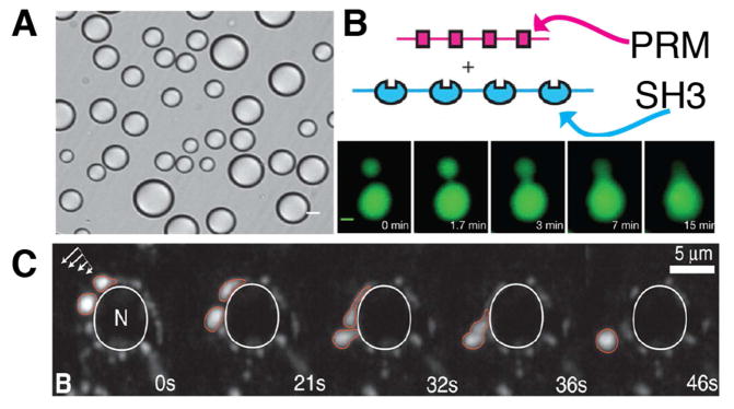
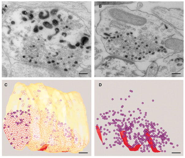
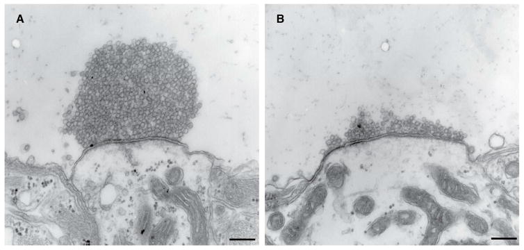
Phase separation in the cytoplasm is emerging as a major principle in intracellular organization. In this process, sets of macromolecules assemble themselves into liquid compartments that are distinct from the surrounding medium but are not delimited by membrane boundaries. Here, we discuss how phase separation, in which a component of one of the two phases is vesicles rather than macromolecules, could underlie the formation of synaptic vesicle (SV) clusters in proximity to presynaptic sites. The organization of SVs into a liquid phase could explain how SVs remain tightly clustered without being stably bound to a scaffold so that they can be efficiently recruited to release site by active zone components.

Keywords: exocytosis; intrinsically disordered region; liquid phase; phase separation; synapse; synapsin 1; synaptic vesicle clusters; synaptic vesicles.


Figures

Figure 1
Figure 1
Synaptic vesicles cluster in the presynaptic nerve terminal of a mouse brain; scale bar 200 nm (unpublished image courtesy of Dr. Yumei Wu, Yale School of Medicine).
Figure 2
Figure 2
Liquid-liquid phase separation underlies the formation of membrane-less organelles. (A) In vitro reconstitutions of liquid-liquid phase separation of two engineered proteins (repeats of SH3 and cognate proline-rich motifs, PRM) that coalesce into droplets, as visualized by differential interference contrast microscopy. (B) Time-lapse imaging of two protein droplets that merge into a single mass; green: Oregon-green labeled SH3 repeats. (A and B are modified from Li et al., 2012). (C) Under shear street (white arrows: direction of induced shear flow) P granules (marked in red), which are RNA-protein complexes, drip along the nuclear envelope (marked in white) (modified from Brangwynne et al., 2009).
Figure 3
Figure 3
Synaptic vesicles intermix within clusters and exclude other organelles. (A and B) Mouse neurons in primary cultures were stimulated for 90 sec with high [K+] in the presence of extracellular horseradish peroxidase (HRP) and fixed after 3 (A) and 30 (B) minutes following HRP washout. Note in (A) the exclusion of large endocytic intermediates from SV clusters, and in both (A and B) the random intermixing of unlabeled SVs with recycled, HRP reaction product-positive vesicles. Scale bar 200 nm (images from Wu et al., 2014). (C and D) 3D Reconstruction of a frog motor endplate stimulated at 30 Hz for 10 s in the presence of FM 1–43, then rested and fixed after 10 minutes. Synaptic vesicles positive for FM 1–43, as shown by photoconversion (purple) are randomly intermixed with unlabeled vesicles. Active zones are shown in red. Scale bar 200 nm (images from Rizzoli and Betz, 2004).
Figure 4
Figure 4
Antibody-mediated perturbation of synapsin abolishes synaptic vesicle cluster. (A and B) Synapses of the giant reticulospinal axon of the lamprey were microinjected with no antibody (A) or with anti-synapsin antibody (B), then stimulated at 1 Hz for 12 minutes, rested for 90 minutes and fixed. Scale bar 200 nm (image from Pieribone et al., 1995).

References

    1. Ackermann F, Waites CL, Garner CC. Presynaptic active zones in invertebrates and vertebrates. EMBO Rep. 2015;16:923–938. - PMC - PubMed
    1. Acuna C, Liu X, Südhof TC. How to Make an Active Zone: Unexpected Universal Functional Redundancy between RIMs and RIM-BPs. Neuron. 2016;91:792–807. - PubMed
    1. Anderson P, Kedersha N. Stress granules: the Tao of RNA triage. Trends in Biochem Sci. 2008;33:141–150. - PubMed
    1. Banani SF, Rice AM, Peeples WB, Lin Y, Jain S, Parker R, Rosen MK. Compositional Control of Phase-Separated Cellular Bodies. Cell. 2016;166:651–663. - PMC - PubMed
    1. Banjade S, Wu Q, Mittal A, Peeples WB, Pappu RV, Rosen MK. Conserved interdomain linker promotes phase separation of the multivalent adaptor protein Nck. Proc Nat Acad Sci USA. 2015;112:6426–35. - PMC - PubMed

LinkOut - more resources