. 2017 Apr 15;424(2):162-180.
doi: 10.1016/j.ydbio.2017.03.004. Epub 2017 Mar 7.

Satellite-like cells contribute to pax7-dependent skeletal muscle repair in adult zebrafish

Affiliations

Satellite-like cells contribute to pax7-dependent skeletal muscle repair in adult zebrafish

Michael A Berberoglu et al. Dev Biol. .

Abstract

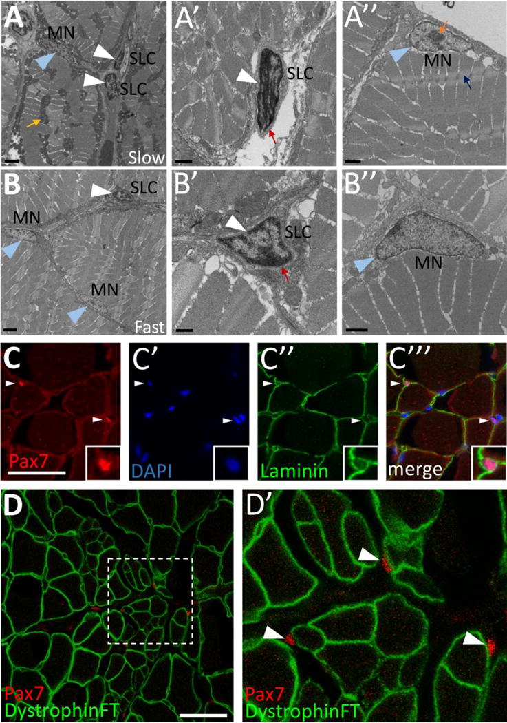
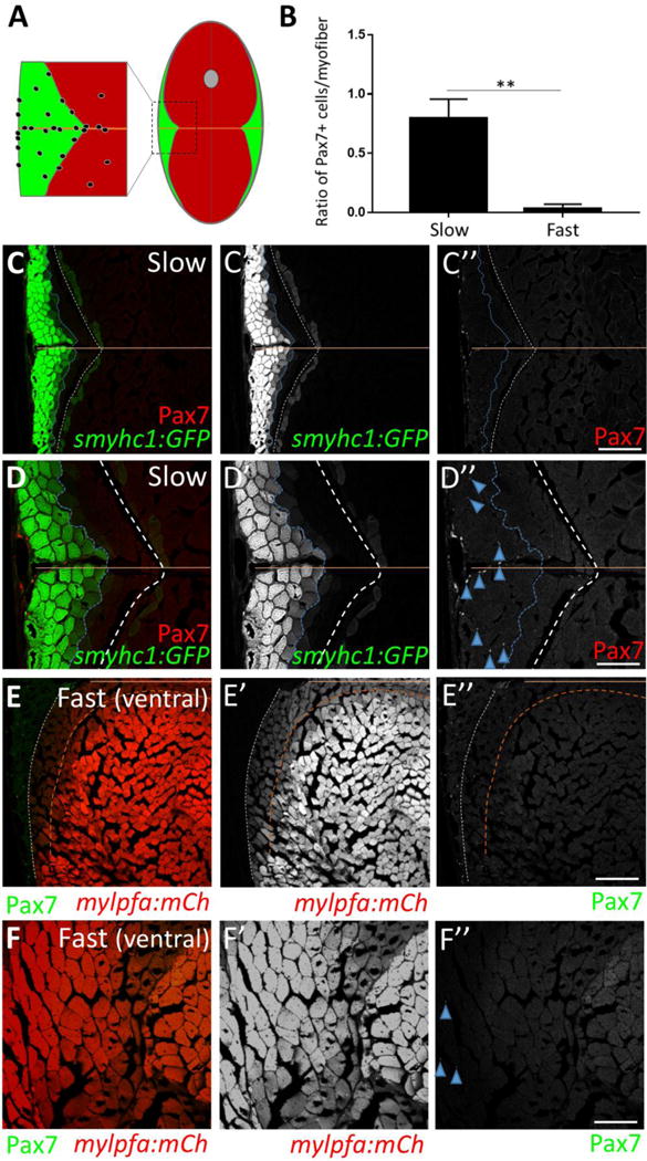
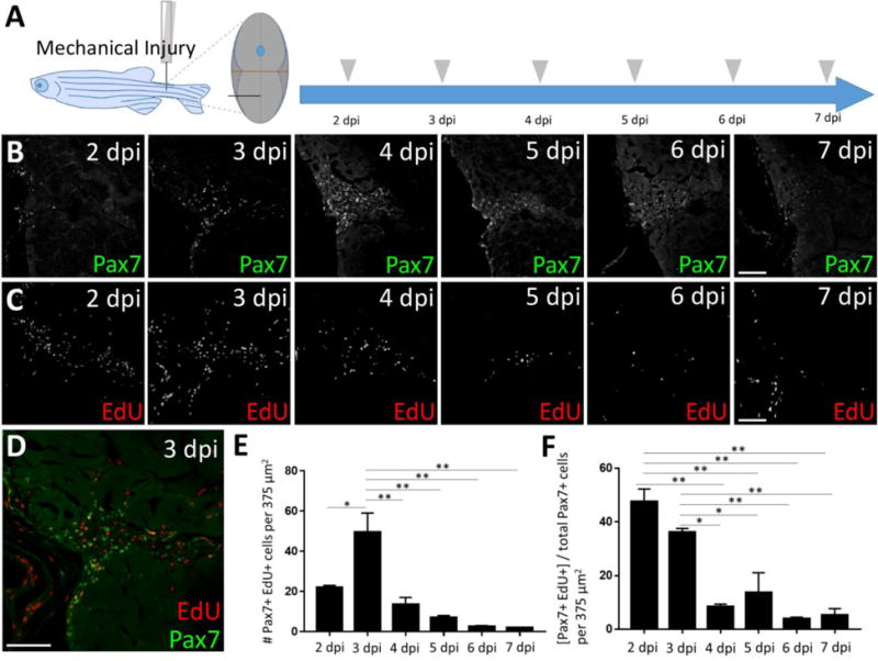
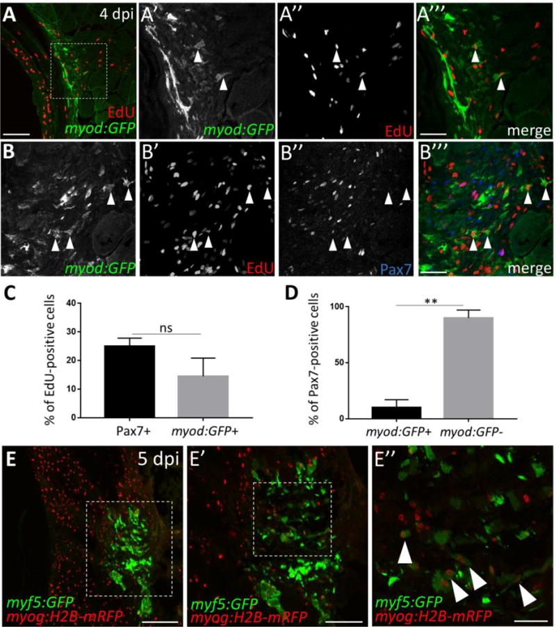
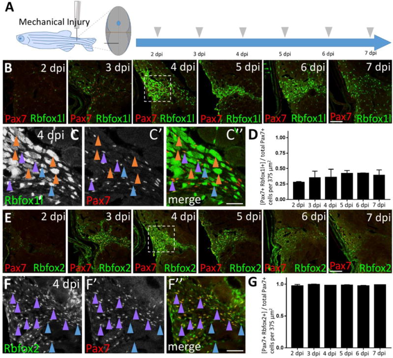
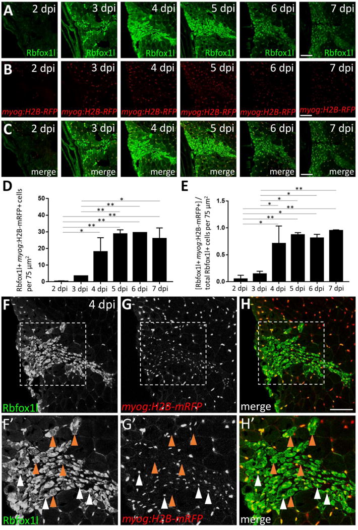
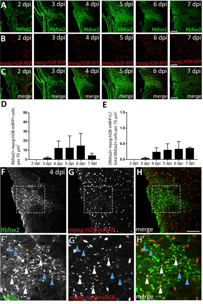
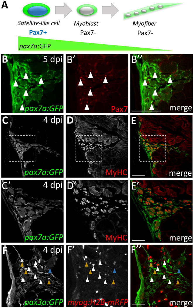
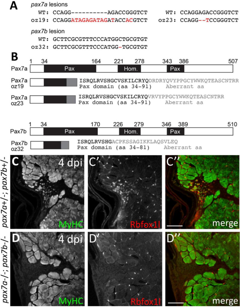
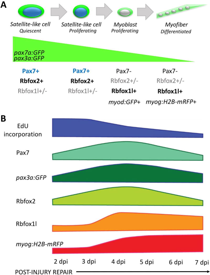
Satellite cells, also known as muscle stem cells, are responsible for skeletal muscle growth and repair in mammals. Pax7 and Pax3 transcription factors are established satellite cell markers required for muscle development and regeneration, and there is great interest in identifying additional factors that regulate satellite cell proliferation, differentiation, and/or skeletal muscle regeneration. Due to the powerful regenerative capacity of many zebrafish tissues, even in adults, we are exploring the regenerative potential of adult zebrafish skeletal muscle. Here, we show that adult zebrafish skeletal muscle contains cells similar to mammalian satellite cells. Adult zebrafish satellite-like cells have dense heterochromatin, express Pax7 and Pax3, proliferate in response to injury, and show peak myogenic responses 4-5 days post-injury (dpi). Furthermore, using a pax7a-driven GFP reporter, we present evidence implicating satellite-like cells as a possible source of new muscle. In lieu of central nucleation, which distinguishes regenerating myofibers in mammals, we describe several characteristics that robustly identify newly-forming myofibers from surrounding fibers in injured adult zebrafish muscle. These characteristics include partially overlapping expression in satellite-like cells and regenerating myofibers of two RNA-binding proteins Rbfox2 and Rbfoxl1, known to regulate embryonic muscle development and function. Finally, by analyzing pax7a; pax7b double mutant zebrafish, we show that Pax7 is required for adult skeletal muscle repair, as it is in the mouse.

Keywords: Muscle injury; Muscle stem cells; Myogenesis; Pax transcription factors; Rbfox RNA-binding proteins.


Figures

Figure 1
Figure 1. Cells with satellite cell features are present in adult zebrafish skeletal muscle
(A–B) Transmission electron microscopy (TEM) on adult zebrafish tail skeletal muscle from slow-twitch (A–A″) and fast-twitch (B–B″) muscle fiber domains. (A) TEM of slow muscle reveals mitochondrial-rich muscle fibers (mustard arrow points to a cluster of dark gray mitochondria), putative satellite-like cells (SLCs; white arrowheads), and myonuclei (MN; blue arrowhead points to a myonucleus). (A′) Higher magnification view of a SLC containing dense heterochromatin (white arrowhead) and surrounded by basal lamina (red arrow). (A″) A myonucleus (MN; blue arrowhead) located within the muscle fiber membrane. Myonuclei are characterized by a lack of dense heterochromatin and often by a prominent nucleolus (orange arrow). An adjacent myofibril is indicated by a blue arrow. (B) TEM of fast muscle reveals fewer mitochondria, a putative SLC (white arrowhead), as well as myonuclei (MN, blue arrowheads). (B′) Higher magnification view of a SLC containing dense heterochromatin (white arrowhead) and surrounded by basal lamina (red arrow). (B″) A myonucleus (MN; blue arrowhead) located within the muscle fiber membrane shows similar characteristics to slow muscle myonuclei. (CC′″) Pax7-positive cells (red; white arrowheads) are localized within the basal lamina of adult zebrafish skeletal muscle (anti-Laminin in green) (inset shows enlarged view of Pax7-positive cell). DAPI labeling confirms Pax7 localization in nuclei. (D–D′) Pax7-positive cells (red; arrowheads) are located outside of the muscle fiber membrane, marked by a Dystrophin FlipTrap transgenic line. (D′) Magnified view of boxed region in D. Scale bar in A, B is 2 μm, in A′, A″, B″ is 1 μm, in B′ is 500 nm, and in C and D is 30 μm.
Figure 2
Figure 2. Adult zebrafish satellite-like cells are concentrated predominantly in slow muscle
(A) Schematic of cross-section through adult zebrafish trunk musculature, depicting fast-twitch (red) and slow-twitch (green) muscle fiber domains and a concentration of SLCs (black dots) within the slow muscle domain. The horizontal myoseptum is indicated by a brown line; the gray circle at the dorsal midline depicts the spinal cord. (B) The number of Pax7-positive cells, normalized to total myofiber number, is significantly higher in slow versus fast muscle; Student’s t-test (p**<0.01). (C–C″) Pax7-positive cells (in red) are more common within slow muscle. Slow muscle fibers are marked with smyhc1-driven GFP (left of the dotted blue line). The boundary between slow and fast muscle fiber domains is marked by a dotted white line. The area between dotted blue and dotted white lines may represent muscle fibers with an intermediate identity. The horizontal myoseptum is indicated by a brown line. (D–D″) Higher magnification view of slow muscle. For quantification shown in B, Pax7-positive cells (D″; examples depicted by blue arrowheads) within the smyhc1:GFP-positive domain (left of the dotted blue line) were counted and normalized to the number of smyhc1:GFP-positive myofibers in the same area. (E–E″) Pax7-positive cells (in green) are rare within ventral fast muscle. Fast (ventral) muscle fibers are marked with mylpfa-driven mCherry (right of the dotted brown line). The area between dotted brown and white lines (weakly expressing mylpfa:mCherry) may represent intermediate muscle fiber identity. A dotted white line separates fast and slow muscle domains. (F–F″) Higher magnification image of fast muscle. For quantification shown in B, Pax7-positive cells (F″; blue arrowheads) within the mylpfa:mCherry-positive domain were counted and normalized to the number of mylpfa:mCherry-positive myofibers in the same area. Scale bar in C” and E” is 150 μm and in D” and F” is 75 μm.
Figure 3
Figure 3. Satellite-like cells in adult zebrafish skeletal muscle proliferate after mechanical injury
(A) Schematic of experimental procedure, in which mechanical injury is performed by a single needle-stick into tail skeletal muscle. The approximate position of needle-stick injury in the ventral myotome is indicated in cross-sectional view. EdU was administered (gray arrowhead) by intraperitoneal injection at each post-injury time-point, allowing for EdU incorporation into S-phase cells, and fish were sacrificed 4 hours later (acute EdU labeling). Tissue was collected at 2, 3, 4, 5, 6, and 7 dpi. (B) Pax7-positive cell numbers increase at the injury site and peak at 4 days post-injury (dpi). (C) Acute EdU labeling indicates robust proliferation at the injury site at 2 and 3 dpi that declines thereafter. (D) Overlap of Pax7 and EdU at 3 dpi indicates satellite-like cell proliferation post-injury. (E) Quantification of Pax7 and EdU double positive cells at each post-injury time-point reveals a peak number of proliferating satellite-like cells at 3 dpi in an 375 μm2 area encompassing the injury site; p*<0.05; p**<0.01. (F) Quantification of the percentage of Pax7-positive cells that are EdU-positive (out of total Pax7-positive cell population) at each post-injury time-point indicates that nearly 50% of Pax7-positive cells are proliferating at 2 dpi, and ~35% are proliferating at 3 dpi. The percentage of Pax7-positive cells that are EdU-positive at later post-injury time-points (4–7 dpi) is significantly less; p*<0.05; p**<0.01. One-way ANOVA followed by Tukey’s multiple comparisons test was performed for statistical analyses. Scale bar in all panels is 75 μm.
Figure 4
Figure 4. Myoblast-like cells, detected using myf5:GFP and myod:GFP transgene expression, are present post-injury in adult zebrafish skeletal muscle
(A) The myod:GFP transgenic line identifies myoblast-like cells at 4 dpi. (A′–A′″) Magnified view of boxed area in A, showing myod:GFP expression (A′), EdU incorporation after an acute EdU pulse (A”), and the overlay (A′″) at 4 dpi. Double-positive cells are indicated by arrowheads. (B–B′″) At 4 dpi, myod:GFP-expressing cells are largely Pax7-negative (examples indicated by arrowheads). (C) Percentage of total proliferating cells (i.e. those that incorporate EdU) at 4 dpi that are Pax7-positive (25%) versus myod:GFP-positive (15%). The percentage of proliferating cells that are Pax7-positive (25%) is similar to that shown in Fig. S3I and is slightly higher than the percentage of proliferating cells that are myod:GFP-positive (15%), although the difference is not statistically significant. (D) Percentage of Pax7-expressing cells at 4 dpi that are myod:GFP-positive (10%) versus myod:GFP-negative (90%), p**<0.01. (E) myf5:GFP-positive cells are present at the injury site at 5 dpi. (E′) Magnified view of boxed region in E. (E”) Magnified view of boxed region in E′ reveals myf5:GFP-positive cells that are myog:H2B-mRFP-positive (arrowheads). Scale bar in A and E′ is 75 μm, in E is 150 μm, and in A′–B′″ and E” is 30 μm.
Figure 5
Figure 5. Satellite-like cells express Rbfox1l and Rbfox2 RNA-binding proteins after mechanical injury of adult zebrafish skeletal muscle
(A) Schematic of experimental procedure, which is identical to that described Fig. 3A. (B) Rbfox1l (green) is robustly expressed beginning at 4 dpi, coinciding with the onset of new myofiber formation. Pax7 expression (overlaid in red) is the same as shown in Fig. 3C; the merge here shows overlap of Pax7 and Rbfox1l expression. The boxed region at 4 dpi is magnified in C–C”. (C–C”) Magnified view of boxed area in B, showing expression of Rbfox1l (C) and Pax7 (C′), and the overlay (C”) at 4 dpi. Examples of Pax7-positive, Rbfox1l-positive cells are indicated by purple arrowheads and of Pax7-negative, Rbfox1l-positive cells by blue arrowheads. Rbfox1l expression is observed in the cytoplasm of presumptive newly-forming myofibers (orange arrowheads indicate examples). Rbfox1l expression in uninjured areas surrounding the injury site appears less uniform in muscle fiber nuclei (compared to Fig. S6 D–D″) as laser intensity was reduced to prevent over-saturation of signals within the injury site. (D) Ratio of Pax7-positive cells that are Rbfox1l-positive at post-injury time-points 2–7 dpi. (E) Onset of Rbfox2 expression (green) at 3 dpi is earlier than Rbfox1l, and closely resembles post-injury Pax7 expression (overlaid in red). The sections are alternating with those in B. The boxed region at 4 dpi is magnified in F–F”. (F–F”) Magnified view of boxed area in C, showing expression of Rbfox2 (F) and Pax7 (F′), and the overlay (F”) at 4 dpi. Examples of Pax7-positive, Rbfox2-positive cells are indicated by purple arrowheads, and of Pax7-negative, Rbfox2-positive cells by blue arrowheads. (G) Ratio of Pax7-positive cells that are Rbfox2-positive at post-injury time-points 2–7 dpi. Scale bar in B and E is 75 μm, and in C–C” and F–F” is 30 μm.
Figure 6
Figure 6. Rbfox1l is expressed predominantly in the nucleus and cytoplasm of newly-forming myofibers during skeletal muscle repair
(A) Injury time-course experiment performed in the myog:H2B-mRFP transgenic line showing Rbfox1l expression at each post-injury time-point. (B) myog:H2B-mRFP expression is readily observed within the injury site by 4 dpi. (C) Overlay of Rbfox1l and myog:H2B-mRFP expression. (D) Total number of Rbfox1l, myog:H2B-mRFP double-positive cells at 2–7 dpi in a 75 μm2 area within the injury site (to exclude Rbfox1l-positive; myog:H2B-mRFP-positive myonuclei within surrounding uninjured fibers). (E) Ratio of Rbfox1l-positive cells that are myog:H2B-mRFP-positive at 2–7 dpi. One-way ANOVA followed by Tukey’s multiple comparisons test was performed for statistical analyses in D and E (p*<0.05; p**<0.01). (F–H) Higher magnification view of 4 dpi injury site shown in A–C. (F′–H′) Area shown is the boxed region in F–H. A majority of Rbfox1l-positive nuclei are also positive for myog:H2B-mRFP (examples indicated by white arrowheads), as expected from the large overlap of these two markers in differentiated muscle outside of the injury site. Rbfox1l expression in uninjured areas surrounding the injury site appears less uniform in muscle fiber nuclei in this figure (compared to Fig. S6 D–D″) as laser intensity was reduced to prevent over-saturation of signals within the injury site. Some newly-forming myofibers that express Rbfox1l in the nucleus and/or cytoplasm are myog:H2B-mRFP-negative (examples indicated by brown arrowheads). Scale bar in all panels is 75 μm.
Figure 7
Figure 7. A subset of Rbfox2-expressing cells are myog:H2B-mRFP-positive during skeletal muscle repair
(A) Injury time-course experiment performed in myog:H2B-mRFP transgenic line showing Rbfox2 expression (on alternating sections with those described in Fig. 6). (B) As shown in Fig. 6B, myog:H2B-mRFP expression is readily observed within the injury site by 4 dpi. (C) Overlay of Rbfox2 and myog:H2B-mRFP expression. (D) Total number of Rbfox2, myog:H2B-mRFP double-positive cells at 2–7 dpi. (E) Ratio of Rbfox2-positive cells that are myog:H2B-mRFP-positive at 2–7 dpi. One-way ANOVA followed by Tukey’s multiple comparisons test was performed for all of the above statistical analyses; differences among time-points are not statistically significant. (F–H) Higher magnification view of 4 dpi injury site shown in A–C. (F′–H′) Area shown is the boxed region in F–H that highlights examples of Rbfox2-expressing cells that are myog:H2B-mRFP-positive (white arrowheads) and myog:H2B-mRFP-negative (blue arrowheads). Scale bar in all panels is 75 μm.
Figure 8
Figure 8. GFP perdurance in the pax7a:GFP transgenic line implicates satellite-like cells as a source of new muscle fibers during injury-induced repair
(A) Schematic diagram depicting Pax7 protein and pax7a:GFP transgene expression in satellite-like cells and myofibers. pax7a:GFP-positive; Pax7-positive cells represent satellite-like cells. Cells that show pax7a:GFP expression within the cytoplasm but are Pax7-negative are likely differentiating or differentiated cells in which pax7a-driven GFP perdures. (B–B″) At 5 dpi, pax7a:GFP and Pax7 double-positive satellite-like cells are present at the injury site (arrowheads); however, GFP expression is also detected in Pax7-negative presumptive newly-forming myofibers. (C–E) At 4 dpi, many cells expressing MyHC (A4.1025), a differentiation marker, also express pax7a:GFP. (C′–E′) Magnified view of boxed region in C–E highlights the overlap of pax7a:GFP and MyHC expression. (F–F″) pax3a:GFP and myog:H2B-mRFP transgene expression overlap in presumptive newly-forming myofibers at 4 dpi (white arrowheads), but pax3a:GFP-positive, myog:H2B-mRFP-negative are also present (mustard arrowheads). A rare presumptive newly-forming myofiber with a centrally-located nucleus is indicated by a blue arrowhead. Scale bar in B–E is 75 μm, in C′, D′, E′ and F–F” is 30 μm.
Figure 9
Figure 9. Pax7 function is required for skeletal muscle repair in adult zebrafish
(A) DNA lesions for pax7a alleles (oz19, oz23) and the pax7b (oz32) allele. Mutant allele sequence is shown below the wild-type sequence. Red letters and dashes indicate the lesion as well as single nucleotide changes near the CRISPR-induced frameshifting mutation. (B) CRISPR-induced frameshifting mutations in exon 2 of pax7a and pax7b cause truncations in Pax7a and Pax7b proteins, respectively, that eliminate much of the first conserved paired domain (Pax) and the entire homeodomain (Hom) and second Pax domain. The pax7aoz19 and pax7aoz23 alleles encode the first 91 of 507 amino acids of the Pax7a protein, as well as an additional 26 and 22 aberrant amino acids, respectively. The pax7boz32 allele encodes the first 81 of 510 amino acids of the Pax7b protein, as well as an additional 19 aberrant amino acids. (C–C”) pax7a+/−; pax7b+/− doubly heterozygous sibling controls show normal muscle repair at 4 dpi, indicated by expression of newly-forming myofiber markers, MyHC (A4.1025) and Rbfox1l. (D–D”) In contrast, pax7a−/−; pax7b−/− double homozygous mutants are severely deficient in newly-forming myofibers at 4 dpi, indicated by the near absence of MyHC (A4.1025) and Rbfox1l expression within the injury site. The pax7aoz23 and pax7boz32 alleles were used in these experiments. Scale bar in all panels is 75 μm.
Figure 10
Figure 10. Model depicting changes in gene expression as satellite-like cells differentiate into new muscle fibers during adult zebrafish skeletal muscle repair
(A) Adult zebrafish satellite-like cells express Pax7 and Rbfox2, and in transgenic pax7a:GFP and pax3a:GFP adults, express GFP. A subset (~37%) express Rbfox1l. During injury-induced repair, some Pax7-expressing satellite cells-like become activated; initially, activated cells proliferate, then down-regulate Pax7 and activate differentiation markers. Although Pax7 protein is largely absent in myoblasts, pax7a-driven and pax3a-driven GFP perdures in pax7a:GFP and pax3a:GFP transgenic lines (green wedge). As cells adopt more differentiated fates, Rbfox1l becomes prominently expressed, as do transgenic markers of myoblasts (myod:GFP) and myofibers (myog:H2B-mRFP). (B) Diagram depicting marker expression and proliferation status at the population level at the injury site at 2–7 dpi. A large number of proliferating (EdU+) cells are present at the injury site at 2 and 3 dpi, some of which are Pax7-positive. This early proliferation of Pax7-positive satellite-like cells appears to lead to an increase in Pax7-positive cell numbers by 4 dpi. pax3a:GFP shows a similar wave of expression that peaks at the time when Pax7-positive cell numbers are highest; however, pax3a:GFP expression is also observed at later post-injury time-points (as is pax7a:GFP expression). Rbfox2 expression largely mirrors that of Pax7, although Rbfox2 is expressed in some myonuclei of new myofibers that no longer express Pax7. Rbfox1l is robustly expressed at the onset of new myofiber formation (3–4 dpi), with peak expression at 4 dpi in both nucleus and cytoplasm of newly-forming fibers. The number of myog:H2B-mRFP-positive cells gradually increase post-injury as myogenic differentiation and new myofiber formation progresses.

References

    1. Asakura A, Komaki M, Rudnicki M. Muscle satellite cells are multipotential stem cells that exhibit myogenic, osteogenic, and adipogenic differentiation. Differentiation. 2001;68:245–253. - PubMed
    1. Baraniak AP, Chen JR, Garcia-Blanco MA. Fox-2 mediates epithelial cell-specific fibroblast growth factor receptor 2 exon choice. Mol Cell Biol. 2006;26:1209–1222. - PMC - PubMed
    1. Bassett DI, Currie PD. The zebrafish as a model for muscular dystrophy and congenital myopathy. Hum Mol Genet. 2003;12:R265–270. - PubMed
    1. Baxendale S, Chen CK, Tang H, Davison C, Hateren LV, Croning MD, Humphray SJ, Hubbard SJ, Ingham PW. Expression screening and annotation of a zebrafish myoblast cDNA library. Gene Expr Patterns. 2009;9:73–82. - PubMed
    1. Bentzinger CF, Wang YX, Rudnicki MA. Building muscle: molecular regulation of myogenesis. Cold Spring Harb Perspect Biol. 2012;4(2) doi:pii: a008342. Review. - PMC - PubMed

MeSH terms

LinkOut - more resources